. 2017 Dec 14;552(7684):258-262.
doi: 10.1038/nature24998. Epub 2017 Nov 13.

A non-canonical Notch complex regulates adherens junctions and vascular barrier function

Affiliations

A non-canonical Notch complex regulates adherens junctions and vascular barrier function

William J Polacheck et al. Nature. .

Abstract

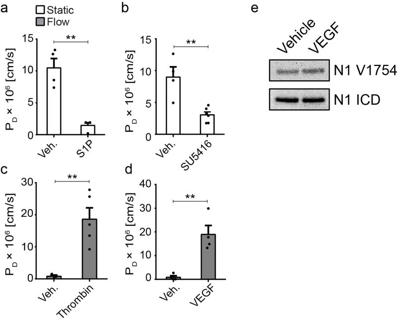
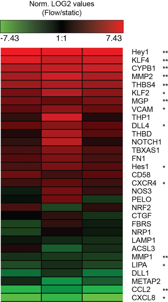
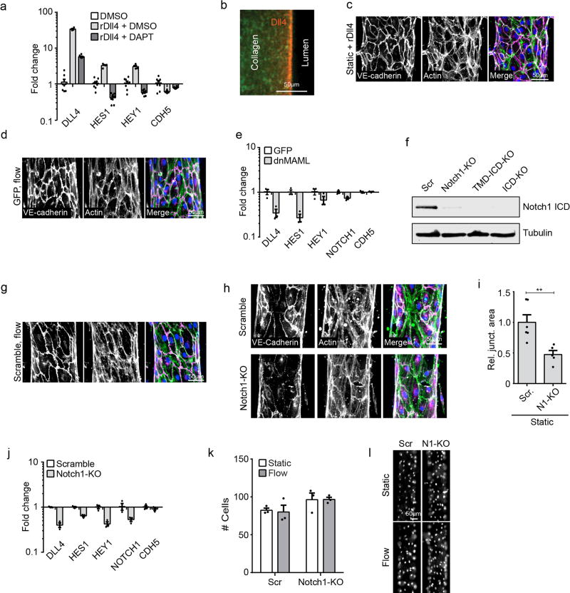
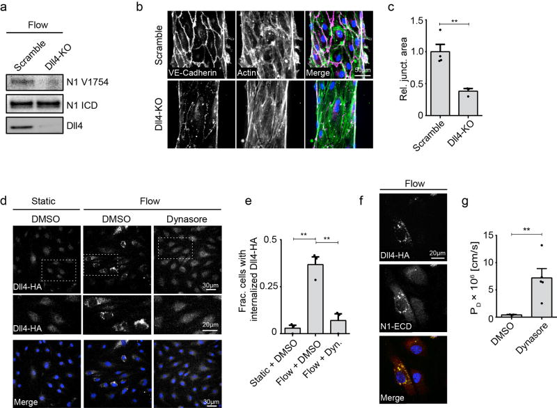
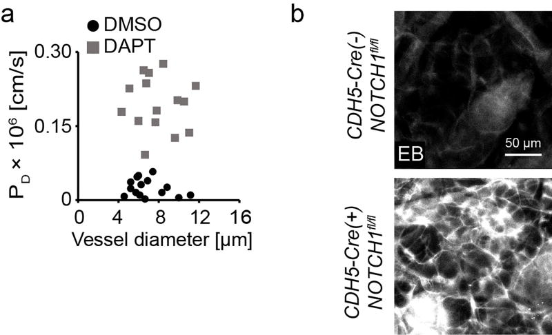
The vascular barrier that separates blood from tissues is actively regulated by the endothelium and is essential for transport, inflammation, and haemostasis. Haemodynamic shear stress plays a critical role in maintaining endothelial barrier function, but how this occurs remains unknown. Here we use an engineered organotypic model of perfused microvessels to show that activation of the transmembrane receptor NOTCH1 directly regulates vascular barrier function through a non-canonical, transcription-independent signalling mechanism that drives assembly of adherens junctions, and confirm these findings in mouse models. Shear stress triggers DLL4-dependent proteolytic activation of NOTCH1 to expose the transmembrane domain of NOTCH1. This domain mediates establishment of the endothelial barrier; expression of the transmembrane domain of NOTCH1 is sufficient to rescue defects in barrier function induced by knockout of NOTCH1. The transmembrane domain restores barrier function by catalysing the formation of a receptor complex in the plasma membrane consisting of vascular endothelial cadherin, the transmembrane protein tyrosine phosphatase LAR, and the RAC1 guanidine-exchange factor TRIO. This complex activates RAC1 to drive assembly of adherens junctions and establish barrier function. Canonical transcriptional signalling via Notch is highly conserved in metazoans and is required for many processes in vascular development, including arterial-venous differentiation, angiogenesis and remodelling. We establish the existence of a non-canonical cortical NOTCH1 signalling pathway that regulates vascular barrier function, and thus provide a mechanism by which a single receptor might link transcriptional programs with adhesive and cytoskeletal remodelling.


Figures

Extended Data Figure 1
Extended Data Figure 1. Known permeability modulating agents regulate barrier function in hEMVs
PD measured in hEMVs under static or flow conditions treated with a, 500nM S1P for 1hr, b, 10 µM SU415 (VEGFR2 inhibitor) overnight, c, 1 U/mL thrombin for 15min, or d, 40ng/mL VEGF for 1hr. e, western blots of cleaved Notch1 ICD (V1754) from lysates of ECs treated with vehicle or 40ng/mL VEGF for 1hr. For all plots, mean ± s.e.m., n≥3 hEMVs, **p<0.01. Exact p and n values available in Figure Source Data, western blot representative of two independent experiments.
Extended Data Figure 2
Extended Data Figure 2. Flow activates expression of mechanotransduction associated gene networks
Relative gene expression measured by qPCR. –ddCT of GOI under flow normalized to static control is plotted as a heatmap. n=3 flow/static qPCR analyses from distinct hEMV sets, each column representative of an independent experiment. *p<0.05, **p<0.01. Exact p and n values available in Figure Source Data.
Extended Data Figure 3
Extended Data Figure 3. Non-transcriptional Notch1 signaling regulates vascular barrier function
a, Gene expression of Notch1 target genes HES1 and HEY1, the Notch1 ligand DLL4, and VE-cadherin (CDH5) measured via qPCR in ECs treated with DAPT or DMSO load control on Dll4-coated and control tissue culture plastic substrates. b, Fluorescent micrograph of rDll4-coated device prior to cell seeding (green – Alexa Fluor 488 Collagen I, red – immunostain of Dll4). c, Fluorescent micrograph of ECs in hEMVs coated with rDll4 prior to cell seeding. d, Micrographs of GFP-infection control cells under flow. e, Gene expression of HES1, HEY1, DLL4, NOTCH1, and VE-cadherin (CDH5) measured via qPCR in ECs expressing dnMAML or infection control (GFP). f, Western blot validation of Notch1 CRISPR lines: Scramble, Notch1-KO, TMD+ICD-KO, and ICD-KO. g, Fluorescent micrographs of CRISPR/Cas9 scramble control cells under flow. h, Fluorescent micrographs of Scramble and Notch1-KO hEMVs under static conditions immunostained for VE-cadherin and labeled with phalloidin (actin). i, Quantification of junctional area measured from VE-cadherin immunostained micrographs. j, Gene expression measured via qPCR in Notch1-KO cells and scramble control cells. k, Quantification of cell number in f.o.v at 10x magnification of Scramble or Notch1-KO hEMV under static and flow conditions. l, Micrographs of nuclei as visualized by DAPI in Scramble or Notch1-KO hEMVs. For all plots, mean ± s.e.m., n≥3 hEMVs, **p<0.01. Exact p and n values available in Figure Source Data, images representative of at least three independent experiments.
Extended Data Figure 4
Extended Data Figure 4. Notch1 is activated in response to shear stress by endocytosis of Dll4
a. ICD cleavage as measured by western blot with an antibody specific to cleaved ICD (N1 V1754) in Scramble and Dll4-KO ECs under flow. b, Fluorescent micrographs of Scramble and Dll4-KO hEMVs under flow conditions immunostained for VE-cadherin and labeled with phalloidin (actin). c, Quantification of junctional area measured from VE-cadherin immunostained micrographs. d, Immunofluorescent micrographs of recombinant Dll4-HA expressing ECs under static + DMSO, flow + DMSO, and flow + Dynasore conditions stained for HA (Dll4-HA) and DAPI. e, Quantification of internalized Dll4-HA in ECs under static + DMSO, flow + DMSO, and flow + Dynasore conditions. Cells with internalized Dll4-HA counted as those with >1 AlexaFluor-488 positive puncta. f, Immunofluorescent micrograph of a Dll4-HA expressing EC under flow stained for HA (Dll4-HA), Notch1 ECD, and DAPI. g, Diffusive permeability of 70kDa dextran measured in cells treated with Dynasore hydrate or DMSO load control and exposed to flow overnight. For all plots, mean ± s.e.m., n≥3 hEMVs, **p<0.01. Exact p and n values available in Figure Source Data, images representative of at least two independent experiments.
Extended Data Figure 5
Extended Data Figure 5. DAPT and Notch1 depletion modulate vascular permeability in vivo
a, Diffusive permeability of dermal vasculature as a function of vessel diameter in the mouse dermal vasculature in mice after 1hr of IV-administered DMSO or DAPT (n = 15 vessels across 3 mice per condition). b, High magnification whole-mount micrographs of Evans Blue in the mouse dermal vasculature. Fluorescent images representative of three independent experiments.
Extended Data Figure 6
Extended Data Figure 6. Notch1 regulates junctional stability through association with VE-cadherin
a, Timelapse images of cells expressing VE-cadherin-mApple in the presence of DAPT or DMSO load control demonstrate that AJs disassemble after 30min of exposure to DAPT, leading to macroscopic intercellular gaps (red arrows). b, Fluorescent micrographs of Notch1 KO cells expressing TMD+ICD-mApple or TMD+ICD V1754G-mApple immunostained for cleaved Notch1 ICD V1754 and DAPI. c, Fluorescent micrographs of TMD-mApple expressed in VE-cadherin knockout or scramble control ECs and immunostained for VE-cadherin. d, Western blot for Notch1 ICD and VE-cadherin in Notch1-KO and VE-cadherin KO ECs. e, Western blot validation of Notch1 rescue constructs: mApple, TMD-mApple, ICD+TMD-mApple, and ICD+TMD V1754G-mApple. f, Immunoprecipitation of VE-cadherin from hMVEC-D cells treated with DMSO or DAPT. Co-immunoprecipitation of mechanosensory complex components was assessed by immunoblotting for Notch1 ICD, Trio, and LAR. g. Western blot of VE-cadherin immunoprecipitations from Notch1 KO cells expressing Notch1-TMD truncation constructs (6, 8, 12 amino acids from the N-terminus) fused to mApple. h, Western blot of VE-cadherin immunoprecipitations from Notch1 KO cells expressing single and dual point mutation Notch1-TMD constructs (within the transmembrane segment of Notch1 TMD) fused to mApple. All images representative of at least three independent experiments.
Extended Data Figure 7
Extended Data Figure 7. Dll4 and the Notch1 mechanosensory complex are critical for increased Rac1 activity in response to shear stress
a, Active Rac1 was isolated with GST-PBD from hMVEC-D cell lysates treated with DMSO and DAPT (20 µM). b, Quantification of western blot band intensity demonstrates a decrease (~30%) in Rac1 activity with DAPT treatment. c, Active Rac1 was isolated using GST-PBD from hMVEC-D cell lysates from static or shear flow conditions. d, Active Rac1 was isolated using GST-PBD from Dll4-KO cell lysates under flow conditions. e, Active Rac1 was isolated using GST-PBD from Notch1 KO, LAR KO, and Trio KO cell lysates under flow conditions. Mean ± s.e.m., n=3 independent lysates, **p<0.01. Exact p and n values available in Figure Source Data, all images representative of at least three independent experiments.
Extended Data Figure 8
Extended Data Figure 8. Notch1 regulates VE-cadherin interacting proteins to form the Notch1 mechanosensory complex
a, Immunoprecipitation of VE-cadherin from scramble and Notch1-KO cells. Co-immunoprecipitation of candidate Notch1-dependent, VE-cadherin effectors was assessed by immunoblotting for VE-PTP, VEGFR2, and LAR (85 kDa P-subunit). b, Immunoprecipitation of the Rac1 GEF Trio from scramble, Notch1-KO, and LAR-KO cells. Immunoblotting for VE-cadherin was used to assess impaired Trio-VE-cadherin co-immunoprecipitation upon depletion of Notch1 or LAR. c, Western blots of VE-cadherin immunoprecipitated from the lysates of lungs from CDH5-Cre(+);NOTCH1fl/fl and control CDH5-Cre(+);NOTCH1fl/fl mice and immunoblotted for LAR. d, Western blots of Trio immunoprecipitated from the lysates of lungs from CDH5-Cre(+);NOTCH1fl/fl and control CDH5-Cre(+);NOTCH1fl/fl mice and immunoblotted for VE-cadherin. e, Western blot of proximal interacting proteins extracted with streptavidin from hMVEC-D cells expressing BirA-HA (BioID) or VE-cadherin-BirA-HA (VE-BioID) that were treated with DMSO, Dll4, or DAPT, immunoblotted for HA and Notch1 ICD. f, Western blot of proximal interacting proteins extracted with streptavidin in hMVEC-D cells expressing VE-cadherin-BirA-HA (VE-BioID) that were treated with DMSO, Dll4, or DAPT, immunoblotted for Trio and LAR. All images representative of at least two independent experiments.
Extended Data Figure 9
Extended Data Figure 9. The Notch1 mechanosensory complex stabilizes cell-cell junctions through activation of Rac1
Flow induces endocytosis of Dll4, triggering the activation and cleavage of Notch1 ICD and ECD, which allows the N1-TMD domain to scaffold the adaptor protein LAR with VE-cadherin and recruit the Rac1 GEF Trio to AJs. The resulting complex activates Rac1, elaborates cortical actin, and stabilizes cell-cell junctions to establish barrier function.
Extended Data Figure 10
Extended Data Figure 10. Supplemental methods
a, Schematic and summary of methods for quantifying vascular permeability in vivo with intravital microscopy. b, Intensity as a function of distance along lines connecting the nucleus centroids of neighboring cells. Blue circles are local maxima used to count the number of stress fibers per unit length, and the shaded region is the area under the peak corresponding to cortical actin and is normalized by the total area under the curve for quantification. Graph representative of three independent experiments.
Figure 1
Figure 1. Notch1 regulates shear stress-induced vascular barrier function
a, Organotypic microfluidic devices of human engineered microvessels (hEMVs) consisting of human ECs (red) in physiologic ECM (green), enabling vessel perfusion at a defined luminal shear stress (inlet and outlet in blue). Inset: 3D reconstruction of hEMVs (red - 70kDa dextran, white – VE-cadherin, green – collagen I). b, Real-time assessment of vascular barrier function in hEMVs cultured statically or under flow (heatmap of fluorescent intensity of 70 kDa dextran, imaging plane indicated by dashed line in a). c, Quantification of the diffusive permeability (PD) of 70 kDa dextran across EC barrier as a function of endothelial wall shear stress. d, Relative gene expression for ECs cultured statically or under flow was quantified with qPCR, and Notch target genes regulated by flow are indicated (each column representative of an independent experiment, full gene panel in Extended Data Fig. 2). e, ICD cleavage in static and flow EC lysates as measured by western blot with an antibody specific to cleaved ICD (N1 V1754). f, PD measured in hEMVs under static or flow conditions in the presence of Notch inhibitor (DAPT) or rDll4-coated collagen (rDll4). g, Fluorescent micrographs of hEMVs immunostained for VE-cadherin (magenta) and labeled with phalloidin (actin - green) and DAPI (nucleus – blue). h, Quantification of junctional area measured from VE-cadherin immunostained micrographs. i, PD for hEMVs cultured statically, under flow, or in the presence of rDll4-coated collagen with ECs expressing dnMAML, GFP infection control, or with N1-KO, Dll4-KO, or scramble control ECs. j, Fluorescent micrographs of actin and VE-cadherin for hEMVs under flow with ECs expressing dnMAML or with N1-KO ECs. k, Quantification of junctional area measured from VE-cadherin immunostained micrographs (micrographs of GFP and scramble controls in Extended Data Fig. 3). For (a-k), n≥3 independent hEMVs, mean ± s.e.m. l, Fluorescence intensity heatmaps of Evan’s blue (EB) dye in the mouse dermal vasculature 30min and 60min after IV co-injection of EB and DAPT or DMSO vehicle control. m, Quantification of PD for EB diffusion into the dermal interstitial space (n = 15 vessels across 3 mice/condition). n, Color image of lungs harvested from mice sacrificed 30 min after IV injection of EB. o, Vascular permeability was quantified by eluting and measuring the concentration of EB in lungs relative to the blood EB concentration, (n=4 age, sex-matched littermates, color coded, two-way ANOVA). p, Whole-mount lung vasculature immunostains showing leaked EB and DAPI. *p<0.05, **p<0.01; exact p and n values available in Figure Source Data, all images representative of at least three independent experiments.
Figure 2
Figure 2. The Notch1 transmembrane domain mediates barrier function through interaction with VE-cadherin
a, A library of endogenous Notch1 truncation mutants and over-expression rescue constructs were generated to examine the key functional domains of Notch1 that regulate barrier function. b, PD for ECs with CRISPR/Cas9-mediated endogenous truncation of Notch1 ICD (ICD-KO) or truncation of the TMD and ICD (TMD-ICD-KO) cultured statically, under flow, or in the presence of rDll4-coated collagen. c, Fluorescent micrographs of VE-cadherin and actin for ICD-KO and TMD-ICD-KO ECs under flow conditions. d, Quantification of junctional area measured from VE-cadherin immunostained micrographs. e, PD for N1-KO ECs expressing TMD-ICD-mApple, TMD-ICD V1754G-mApple, TMD-mApple, or mApple infection control cultured statically, under flow, or in the presence of rDll4-coated collagen. f, Fluorescent micrographs of VE-cadherin (magenta), actin (green), and DAPI (blue) in static N1-KO cells expressing TMD-mApple or mApple infection control. g, Quantification of junctional area measured from VE-cadherin immunostained micrographs. h, PD for static N1-KO cells expressing TMD-ICD-mApple or ICD-mApple exposed to DAPT or DMSO load control. i, Immunofluorescent images of Notch1-KO cells expressing either mApple or TMD-mApple, co-stained for VE-cadherin. Co-localization of Notch1 TMD and VE-cadherin (red arrow) is lost at free edges (blue arrow). j, Immunoprecipitation of VE-cadherin and N-Cadherin from Notch1-KO cells expressing either mApple or TMD-mApple. Immunoblotting with a RFP antibody identified co-immunoprecipiting TMD-mApple. For all plots, mean ± s.e.m., n≥3 independent hEMVs, *p<0.05, **p<0.01. Exact p and n values available in Figure Source Data, all images representative of at least three independent experiments.
Figure 3
Figure 3. Notch1 assembles a mechanosensory junctional complex involving LAR, Trio, and Rac
a, Fluorescent micrographs of phalloidin stained ECs. b, Intensity of cortical actin at cell-cell junctions and the number of stress fibers per micron quantified from phalloidin stained ECs (n=3 hEMVs). c, Active Rac1 was isolated from scramble and Notch1-KO cell lysates using a recombinant p21-binding domain of Pak1 (GST-PBD). d, Quantification of western blot band intensity revealed a decrease in Rac1 (~50%) after knockout of Notch1 (n=3 independent lysates). e, Active Rac1 was isolated using GST-PBD from Notch1-KO cell lysates expressing mApple or TMD-mApple. f, Quantification of western blot band intensity revealed an increase in Rac1 (~45%) with expression of TMD-mApple (n=3 independent lysates). g, PD of Notch1-KO ECs expressing TMD-mApple or mApple control in the presence of 50 µM NSC 23766, a Rac1 inhibitor, or vehicle control. h, Immunoprecipitation of VE-cadherin and the Rac1 GEF Trio from ECs cultured statically or under flow. Co-immunoprecipitation of mechanosensory complex proteins was assessed by immunoblotting for Notch1-ECD, Notch1-ICD, LAR (85 kDa P-subunit), and VE-cadherin. i, Immunoprecipitation of VE-cadherin or Trio from scramble and Notch1-KO ECS cultured under flow. Co-immunoprecipitation of mechanosensory complex constituents was assessed by immunoblotting for VE-cadherin, LAR, and Trio. j, PD of LAR-KO and Trio-KO ECs cultured under static or flow conditions or in the presence of rDll4-coated collagen. k, Micrographs of Trio-KO and LAR-KO cells under flow conditions (VE-cadherin – magenta, actin – green, DAPI – blue). l, Quantification of junctional area measured from VE-cadherin immunostained micrographs. m, Immunoprecipitation of VE-cadherin and Trio from Notch1-KO cells expressing TMD-mApple or mApple. Immunoblotting with LAR, VE-cadherin, and RFP antibodies was used to assess co-immunoprecipitation of mechanosensory complex upon expression of TMD. n. PD for scramble, N1KO, LAR-KO, and Trio-KO cells cultured under static conditions expressing TMD-mApple or mApple infection control. For (g,j,n), n≥3 hEMVs. All plots mean ± s.e.m., *p<0.05, **p<0.01. Exact p and n values available in Figure Source Data, all images representative of at least three independent experiments.

Comment in

References

    1. Mehta D, Malik AB. Signaling mechanisms regulating endothelial permeability. Physiol. Rev. 2006;86:279–367. - PubMed
    1. Chiu J-J, Chien S. Effects of dsisturbed flow on vascular endothelium : pathophysiological basis and clinical perspectives. Physiol Rev. 2011;91:327–387. - PMC - PubMed
    1. Lawson ND, et al. Notch signaling is required for arterial-venous differentiation during embryonic vascular development. 2001;3683:3675–3683. - PubMed
    1. Hellström M, et al. Dll4 signalling through Notch1 regulates formation of tip cells during angiogenesis. Nature. 2007;445:776–780. - PubMed
    1. Krebs LT, et al. Notch signaling is essential for vascular morphogenesis in mice. Genes Dev. 2000;14:1343–1352. - PMC - PubMed

Publication types

MeSH terms