. 2017 Nov 21;12(11):e0188520.
doi: 10.1371/journal.pone.0188520. eCollection 2017.

Fasciola spp: Mapping of the MF6 epitope and antigenic analysis of the MF6p/HDM family of heme-binding proteins

Affiliations

Fasciola spp: Mapping of the MF6 epitope and antigenic analysis of the MF6p/HDM family of heme-binding proteins

Victoria Martínez-Sernández et al. PLoS One. .

Abstract

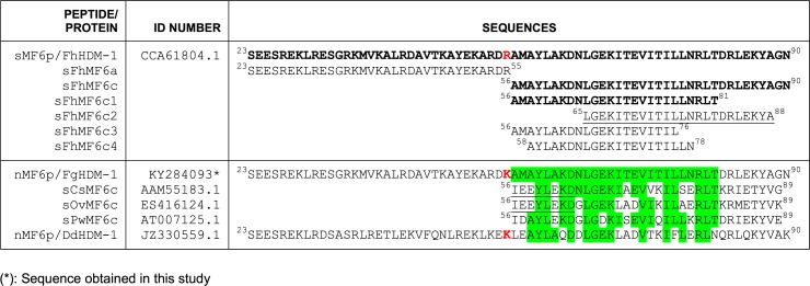
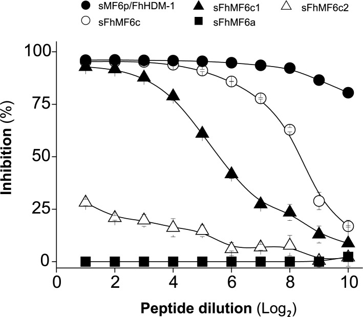
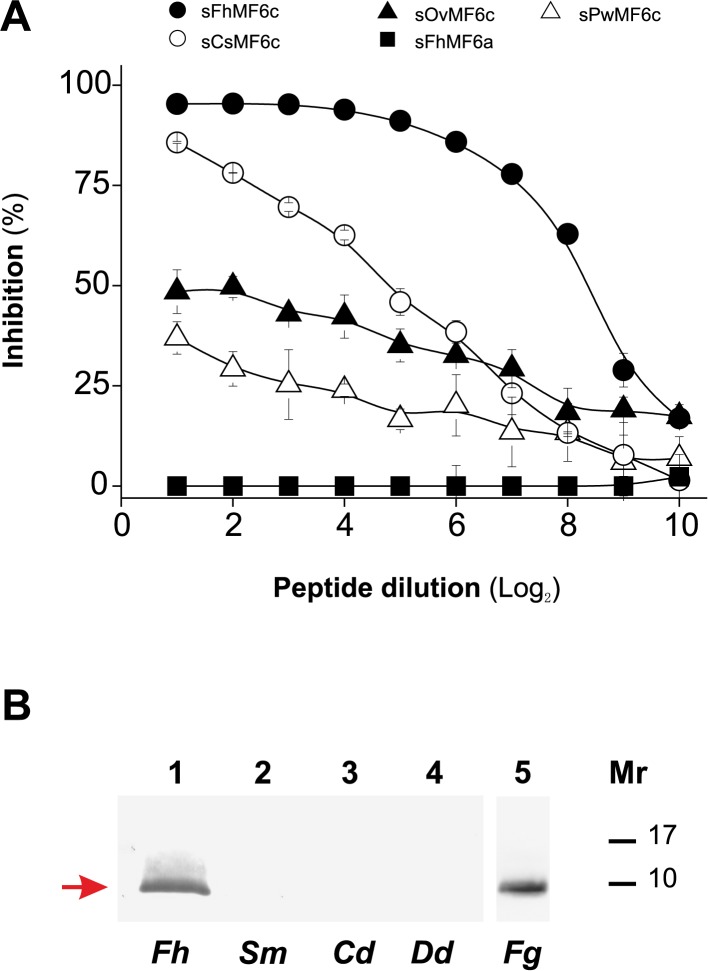
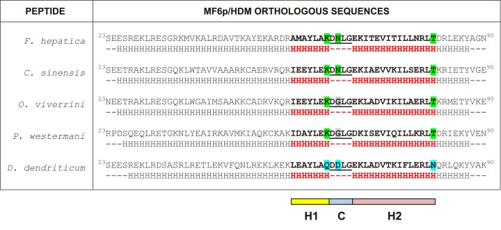
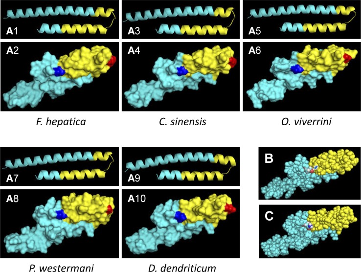
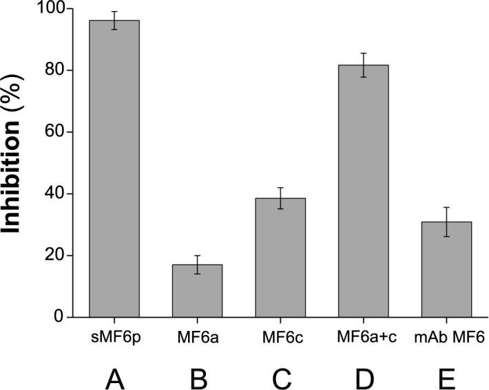
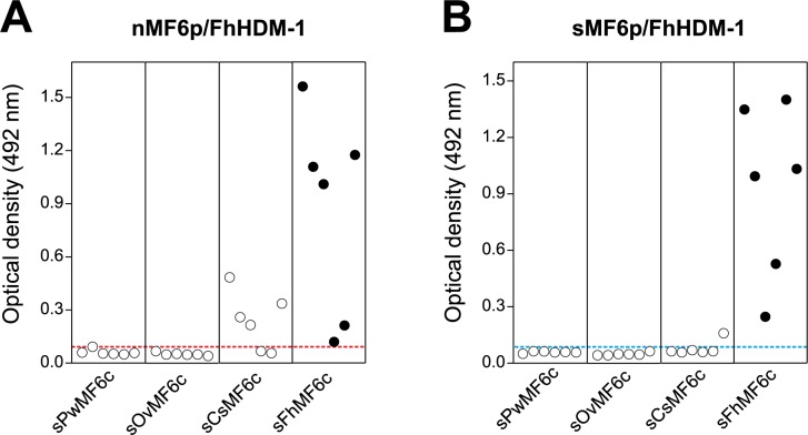
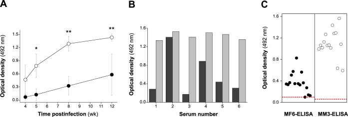
MF6p/FhHDM-1 is a small cationic heme-binding protein which is recognized by the monoclonal antibody (mAb) MF6, and abundantly present in parenchymal cells and secreted antigens of Fasciola hepatica. Orthologs of this protein (MF6p/HDMs) also exist in other causal agents of important foodborne trematodiasis, such as Clonorchis sinensis, Opisthorchis viverrini and Paragonimus westermani. Considering that MF6p/FhHDM-1 is relevant for heme homeostasis in Fasciola and was reported to have immunomodulatory properties, this protein is expected to be a useful target for vaccination. Thus, in this study we mapped the epitope recognized by mAb MF6 and evaluated its antigenicity in sheep. The sequence of the MF6p/FhHDM-1 ortholog from F. gigantica (MF6p/FgHDM-1) was also reported. By means of ELISA inhibitions with overlapping synthetic peptides, we determined that the epitope recognized by mAb MF6 is located within the C-terminal moiety of MF6p/FhHDM-1, which is the most conserved region of MF6p/HDMs. By immunoblotting analysis of parasite extracts and ELISA inhibitions with synthetic peptides we also determined that mAb MF6 reacted with the same intensity with F. hepatica and F. gigantica, and in decreasing order of intensity with C. sinensis, O.viverrini and P. westermani orthologs. On the contrary, mAb MF6 showed no reactivity against Dicrocoelium dendriticum and Schistosoma mansoni. The study of the recognition of peptides covering different regions of MF6p/FhHDM-1 by sera from immunized sheep revealed that the C-terminal moiety is the most antigenic, thus being of potential interest for vaccination. We also demonstrated that the production of antibodies to MF6p/FhHDM-1 in sheep infected by F. hepatica occurs relatively early and follows the same pattern as those produced against L-cathepsins.


Conflict of interest statement

Competing Interests: The authors have declared that no competing interests exist.

Figures

Fig 1
Fig 1. Alignment of F. hepatica MF6p/FhHDM-1 protein with whole mature sequences and truncated fragments of protein orthologs from other trematodes with sequence homology.
Aligned sequences from F. hepatica (MF6p/FhHDM-1), F. gigantica (MF6p/FgHDM-1), D. dendriticum (MF6p/DdHDM-1), F. hepatica truncated peptides (sFhMF6a, sFhMF6c, sFhMF6c1-4), and truncated peptides corresponding to the C-terminal region from C. sinensis (sCsMF6c), O. viverrini (sOvMF6c) and P. westermani (sPwMF6c) are shown. Synthetic or native (from whole parasite extracts) peptide/proteins used in the study are indicated by adding, respectively, an “s” or an “n” before the corresponding name. The sequences of MF6p/FhHDM-1 recognized by mAb MF6 are highlighted in bold. Amino acid residues in common with the MF6 epitope (residues 56–81) are shown in green. The unique amino acid difference between the F. hepatica and F. gigantica protein is indicated in red color. The truncated fragment corresponding to the heme-binding region of MF6p/FhHDM-1 is underlined.
Fig 2
Fig 2. Determination of the region of MF6p/FhHDM-1 recognized by mAb MF6 in competitive ELISA.
ELISA plates coated with sMF6p/FhHDM-1 were incubated with mAb MF6 (diluted 1/200,000) previously incubated with twofold dilutions of the synthetic peptides sMF6p/FhHDM-1 (control for maximal inhibition; filled circles), sFhMF6c (residues 56A-90N; unfilled circles), sFhMF6c1 (residues 56A-81T; filled triangles), sFhMF6c2 (residues 65L-88A; unfilled triangles), and sFhMF6a (residues 23S-55R; filled squares). Peptides sFhMF6c3 and sFhMF6c4 produced the same negative result as sFhMF6a, but for the sake of simplification were not represented here. Data are expressed as percentage inhibition of mAb MF6 by each peptide and are the mean values ± SD of duplicate wells.
Fig 3
Fig 3. Analysis of the potential cross-reactivity of mAb MF6 with MF6p/FhHDM-1 orthologs from other trematodes.
A) Competitive ELISA with synthetic peptides corresponding to the C-terminal region of MF6p/FhHDM-1 orthologs. ELISA plates coated with sMF6p/FhHDM-1 were incubated with mAb MF6 (diluted 1/200,000) previously incubated with twofold dilutions of sFhMF6c (residues 56A-90N; control for maximal inhibition; filled circles) from F. hepatica, sCsMF6c (residues 56I-89G; unfilled circles) from C. sinensis, sOvMF6c (residues 56I-89K; filled triangles) from O. viverrini, sPwMF6c (residues 56I-89E; unfilled triangles) from P. westermani, and sFhMF6a (residues 23S-55R; filled squares) as a negative control. Data are expressed as percentage inhibition of mAb MF6 by each peptide and are the mean values ± SD of duplicate wells. B) Immunoblotting analysis showing the recognition by mAb MF6 of several natural extracts from different trematodes. Lane 1: Fh (F. hepatica); lane 2: Sm (S. mansoni); lane 3: Cd (C. daubneyi); lane 4: Dd (D. dendriticum); lane 5: Fg (F. gigantica).
Fig 4
Fig 4. Comparison of the predicted secondary structure of MF6p/FhHDM-1 orthologs from different trematodes.
Complete sequences of the mature MF6p/HDM proteins from F. hepatica, C. sinensis, O. viverrini, P. westermani and D. dendriticum (residues 23–90). The α-helix (H) and coil regions (C) predicted by J-Pred 4 bioinformatics tool are also represented. The regions H1, C and H2 correspond to the α-helix/coil/α-helix region covering the MF6 epitope. The residue substitutions corresponding to the putative relevant positions 62K, 64N and 81T of MF6p/FhHDM-1 for MF6 binding are shown in different colors.
Fig 5
Fig 5. Three-dimensional (3D) structure predictions of MF6p/FhHDM-1 orthologs from different trematodes.
A) Predicted PDB structures for MF6p/FhHDM-1 orthologs from F. hepatica, C. sinensis, O. viverrini, P. westermani and D. dendriticum using the Quark Online ab initio protein folding and protein structure prediction bioinformatics tool. For better visualization, each 3D structure was represented as a cartoon (odd numbers) or as a surface (even numbers). The overall surface structures are shown in cyan, the area corresponding to the MF6-recognized epitope is represented in yellow and the positions corresponding to 64N and 81T in the sequence of F. hepatica, assumed to be highly relevant for MF6 epitope conformation, are shown in red and blue, respectively. B) Theoretical 3D structure of the MF6p/FhHDM-1 mature protein (represented as balls) showing the overall structure of the MF6-recognized epitope (shaded in yellow) and the position of the N-terminal 81T residue (colored atoms; red = oxygen; white = hydrogen; cyan = carbon). The arrow shows the position of the hydroxyl group of the 81T residue. C) Theoretical 3D structure of the MF6p/FhHDM-1 mature protein ortholog from D. dendriticum (represented as balls) highlighting the equivalent region to the MF6-recognized epitope (shaded in yellow) and the position of the N-terminal 81N residue in substitution of 81T in F. hepatica (colored atoms; red = oxygen, see arrow; white = hydrogen; cyan = carbon). All 3D representations of the PDB models obtained with the Quark Online bioinformatics tool were elaborated using the PyMOL Molecular Graphics System.
Fig 6
Fig 6. Analysis of the antigenicity of the MF6p/FhHDM-1 protein in sheep.
The antibody response from a group of 6 sheep immunized with nMF6p/FhHDM-1 in Quil-A was tested in a competitive ELISA. The experiment was carried out with ELISA plates coated with sMF6p/FhHDM-1 and each serum (diluted at an optimal dilution between 1/400 and 1/6,000) was incubated in duplicate in the absence (control for maximal signal) or in the presence of 5 μM of sMF6p/FhHDM-1 (control for maximal inhibition), sFhMF6a, sFhMF6c, or sFhMF6a plus sFhMF6c. The data shown also include the inhibition achieved with sheep sera in the presence of a large excess of mAb MF6, in order to measure the antigenicity attributable to the MF6 epitope. Data are expressed as percentage inhibition of sheep sera by the protein/peptides or mAb MF6 and are the mean values ± SD of all sheep. The mean OD for the adjusted sera without inhibitor was OD = 0.69 ± 0.07. Abbreviations: sMF6p = sMF6p/FhHDM-1; MF6a = sFhMF6a; MF6c = sFhMF6c; MF6a+c = sFhMF6a plus sFhMF6c. The Tukey-Kramer multiple comparison test revealed statistical differences between all groups (p<0.05 for groups C and D; p<0.01 for the remaining groups).
Fig 7
Fig 7. Analysis of the cross-reactivity of the F. hepatica MF6p/FhHDM-1 protein with orthologs from other trematodes.
Serum samples from sheep immunized with either native (A) or synthetic (B) MF6p/FhHDM-1 were tested in indirect ELISA with plates coated with the peptides sFhMF6c (F. hepatica), sCsMF6c (C. sinensis), sOvMF6c (O. viverrini) or sPwMF6c (P. westermani). Each point in the figure represents the mean OD obtained for one individual serum after subtracting the NSB value (OD = 0.05 ± 0.01). The dotted lined represents a theoretical cut-off value of OD = 0.1. All sera from each category were tested simultaneously by duplicate in the same plate. The intra-assay variation coefficients obtained for each serum did not exceed 7% of the mean OD.
Fig 8
Fig 8. Comparative study of the antibody response obtained in sheep infected with F. hepatica against nMF6p/FhHDM-1 and L-cathepsins in capture ELISAs.
A) Mean antibody response to nMF6p/FhHDM-1 (filled circles) and to L-cathepsins (unfilled circles) of infected sheep (n = 6) along the experimental infection with F. hepatica. Vertical bars represent the SD of the mean for each value. The Tukey-Kramer multiple comparison test was used to compare the antibody responses of sheep measured by MF6-ELISA and MM3-ELISA. (*): significant differences at p <0.01. (**): significant differences at p <0.001. B) Antibody response against nMF6p/FhHDM-1 and L-cathepsins in individual sheep experimentally infected with F. hepatica measured at week 12 p.i. C) Individual antibody response of naturally-infected sheep to nMF6p/FhHDM-1 (filled circles) and to L-cathepsins (unfilled circles). Each point in the figure represents the mean OD obtained for each individual serum after subtracting the corresponding NSB value (OD = 0.06 ± 0.01). Horizontal dotted lines show the cut-off values for MF6-ELISA (theoretical OD = 0.1) and MM3-SERO ELISA (OD = 0.074) [11]. The Mann-Whitney test revealed statistical differences between both groups (p <0.0001). Both ELISAs were previously calibrated using serial dilutions of F. hepatica ESAs and FITC-labeled mAb MF6 or mAb MM3 and HRP-conjugated anti-FITC antibodies as detection system. All sera from each category were tested simultaneously by duplicate in the same plate. The intra-assay variation coefficients obtained for each serum in MF6-ELISA and MM3-ELISA did not exceed 7% of the mean OD.

Similar articles

Cited by

References

    1. Keiser J, Utzinger J. Emerging foodborne trematodiasis. Emerg Infect Dis. 2005;11:1507–1514. doi: 10.3201/eid1110.050614 - DOI - PMC - PubMed
    1. Mas-Coma S, Bargues MD, Valero MA. Fascioliasis and other plant-borne trematode zoonoses. Int J Parasitol. 2005;35:1255–1278. doi: 10.1016/j.ijpara.2005.07.010 - DOI - PubMed
    1. Mas-Coma S, Valero MA, Bargues MD. Fascioliasis. Adv Exp Med Biol. 2014;766:77–114. doi: 10.1007/978-1-4939-0915-5_4 - DOI - PubMed
    1. Valero MA, Bargues MD, Khoubbane M, Artigas P, Quesada C, Berinde L, et al. Higher physiopathogenicity by Fasciola gigantica than by the genetically close F. hepatica: experimental long-term follow-up of biochemical markers. Trans R Soc Trop Med Hyg. 2016;110:55–66. doi: 10.1093/trstmh/trv110 - DOI - PubMed
    1. Charlier J, Duchateau L, Claerebout E, Williams D, Vercruysse J. Associations between anti-Fasciola hepatica antibody levels in bulk-tank milk samples and production parameters in dairy herds. Prev Vet Med. 2007;78:57–66. doi: 10.1016/j.prevetmed.2006.09.010 - DOI - PubMed

MeSH terms