. 2019 Jan 1;29(1):54-69.
doi: 10.1093/cercor/bhx303.

Astrocyte-Specific Deletion of Sox2 Promotes Functional Recovery After Traumatic Brain Injury

Affiliations

Astrocyte-Specific Deletion of Sox2 Promotes Functional Recovery After Traumatic Brain Injury

Chunhai Chen et al. Cereb Cortex. .

Abstract

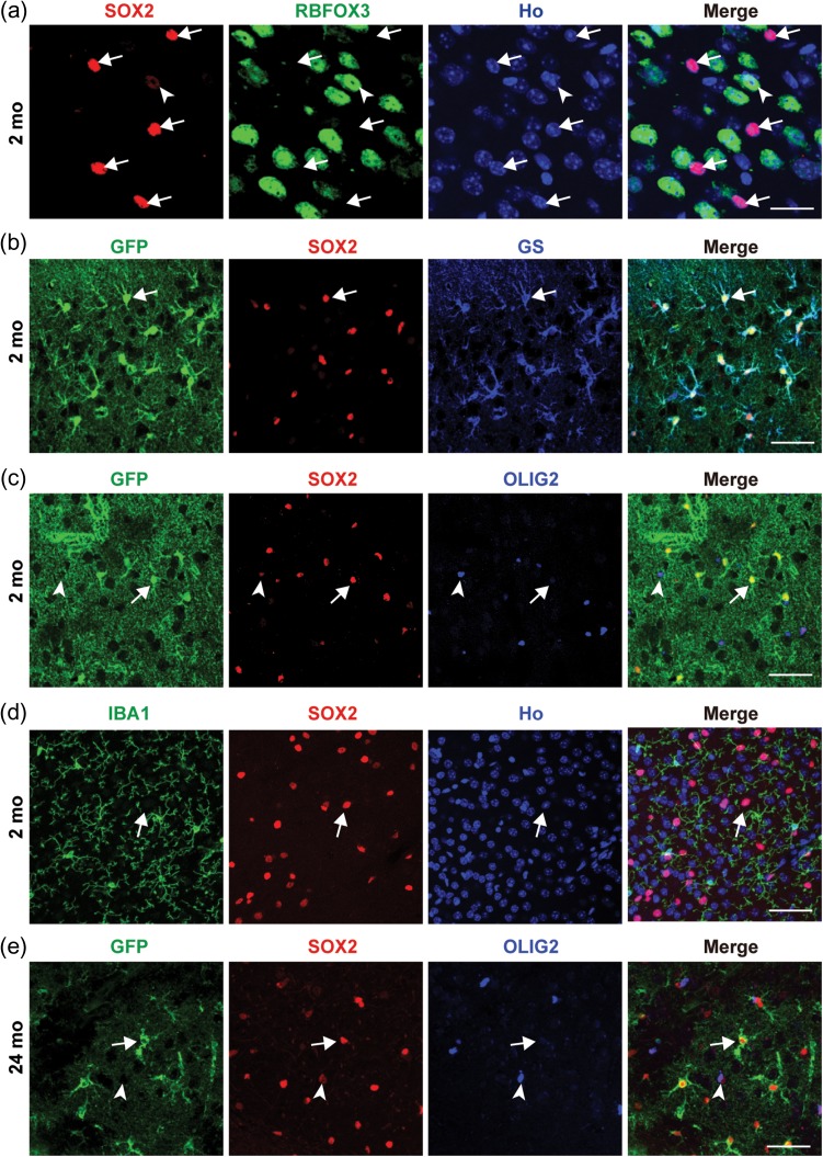
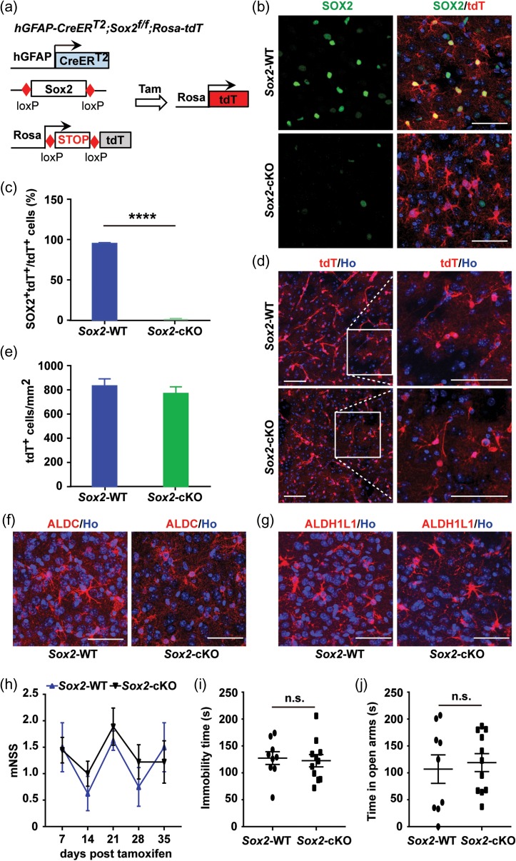
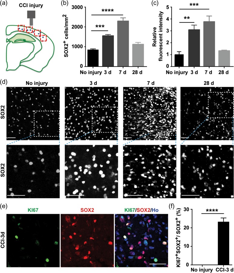
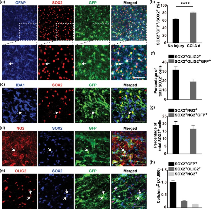
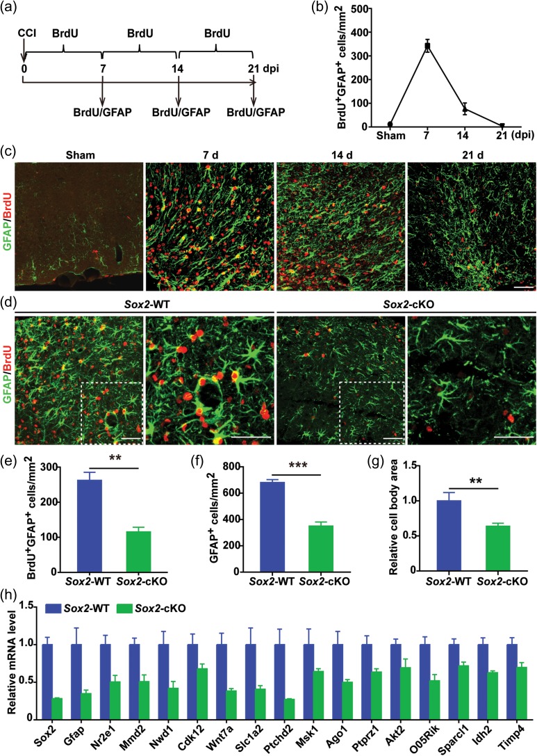
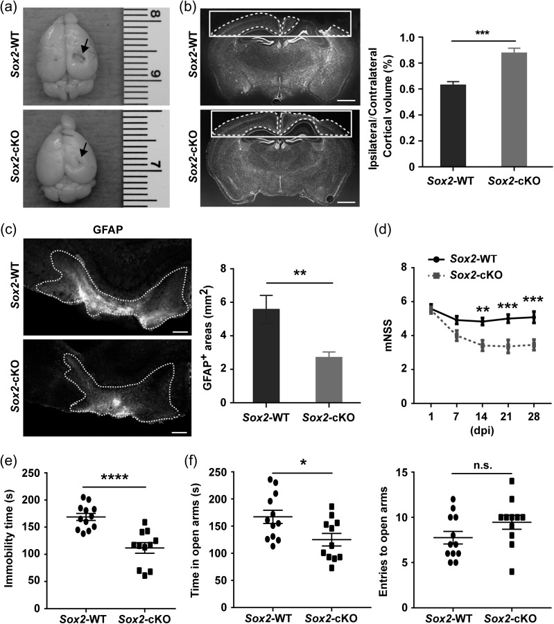
Injury to the adult brain induces activation of local astrocytes, which serves as a compensatory response that modulates tissue damage and recovery. However, the mechanism governing astrocyte activation during brain injury remains largely unknown. Here we provide in vivo evidence that SOX2, a transcription factor critical for stem cells and brain development, is also required for injury-induced activation of adult cortical astrocytes. Genome-wide chromatin immunoprecipitation-seq analysis of mouse cortical tissues reveals that SOX2 binds to regulatory regions of genes associated with signaling pathways that control glial cell activation, such as Nr2e1, Mmd2, Wnt7a, and Akt2. Astrocyte-specific deletion of Sox2 in adult mice greatly diminishes glial response to controlled cortical impact injury and, most unexpectedly, dampens injury-induced cortical loss and benefits behavioral recovery of mice after injury. Together, these results uncover an essential role of SOX2 in somatic cells under pathological conditions and indicate that SOX2-dependent astrocyte activation could be targeted for functional recovery after traumatic brain injury.


Figures

Figure 1.
Figure 1.
SOX2 expression in adult mouse cortex. (a) Rare and weak SOX2 expression in cortical neurons (indicated by an arrowhead). Non-neuronal expression is generally much stronger (indicated by arrows). (b) SOX2 expression in cortical astrocytes of Aldh1l1-EGFP mice. Astrocytes were identified by GS and the reporter GFP. Arrows show a representative GFP+GS+SOX2+ cell. mo, month. (c) SOX2 expression in cortical OLIG2+ oligodendrocytes or oligodendrocyte precursors of Aldh1l1-EGFP mice. Arrows show a representative SOX2highOLIG2GFP+ cell, whereas arrowheads show a representative SOX2lowOLIG2+GFP cell. SOX2 expression is always weaker in OLIG2+ cells. (d) SOX2 is not expressed in IBA1+ microglia. A representative SOX2+IBA1 cell is indicated by an arrow. (e) SOX2 expression in cortical GFP+ astrocytes (indicated by an arrow, SOX2highOLIG2GFP+) or OLIG2+ cells (indicated by an arrowhead, SOX2lowOLIG2+GFP) of 24-month-old Aldh1l1-EGFP mice. Scale bars: 25 μm (a) and 50 μm (be).
Figure 2.
Figure 2.
ChIP-seq analysis to reveal SOX2-regulated genes in the adult mouse cortex. (a) The predominant SOX2-bound motifs identified by genome-wide ChIP-seq analysis. (b) Genome-wide distribution of SOX2-binding peaks. (c) Representative genes directly targeted by SOX2. ChIP-seq was performed in triplicates using independent cortical tissues. (d) Highly enriched Gene Ontology terms by Wikipathway analysis.
Figure 3.
Figure 3.
Lack of overt effects of SOX2-deletion on adult quiescent astrocytes. (a) An inducible approach to specifically delete Sox2 in adult astrocytes. tdT, tdTomato. Tam, tamoxifen. (b) Representative confocal images showing deletion of SOX2 in tdT-traced astrocytes. (c) Quantification showing robust deletion of SOX2 in cortical astrocytes (means ± S.E.M., n = 3 mice, ****P < 0.0001 by t-test). (d) Morphology and density of cortical astrocytes traced by tdT with or without Sox2-deletion. Enlarged views of the boxed regions are also shown. (e) Quantification of astrocyte density in the adult cortex (means ± S.E.M.; n = 3 mice). (f, g) Morphology of cortical astrocytes examined by staining of the astrocyte-specific markers ALDC and ALDH1L1. (h) Overall neurological function determined by mNSS (means ± S.E.M.; n = 9 and 11 for WT and cKO mice, respectively; no significant difference between groups by two-way ANOVA analysis). (i) Tail suspension test to examine depression-like behavior (means ± S.E.M.; n = 9 and 11 for WT and cKO mice, respectively; n.s., not significant by t-test). (j) Elevated plus maze test to examine risk-taking and anxiety-like behavior (means ± S.E.M.; n = 9 and 11 for WT and cKO mice, respectively; n.s., not significant by t-test). Scale bars: 50 μm.
Figure 4.
Figure 4.
TBI-induced upregulation of SOX2 expression. (a) A schematic diagram showing controlled cortical impact (CCI)-induced TBI and counting area (red colored box) for quantification. (b) Density of SOX2+ cells surrounding the injured cortex (means ± S.E.M., n = 3-5 mice; F = 42.82 and P < 0.0001 by one-way ANOVA; ***P = 0.0004 and ****P = 0.0001 by post hoc Tukey’s test). (c) Enhanced SOX2 expression in cells surrounding the injured cortex (means ± S.E.M., n = 3 mice; F = 14.71 and P = 0.0004 by one-way ANOVA; **P = 0.0032 and ***P = 0.0004 by post hoc Tukey’s test). (d) Representative confocal images of SOX2-staining in the injured cortex. Enlarged views of the boxed regions are also shown. d, days post CCI injury. (e, f) Representative images and quantification data of proliferating SOX2+ cells surrounding the injury core at 3 days postinjury (dpi) (means ± S.E.M., n = 3 mice, ****P < 0.0001 by t-test). Scale bars: 50 μm.
Figure 5.
Figure 5.
Broader SOX2 expression in reactive cortical glial cells. (a) Robust SOX2 expression in reactive cortical astrocytes, which are marked by GFAP and GFP expression in mGfap-Cre;Rosa-YFP mice at 3 dpi. (b) A predominant and increased SOX2 expression in reactive astrocytes (means ± S.E.M., n = 3 mice, ****P < 0.0001 by t-test). (c) Lack of SOX2 in reactive IBA1+ microglia/macrophages. Immunohistochemistry was performed on cortical regions of mGfap-Cre;Rosa-YFP mice at 3 dpi. (d) SOX2 expression in reactive NG2+ glia. Astrocytes were traced by GFP in mGfap-Cre;Rosa-YFP mice at 3 dpi. (e) SOX2 expression in OLIG2+ cells. OLIG2+ expression is generally weaker in SOX2+ reactive astrocytes. (f, g) Relative cellular distribution of SOX2+ cells in the adult cortex after TBI (means ± S.E.M., n = 3 mice). (h) Cell density of SOX2-expressing reactive glial cells (means ± S.E.M., n = 3 mice). Scale bars: 50 μm.
Figure 6.
Figure 6.
Astrocyte activation requires SOX2. (a) A strategy for analysis of astrocyte activation post CCI-induced injury. Immunohistochemistry was performed at the indicated time points. dpi, days postinjury. (b) Quantification of reactive astrocytes during a time course. Cells were counted surrounding the injured cortex (means ± S.E.M., n = 4 mice). (c) Representative images of activated cortical astrocytes indicated by BrdU-incorporation and GFAP staining. (d) Confocal images showing reduced proliferation of cortical astrocytes in mice with Sox2-deletion. Enlarged views of the boxed regions are shown on the respective right panels. (e) Quantification of proliferating cortical astrocytes at 7 dpi (means ± S.E.M., n = 4 mice for each genotype, **P = 0.0049 by t-test). (f) Quantification of total activated cortical astrocytes indicated by GFAP staining at 7 dpi (means ± S.E.M., n = 4 mice for each genotype, **P = 0.0009 by t-test). (g) Quantification of cell body areas of GFAP+ cells at 7 dpi (means ± S.E.M., n = 4 mice, **P < 0.01 by t-test). (h) Expression of SOX2 targeted genes from isolated adult cortical astrocytes 3 days after CCI (means ± S.E.M.; n = 4 mice; P < 0.05 for each of the genes by t-test). Scale bars: 50 μm.
Figure 7.
Figure 7.
Sox2-deletion ameliorates TBI-induced functional impairments. (a) Whole brain overviews of the indicated mice at 2 months after CCI. The lesioned brain areas are indicated by arrows. (b) Quantification of lesion size. Coronal brain sections showing areas for quantification (means ± S.E.M.; n = 5 mice for each genotype; ***P < 0.001 by t-test). Scale bars: 1 mm. (c) Quantification of cortical areas with reactive astrogliosis. Coronal brain sections with GFAP staining showing areas for quantification. The regions with reactive gliosis are outlined by the dashed lines (means ± S.E.M.; n = 6 mice for each genotype; **P = 0.0098 by t-test). Scale bars: 100 μm. (d) mNSS showing TBI-induced overall neurological impairments (means ± S.E.M.; n = 12 and 11 for WT and cKO mice, respectively). Data were analyzed by two-way ANOVA and post hoc Tukey’s test (genotype effect: F = 40.44, P < 0.0001; time effect: F = 8.758, P < 0.0001; effect of genotype × time, F = 2.721, P = 0.033; **P < 0.01 and ***P < 0.001). (e) Tail suspension test to examine depression-like behavior (means ± S.E.M.; n = 12 and 11 for WT and cKO mice, respectively; ****P < 0.0001 by t-test). (f) Elevated plus maze test to examine risk-taking irrational behavior (means ± S.E.M.; n = 12 and 11 for WT and cKO mice, respectively; *P = 0.02 by t-test; n.s., not significant).

References

    1. Adhikari A, Topiwala MA, Gordon JA. 2011. Single units in the medial prefrontal cortex with anxiety-related firing patterns are preferentially influenced by ventral hippocampal activity. Neuron. 71:898–910. - PMC - PubMed
    1. Anderson MA, Burda JE, Ren Y, Ao Y, O’Shea TM, Kawaguchi R, Coppola G, Khakh BS, Deming TJ, Sofroniew MV. 2016. Astrocyte scar formation aids central nervous system axon regeneration. Nature. 532:195–200. - PMC - PubMed
    1. Avilion AA, Nicolis SK, Pevny LH, Perez L, Vivian N, Lovell-Badge R. 2003. Multipotent cell lineages in early mouse development depend on SOX2 function. Genes Dev. 17:126–140. - PMC - PubMed
    1. Bani-Yaghoub M, Tremblay RG, Lei JX, Zhang D, Zurakowski B, Sandhu JK, Smith B, Ribecco-Lutkiewicz M, Kennedy J, Walker PR, et al. . 2006. Role of Sox2 in the development of the mouse neocortex. Dev Biol. 295:52–66. - PubMed
    1. Benner EJ, Luciano D, Jo R, Abdi K, Paez-Gonzalez P, Sheng H, Warner DS, Liu C, Eroglu C, Kuo CT. 2013. Protective astrogenesis from the SVZ niche after injury is controlled by Notch modulator Thbs4. Nature. 497:369–373. - PMC - PubMed

Publication types

Substances