Review
. 2017 Nov 20;43(4):387-401.
doi: 10.1016/j.devcel.2017.10.001.

Skin and Its Regenerative Powers: An Alliance between Stem Cells and Their Niche

Affiliations
Review

Skin and Its Regenerative Powers: An Alliance between Stem Cells and Their Niche

Kevin Andrew Uy Gonzales et al. Dev Cell. .

Abstract

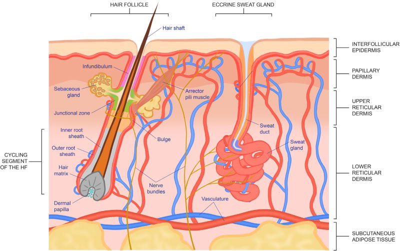
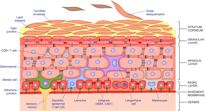
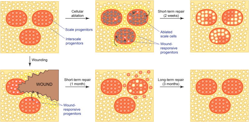
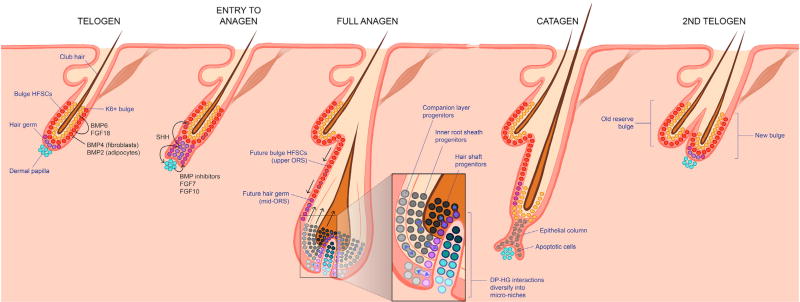
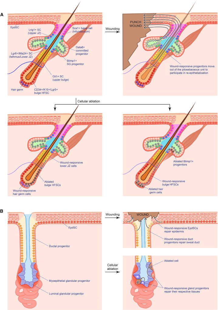
Tissues have a natural capacity to replace dying cells and to heal wounds. This ability resides in resident stem cells, which self-renew, preserve, and repair their tissue during homeostasis and following injury. The skin epidermis and its appendages are subjected to daily assaults from the external environment. A high demand is placed on renewal and regeneration of the skin's barrier in order to protect the body from infection and dehydration and to heal wounds. This review focuses on the epithelial stem cells of skin, where they come from, where they reside, and how they function in normal homeostasis and wound repair.

Keywords: cellular plasticity; epidermis; hair follicle; homeostasis; regeneration; skin stem cells; stem cell niche; wound healing.


Figures

Figure 1
Figure 1. Structure and Components of the Skin
Figure 2
Figure 2. The Epidermal Progenitor Niche
The epidermis is a stratified squamous epithelium. It is divided into four main layers that are distinguished morphologically according to the differentiation status of the keratinocytes as they cease to proliferate and move upward to produce the skin’s barrier. Keratinocytes within the basal layer experience a unique niche distinguished by their contact with the basement membrane, composed of extracellular matrix components and growth factors, contributed by both the epidermis and underlying dermis. This feature maintains their proliferative status. By contrast, keratinocytes that have exited the basal layer embark upon a terminal differentiation program, culminating in the production of dead squames that are sloughed from the skin surface and replaced by inner cells moving upward. Immune cells, mechanosensory cells, and melanocytes also populate discrete layers of the epidermis, reflective of their as yet poorly understood, but vital cross-talk with the keratinocytes and with the microbiota at the skin surface.
Figure 3
Figure 3. Skin Morphogenesis
Shortly after gastrulation, the skin begins as a single layer of epidermal progenitors that divide exclusively parallel to the basement membrane underneath. Within several days, divisions become first oblique and then more perpendicular, leading to asymmetric fates, stratification, and differentiation of the epidermis. During this time, hair placodes also form from gathering WNThigh cells within the basal layer. When these cells begin to divide, they do so perpendicular to the basement membrane, leading to asymmetrically fated daughters. WNThigh cells produce SHH, but only neighboring cells appear to respond. SHH prompts the mesenchyme underneath to organize into a dermal condensate and produce BMP inhibitors. SHH also prompts the overlying daughter cells that lose contact with the basement membrane to dampen WNT signaling and divide symmetrically. These WNTlow daughters will generate the outer root sheath (ORS) which develops a niche (bulge) of stem cells, while the WNThigh daughters generate the inner root sheath (IRS) and hair shaft (HS). DP, dermal papilla; SG, sebaceous gland.
Figure 4
Figure 4. Unified Model for the Dynamics of Epidermal Homeostasis
The basal layer is composed of progenitors that divide parallel to the basement membrane. Division rates (number of divisions in a given amount of time) vary significantly across different body locations and are not coupled with fate (whether daughter cells retain a proliferative [P] status or differentiate [D] and delaminate). Instead, fate determination appears to be mostly stochastic, although outcome probabilities also vary in different skin locales. In contrast to the hair follicle or to human epidermis, label-retaining cells (LRCs) with properties of long-lived stem cells have only been found in the interscale regions of mouse tail. That said, some rapidly proliferating progenitors persist for up to a year in other body sites, including back skin, and thus merit assignment as bona fide EpdSCs.
Figure 5
Figure 5. Wound Response from Scale and Interscale EpdSCs
Cellular (laser) ablation and wounding induce a re-epithelialization response from all nearby EpdSCs, regardless of origin. However, only local cells contribute to long-term homeostasis of the injured site. Thus, interscale contribution to the injured scale region is only transient and vice versa. Diagrams represent a top-down view of EpdSCs of the tail/back interfollicular epidermis.
Figure 6
Figure 6. The Hair Cycle
During the resting phase (telogen), bulge HFSCs are kept inactive by BMP and FGF18 signals from the neighboring K6+ bulge and from nearby fibroblasts and adipocytes. BMP inhibitors and pro-activating FGFs from the dermal papilla (DP) overcome the inhibitory cues, leading to entry into anagen. At the base of the bulge, some hair germ (HG) cells become WNThigh multipotent progenitors, which express SHH. SHH triggers bulge HFSCs to divide symmetrically and grow the outer root sheath (ORS), which then pushes the signaling center away, returning the bulge to quiescence, and then progressively returning the ORS cells to quiescence in a cascade as it grows downward. Progenitors that maintain contact with the DP continue to produce SHH, which fuels the DP to elevate signaling and expand the multipotent progenitor pool. As the hair bulb grows, it envelops the DP. Interactions at the elaborated interface between the DP and hair bulb establish micro-niches, each of which harbors unipotent progenitors that produce the seven concentric differentiating layers of the hair shaft and its channel, or inner root sheath (IRS). By mechanisms still unclear, these progenitors exhaust their proliferative capacity and an apoptotic phase (catagen) ensues, during which the follicle is degenerated and restored back to its resting size as it enters the next telogen. Note that some cells within the upper/mid ORS are spared, and these become the new bulge and new HG for the next hair cycle.
Figure 7
Figure 7. Wound Response from Hair Follicle and Sweat Gland Progenitors
(A) Different niches within the pilosebaceous unit house various progenitors that all display plasticity and contribute to wound-induced re-epithelialization, albeit at varying degrees. Ablation of these populations likewise trigger neighbor progenitors to refill the vacated niche. (B) In contrast, the sweat gland displays stricter compartmentalization in terms of injury response. Wounding of the sweat gland orifice only triggers duct (but not gland) progenitors to reconstruct the sweat duct. Similarly, ablation of specific gland progenitors only activates the same type of progenitor for repair. SC, stem cell; JZ, junctional zone; SG, sebaceous gland.

Similar articles

Cited by

References

    1. Adam RC, Yang H, Rockowitz S, Larsen SB, Nikolova M, Oristian DS, Polak L, Kadaja M, Asare A, Zheng D, et al. Pioneer factors govern super-enhancer dynamics in stem cell plasticity and lineage choice. Nature. 2015;521:366–370. - PMC - PubMed
    1. Ahtiainen L, Lefebvre S, Lindfors PH, Renvoise E, Shirokova V, Vartiainen MK, Thesleff I, Mikkola ML. Directional cell migration, but not proliferation, drives hair placode morphogenesis. Dev. Cell. 2014;28:588–602. - PubMed
    1. Ali N, Zirak B, Rodriguez RS, Pauli ML, Truong HA, Lai K, Ahn R, Corbin K, Lowe MM, Scharschmidt TC, et al. Regulatory T cells in skin facilitate epithelial stem cell differentiation. Cell. 2017;169:1119–1129. - PMC - PubMed
    1. Ansell DM, Kloepper JE, Thomason HA, Paus R, Hardman MJ. Exploring the “hair growth-wound healing connection”: anagen phase promotes wound re-epithelialization. J. Invest. Dermatol. 2011;131:518–528. - PubMed
    1. Aragona M, Dekoninck S, Rulands S, Lenglez S, Mascre G, Simons BD, Blanpain C. Defining stem cell dynamics and migration during wound healing in mouse skin epidermis. Nat. Commun. 2017;8:14684. - PMC - PubMed

Publication types