

Spatial resolution measurements by Radia diagnostic software with SEDENTEXCT image quality phantom in cone beam CT for dental use
- PMID: 29161903
- PMCID: PMC6047630
- DOI: 10.1259/dmfr.20170307
Spatial resolution measurements by Radia diagnostic software with SEDENTEXCT image quality phantom in cone beam CT for dental use
Abstract
Objectives: We aimed to employ the Radia diagnostic software with the safety and efficacy of a new emerging dental X-ray modality (SEDENTEXCT) image quality (IQ) phantom in CT, and to evaluate its validity.
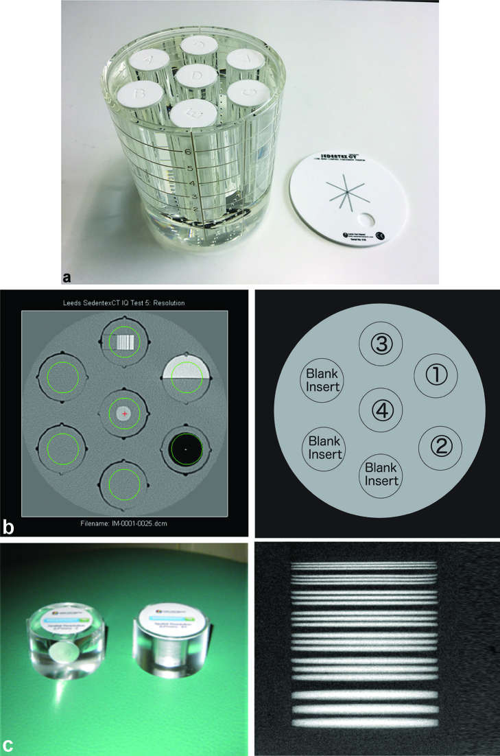
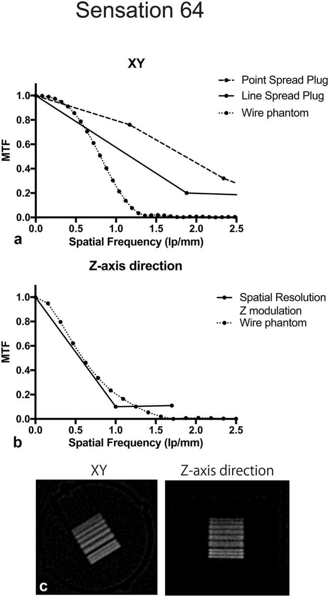
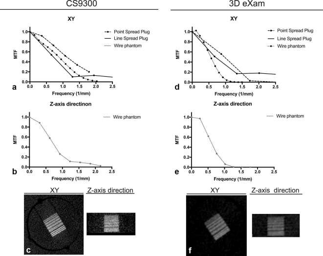
Methods: The SEDENTEXCT IQ phantom and Radia diagnostic software were employed. The phantom was scanned using one medical full-body CT and two dentomaxillofacial cone beam CTs. The obtained images were imported to the Radia software, and the spatial resolution outputs were evaluated. The oversampling method was employed using our original wire phantom as a reference. The resultant modulation transfer function (MTF) curves were compared. The null hypothesis was that MTF curves generated using both methods would be in agreement. One-way analysis of variance tests were applied to the f50 and f10 values from the MTF curves. The f10 values were subjectively confirmed by observing the line pair modules.
Results: The Radia software reported the MTF curves on the xy-plane of the CT scans, but could not return f50 and f10 values on the z-axis. The null hypothesis concerning the reported MTF curves on the xy-plane was rejected. There were significant differences between the results of the Radia software and our reference method, except for f10 values in CS9300. These findings were consistent with our line pair observations.
Conclusions: We evaluated the validity of the Radia software with the SEDENTEXCT IQ phantom. The data provided were semi-automatic, albeit with problems and statistically different from our reference. We hope the manufacturer will overcome these limitations.
Conflict of interest statement
Figures
Similar articles
-
Image quality assessment of three cone beam CT machines using the SEDENTEXCT CT phantom.Dentomaxillofac Radiol. 2013;42(8):20120445. doi: 10.1259/dmfr.20120445. Dentomaxillofac Radiol. 2013. PMID: 23956235 Free PMC article.
-
Modulation transfer function evaluation of cone beam computed tomography for dental use with the oversampling method.Dentomaxillofac Radiol. 2010 Jan;39(1):28-32. doi: 10.1259/dmfr/27069629. Dentomaxillofac Radiol. 2010. PMID: 20089741 Free PMC article.
-
A quality assurance framework for the fully automated and objective evaluation of image quality in cone-beam computed tomography.Med Phys. 2014 Mar;41(3):031901. doi: 10.1118/1.4863507. Med Phys. 2014. PMID: 24593719
-
Relationship between physical factors and subjective image quality of cone-beam computed tomography images according to diagnostic task.Oral Surg Oral Med Oral Pathol Oral Radiol. 2015 Mar;119(3):357-65. doi: 10.1016/j.oooo.2014.11.010. Epub 2014 Dec 6. Oral Surg Oral Med Oral Pathol Oral Radiol. 2015. PMID: 25592866
-
Comparison of methods for acceptance and constancy testing in dental cone-beam computed tomography.Rofo. 2015 Apr;187(4):283-90. doi: 10.1055/s-0034-1385333. Epub 2014 Nov 12. Rofo. 2015. PMID: 25389669
Cited by
-
Optimization of exposure parameters and relationship between subjective and technical image quality in cone-beam computed tomography.Imaging Sci Dent. 2019 Jun;49(2):139-151. doi: 10.5624/isd.2019.49.2.139. Epub 2019 Jun 24. Imaging Sci Dent. 2019. PMID: 31281791 Free PMC article.
-
[Design and optimization of a cone-beam CT system for extremity imaging].Nan Fang Yi Ke Da Xue Xue Bao. 2018 Nov 30;38(11):1331-1337. doi: 10.12122/j.issn.1673-4254.2018.11.09. Nan Fang Yi Ke Da Xue Xue Bao. 2018. PMID: 30514681 Free PMC article. Chinese.
-
Imaging of mandibular fractures: a pictorial review.Insights Imaging. 2020 Feb 19;11(1):30. doi: 10.1186/s13244-020-0837-0. Insights Imaging. 2020. PMID: 32076873 Free PMC article. Review.
References
-
- Project S. Radiation protection n°172: cone beam CT for dental and maxillofacial radiology. Luxembourg: The British Institute of Radiology.; 2012.
Publication types
MeSH terms
LinkOut - more resources
Full Text Sources
Other Literature Sources
Research Materials
Miscellaneous
