Multilevel lumbar spine infection due to poor dentition in an immunocompetent adult: a case report
- PMID: 29162118
- PMCID: PMC5698996
- DOI: 10.1186/s13256-017-1492-z
Multilevel lumbar spine infection due to poor dentition in an immunocompetent adult: a case report
Abstract
Background: Although spinal infections have been reported following dental procedures, development of a spinal infection attributed to poor dentition without a history of a dental procedure in an immunocompetent adult has not been previously reported. Here we provide a case report of a multilevel lumbar spine infection that developed in an immunocompetent adult with poor dentition.
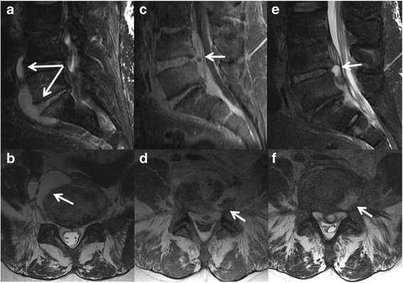
Case presentation: A 63-year-old white male man with past medical history of hypertension presented to a hospital emergency department with a 4-month history of progressively worsening low back pain. A musculoskeletal examination demonstrated diffuse tenderness in his lumbar spine area and the results of a neurological examination were within normal limits. Computed tomography and magnetic resonance imaging of his lumbar spine demonstrated a prevertebral and presacral fluid collection ventral to the L4 to L5 and L5 to S1 interspaces. Blood cultures grew pan-sensitive Streptococcus intermedius in four of four bottles within 45 hours. Using computed tomography guidance, three core biopsies of the L4 to L5 interspace were taken and subsequent cultures were positive for Streptococcus intermedius. He reported that his last episode of dental care occurred more than 20 years ago and a dental panoramic radiograph demonstrated significant necrotic dentition. Ten teeth were extracted and the necrotic dentition was assumed to be the most likely source of infection. On hospital dismissal, he received a 12-week course of intravenously administered ceftriaxone followed by an 8-week course of orally administered cefadroxil pending repeat imaging.
Conclusions: This case report demonstrates the importance of determining the source of infection in a patient with a spontaneous spinal infection. Even in the absence of a recent dental procedure, dentition should be considered a possible source of infection in an immunocompetent patient who presents with a spontaneous spinal infection.
Keywords: Dentition; Epidural abscess; Immunocompetent; Lumbar spine.
Conflict of interest statement
Ethics approval and consent to participate
The Mayo Clinic Institutional Review Board (IRB) acknowledged that the case report did not require IRB review based on submitted responses to an IRB application.
Consent for publication
Written informed consent was obtained from the patient for publication of this case report and any accompanying images. A copy of the written consent is available for review by the Editor-in-Chief of this journal.
Competing interests
The authors declare that they have no competing interests.
Publisher’s Note
Springer Nature remains neutral with regard to jurisdictional claims in published maps and institutional affiliations.
Figures


Similar articles
-
First report on treating spontaneous infectious spondylodiscitis of lumbar spine with posterior debridement, posterior instrumentation and an injectable calcium sulfate/hydroxyapatite composite eluting gentamicin: a case report.J Med Case Rep. 2016 Dec 12;10(1):349. doi: 10.1186/s13256-016-1125-y. J Med Case Rep. 2016. PMID: 27955704 Free PMC article.
-
Rare cause of back pain: Staphylococcus aureus vertebral osteomyelitis complicated by recurrent epidural abscess and severe sepsis.BMJ Case Rep. 2016 Dec 13;2016:bcr2016217111. doi: 10.1136/bcr-2016-217111. BMJ Case Rep. 2016. PMID: 27965310 Free PMC article.
-
Low back pain after a dental procedure: a case of Streptococcus viridans vertebral osteomyelitis.BMJ Case Rep. 2016 Jun 7;2016:bcr2016216087. doi: 10.1136/bcr-2016-216087. BMJ Case Rep. 2016. PMID: 27268493 Free PMC article.
-
Cervical Facet Joint Infection and Associated Epidural Abscess with Streptococcus intermedius from a Dental Infection Origin A Case Report and Review.Bull Hosp Jt Dis (2013). 2016 Sep;74(3):237-43. Bull Hosp Jt Dis (2013). 2016. PMID: 27620549 Review.
-
L5-S1 Achromobacter xylosoxidans infection secondary to oxygen-ozone therapy for the treatment of lumbosacral disc herniation: a case report and review of the literature.Spine (Phila Pa 1976). 2014 Mar 15;39(6):E413-6. doi: 10.1097/BRS.0000000000000195. Spine (Phila Pa 1976). 2014. PMID: 24384664 Review.
Cited by
-
Trends in infectious spondylitis from 2000 to 2020.Eur Spine J. 2024 Aug;33(8):3154-3160. doi: 10.1007/s00586-024-08286-7. Epub 2024 May 1. Eur Spine J. 2024. PMID: 38693341
-
Osteomyelitis caused by Streptococcus intermedius in immunocompetent adults - a case report and systematic literature review.Eur J Clin Microbiol Infect Dis. 2023 Sep;42(9):1055-1061. doi: 10.1007/s10096-023-04640-7. Epub 2023 Jul 20. Eur J Clin Microbiol Infect Dis. 2023. PMID: 37468663 Free PMC article.
-
Streptococcus anginosus spondylodiscitis causing incapacitating back pain in an immunocompetent patient: A case report.SAGE Open Med Case Rep. 2024 Aug 16;12:2050313X241272606. doi: 10.1177/2050313X241272606. eCollection 2024. SAGE Open Med Case Rep. 2024. PMID: 39161921 Free PMC article.
-
A case report of Klebsiella aerogenes-caused lumbar spine infection identified by metagenome next-generation sequencing.BMC Infect Dis. 2022 Jul 15;22(1):616. doi: 10.1186/s12879-022-07583-0. BMC Infect Dis. 2022. PMID: 35840919 Free PMC article.
-
Concomitant pyogenic spondylodiscitis and empyema following tongue cancer resection and wisdom tooth extraction: A case report and literature review.Medicine (Baltimore). 2024 Jul 26;103(30):e39087. doi: 10.1097/MD.0000000000039087. Medicine (Baltimore). 2024. PMID: 39058851 Free PMC article. Review.
References
Publication types
MeSH terms
Substances
LinkOut - more resources
Full Text Sources
Other Literature Sources
Medical
Miscellaneous

