Comparative Study
. 2017 Nov 21;19(1):91.
doi: 10.1186/s12968-017-0407-x.

Prospective comparison of novel dark blood late gadolinium enhancement with conventional bright blood imaging for the detection of scar

Affiliations
Comparative Study

Prospective comparison of novel dark blood late gadolinium enhancement with conventional bright blood imaging for the detection of scar

Rohin Francis et al. J Cardiovasc Magn Reson. .

Abstract

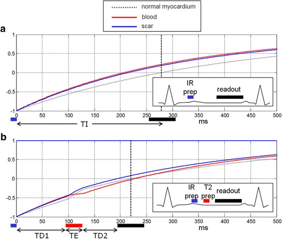
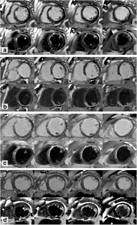
Background: Conventional bright blood late gadolinium enhancement (bright blood LGE) imaging is a routine cardiovascular magnetic resonance (CMR) technique offering excellent contrast between areas of LGE and normal myocardium. However, contrast between LGE and blood is frequently poor. Dark blood LGE (DB LGE) employs an inversion recovery T2 preparation to suppress the blood pool, thereby increasing the contrast between the endocardium and blood. The objective of this study is to compare the diagnostic utility of a novel DB phase sensitive inversion recovery (PSIR) LGE CMR sequence to standard bright blood PSIR LGE.

Methods: One hundred seventy-two patients referred for clinical CMR were scanned. A full left ventricle short axis stack was performed using both techniques, varying which was performed first in a 1:1 ratio. Two experienced observers analyzed all bright blood LGE and DB LGE stacks, which were randomized and anonymized. A scoring system was devised to quantify the presence and extent of gadolinium enhancement and the confidence with which the diagnosis could be made.

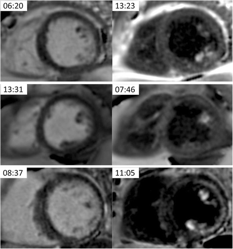
Results: A total of 2752 LV segments were analyzed. There was very good inter-observer correlation for quantifying LGE. DB LGE analysis found 41.5% more segments that exhibited hyperenhancement in comparison to bright blood LGE (248/2752 segments (9.0%) positive for LGE with bright blood; 351/2752 segments (12.8%) positive for LGE with DB; p < 0.05). DB LGE also allowed observers to be more confident when diagnosing LGE (bright blood LGE high confidence in 154/248 regions (62.1%); DB LGE in 275/324 (84.9%) regions (p < 0.05)). Eighteen patients with no bright blood LGE were found to have had DB LGE, 15 of whom had no known history of myocardial infarction.

Conclusions: DB LGE significantly increases LGE detection compared to standard bright blood LGE. It also increases observer confidence, particularly for subendocardial LGE, which may have important clinical implications.

Keywords: Bright blood; Dark blood; Inversion recovery; Late gadolinium enhancement; Myocardial infarction; PSIR.


Conflict of interest statement

Ethics approval and consent to participate

All ethics were approved by the UCL/UCLH Joint Committees on the Ethics of Human Research Committee, and all participants provided written informed consent.

Consent for publication

Written informed consent was obtained from patients for publication of their individual details and accompanying images in this manuscript. The consent form is held on record and is available for review by the Editor-in-Chief.

Competing interests

The authors declare that they have no competing interests.

Publisher’s Note

Springer Nature remains neutral with regard to jurisdictional claims in published maps and institutional affiliations.

Figures

Fig. 1
Fig. 1
a inversion recovery (IR) for bright blood LGE in case with scar signal (blue) less than blood (red) resulting in poor contrast. b IR for dark blood (DB) late gadolinium enhancement (LGE) using combined IR and T2 preparation to shift the null time of blood relative to the normal myocardium. In this case the delays are chosen such that the blood signal (red) is less than the myocardium (dashed grey) resulting in dark blood using phase sensitive inversion recovery (PSIR) reconstruction, which preserves the signal polarity. Inversion times to null the normal myocardium are depicted by vertical dashed lines. The loss in signal-to-noise ratio (SNR) due to the T2 preparation is mitigated by increased respiratory motion corrected averaging (MOCO) [15]
Fig. 2
Fig. 2
Total number of left ventricular (LV) segments identified (left) and diagnostic confidence when diagnosing late gadolinium enhancement (LGE) (right) using both bright blood and dark blood (DB) imaging. The time after gadolinium administration is recorded for each sequence (minutes:seconds)
Fig. 3
Fig. 3
Consecutive short axis slices from four patients. In each panel, bright blood late gadolinium enhancement (LGE) sequences appear above dark blood (DB) LGE. The time after gadolinium administration is recorded for each sequence (minutes:seconds). Patient A: DB LGE reveals a large left anterior descending (LAD) territory infarct, the size and borders of which are not clearly delineated on bright blood imaging. Bright blood 14:43, DB 21:21. Patient B: Inferior infarction can be seen on the bright blood images but it is difficult to appreciate if the basal slice is affected or to see the endomyocardial border at all. DB images reveal segments showing LGE and the myocardial structure. Bright blood 10:31, DB 16:07. Patient C: Bright blood images suggest anterior LGE but it is difficult to quantify accurately. DB LGE shows the extent of an anterior infarct. Bright blood 8:13, DB 16:14. Patient D: Poor contrast between scar and the blood pool masks antero-lateral scar, seen easily with DB LGE. Bright 6:59, DB 12:12
Fig. 4
Fig. 4
Paired SA slices from 3 patients showing papillary muscle late gadolinium enhancement (LGE). In the top left corner of each image is the time after administration of gadolinium (minutes:seconds)
Fig. 5
Fig. 5
Examples of 4 patients that underwent bright blood late gadolinium enhancement (LGE) first and dark blood (DB) LGE second (left column) and 4 additional patients that underwent the sequences in the opposite order (right column). The bright blood LGE, for either acquisition order, exhibits a range of contrast between the subendocardial scar and adjacent blood pool, from good (top rows) to poor (bottom rows) contrast, whereas the DB LGE had excellent contrast in all cases. In the top left corner of each image is the time after administration of gadolinium (minutes:seconds)

References

    1. Kellman P, Chung YC, Simonetti OP, McVeigh ER, Arai AE. Multi-contrast delayed enhancement provides improved contrast between myocardial infarction and blood pool. J Magn Reson Imaging. 2005;22(5):605–613. doi: 10.1002/jmri.20426. - DOI - PMC - PubMed
    1. Burns RJ, Gibbons RJ, Yi Q, Roberts RS, Miller TD, Schaer GL, et al. The relationships of left ventricular ejection fraction, end-systolic volume index and infarct size to six-month mortality after hospital discharge following myocardial infarction treated by thrombolysis. J Am Coll Cardiol. 2002;39(1):30–36. doi: 10.1016/S0735-1097(01)01711-9. - DOI - PubMed
    1. Bulluck H, Yellon DM, Hausenloy DJ. Reducing myocardial infarct size: challenges and future opportunities. Heart. 2016;102(5):341–348. doi: 10.1136/heartjnl-2015-307855. - DOI - PMC - PubMed
    1. Desch S, Eitel I, de Waha S, Fuernau G, Lurz P, Gutberlet M, et al. Cardiac magnetic resonance imaging parameters as surrogate endpoints in clinical trials of acute myocardial infarction. Trials. 2011;12:204. doi: 10.1186/1745-6215-12-204. - DOI - PMC - PubMed
    1. Vergara GR, Marrouche NF. Tailored management of atrial fibrillation using a LGE-MRI based model: from the clinic to the electrophysiology laboratory. J Cardiovasc Electrophysiol. 2011;22(4):481–487. doi: 10.1111/j.1540-8167.2010.01941.x. - DOI - PubMed

Publication types

MeSH terms