. 2017 Nov 21;14(1):229.
doi: 10.1186/s12985-017-0895-1.

Geraniin extracted from the rind of Nephelium lappaceum binds to dengue virus type-2 envelope protein and inhibits early stage of virus replication

Affiliations

Geraniin extracted from the rind of Nephelium lappaceum binds to dengue virus type-2 envelope protein and inhibits early stage of virus replication

Siti Aisyah Abdul Ahmad et al. Virol J. .

Abstract

Background: The rapid rise and spread in dengue cases, together with the unavailability of safe vaccines and effective antiviral drugs, warrant the need to discover and develop novel anti-dengue treatments. In this study the antiviral activity of geraniin, extracted from the rind of Nephelium lappaceum, against dengue virus type-2 (DENV-2) was investigated.

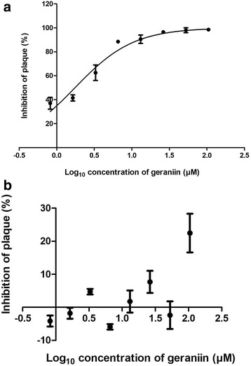
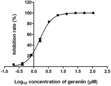
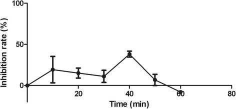
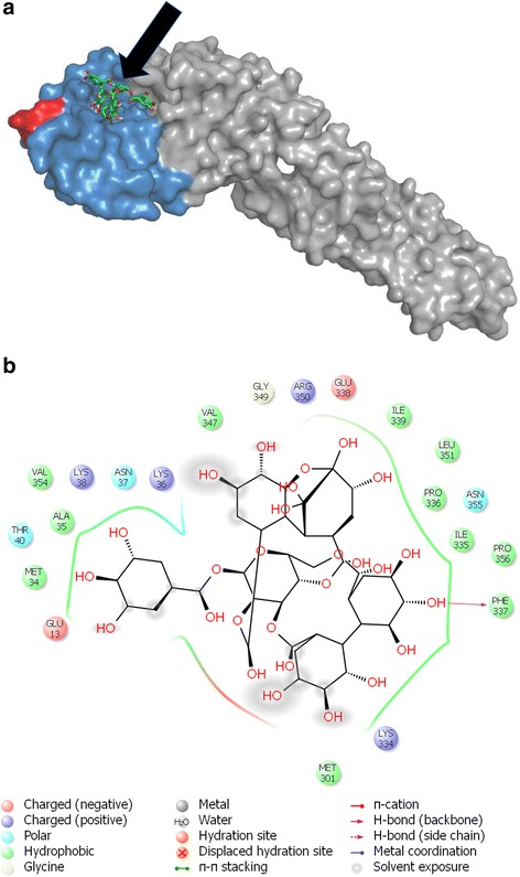
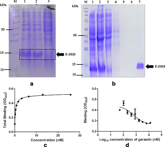
Methods: Geraniin was prepared from Nephelium lappaceum rind by reverse phase C-18 column chromatography. Cytotoxicity of geraniin towards Vero cells was evaluated using MTT assay while IC50 value was determined by plaque reduction assay. The mode-of-action of geraniin was characterized using the virucidal, attachment, penetration and the time-of-addition assays'. Docking experiments with geraniin molecule and the DENV envelope (E) protein was also performed. Finally, recombinant E Domain III (rE-DIII) protein was produced to physiologically test the binding of geraniin to DENV-2 E-DIII protein, through ELISA competitive binding assay.

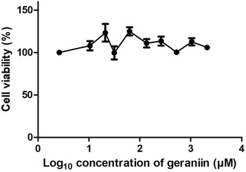
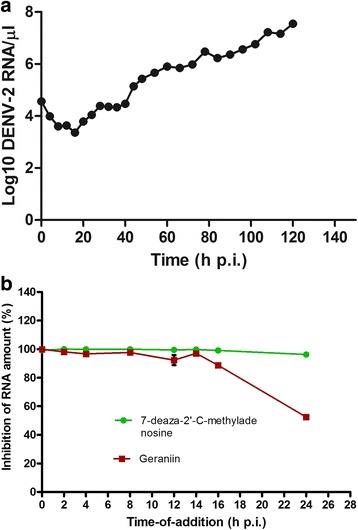
Results: Cytotoxicity assay confirmed that geraniin was not toxic to Vero cells, even at the highest concentration tested. The compound exhibited DENV-2 plaque formation inhibition, with an IC50 of 1.75 μM. We further revealed that geraniin reduced viral infectivity and inhibited DENV-2 from attaching to the cells but had little effect on its penetration. Geraniin was observed to be most effective when added at the early stage of DENV-2 infection. Docking experiments showed that geraniin binds to DENV E protein, specifically at the DIII region, while the ELISA competitive binding assay confirmed geraniin's interaction with rE-DIII with high affinity.

Conclusions: Geraniin from the rind of Nephelium lappaceum has antiviral activity against DENV-2. It is postulated that the compound inhibits viral attachment by binding to the E-DIII protein and interferes with the initial cell-virus interaction. Our results demonstrate that geraniin has the potential to be developed into an effective antiviral treatment, particularly for early phase dengue viral infection.

Keywords: Antiviral; DENV-2 E protein; Geraniin; Nephelium lappaceum.


Conflict of interest statement

Ethics approval and consent to participate

Not applicable.

Consent for publication

Not applicable.

Competing interests

The authors declare that they have no competing interests.

Publisher’s Note

Springer Nature remains neutral with regard to jurisdictional claims in published maps and institutional affiliations.

Figures

Fig. 1
Fig. 1
Cytotoxicity assay of geraniin. Geraniin was not toxic to Vero cells even at the highest concentration tested; hence validate its use in the antiviral assays. Data are shown as the mean ± SEM of triplicate from one independent experiment
Fig. 2
Fig. 2
Inhibitory effect of geraniin against DENV-2. a A dose response curve showing concentration-dependent inhibition of DENV-2 plaque formation when geraniin was incubated with DENV-2 before adsorption. b A dose-response curve indicating that geraniin caused no inhibition towards DENV-2 plaque formation when it was incubated with cells before adsorption. Percentage of plaque inhibition was calculated, and the data was incorporated into the GraphPad Prism 5 software to obtain the IC50 values. Data represent mean values ±SEM
Fig. 3
Fig. 3
Effect of geraniin on the attachment of DENV-2 to Vero cells. Vero monolayer was pre-chilled at 4 °C for 1 h. DENV-2 was inoculated to Vero monolayer in the absence (virus control) or presence of geraniin and incubated at 4 °C for another 3 h. The dose-response curve indicates the inhibition of DENV-2 attachment to Vero cells by geraniin. Data represent mean values ±SEM
Fig. 4
Fig. 4
Effect of geraniin on the penetration of DENV-2 into Vero cells. Vero monolayer was pre-chilled at 4 °C for 1 h and then infected with DENV-2 at 4 °C for another 3 h. After 3 h incubation, geraniin was added. At 10 min interval, extracellular virus was killed by PBS at pH 3. PBS at pH 11 was then added to neutralize acidic PBS. The graph of percentage of DENV-2 plaque inhibition was plotted against time taken for penetration. Data represent mean values ±SEM
Fig. 5
Fig. 5
Effect of geraniin on the time of addition. a DENV-2 Replication in Vero cells. Vero cells were infected with 6.74 × 104 PFU/ml DENV-2. At selected intervals, intracellular RNA was isolated and DENV-2 RNA levels were quantified using SYBR Green dye-based qRT-PCR assay. Plotted absolute number of viral RNA copy in log scale per μl of RNA used for qRT-PCR. (b) Geraniin or the control compound was added to DENV-2 infected Vero cells at different time points after infection. Addition of compounds was conducted up to 24 h before the cells were harvested at 48 h p.i. Samples were subjected to RNA extraction and later to two-step qRT-PCR. Absolute quantification of viral load in treated samples was conducted using the standard curve method. Percentage of inhibition was calculated based on the amount of viral RNA in treated samples compared with untreated controls. Geraniin was effective in inhibiting DENV-2 RNA synthesis when it was added at earlier time-points post-infection. Data represent mean values ±SEM
Fig. 6
Fig. 6
Molecular Docking. a Binding pose of geraniin’s molecular structure on DENV-2 E protein (PDB entry: 1OKE). Arrow indicates the molecular structure of geraniin. Domain III is marked blue and a putative receptor-binding loop in domain III is marked red. b Interaction of geraniin with the amino acid residues of DENV-2 E protein as shown by docking. Geraniin binds at domain III of E protein which ranges from amino acid residues 296–394, by forming a hydrogen bond with the amino acid residues of the E protein
Fig. 7
Fig. 7
Binding of geraniin to recombinant DENV-2 E Domain III protein. a SDS-PAGE gel showing expression of DENV-2 E-DIII protein. Lane M indicates the 10 kDa protein ladder, Lane 1- protein induced with IPTG; Lane 2- protein induced with IPTG; Lane 3- uninduced protein. Size of E-DIII protein is 15 kDa. b SDS-PAGE gel showing purification of DENV-2 E-DIII protein. Lane M-10 kDa protein ladder; Lanes; 1-pellet, 2-supernatant, 3-flow-through, 4-wash 1, 5-wash 2, 6-wash 3, 7-eluent. A single band at the expected size indicated that the recombinant protein obtained was pure. c The saturation binding curve obtained when recombinant E-DIII protein was coated onto ELISA plate and was allowed to bind to different concentrations of monoclonal antibody, clone 5j122 (MAB8901) before the signal was detected using an AP conjugated secondary antibody. The Kd value of the mAb was determined from the curve by nonlinear regression using GraphPad Prism 5 software. d The competitive binding curve was obtained through non-linear regression analysis using GraphPad Prism 5 software. The same monoclonal antibody (MAB8901) was used at one constant concentration of 3.33 nM while various concentrations of geraniin was used to test the ability of geraniin to bind to the recombinant E-DIII protein, given that the Kd value of the mAb was known. Data represent mean values ±SEM

References

    1. Bhatt S, et al. The global distribution and burden of dengue. Nature. 2013;496(7446):504–507. doi: 10.1038/nature12060. - DOI - PMC - PubMed
    1. Gubler, D.J., Dengue Viruses, in Encyclopedia of Virology (Third Edition), B.W.J. Mahy and M.H.V.V. Regenmortel, Editors. 2008, Academic Press: Oxford. p. 5–14.
    1. World Health Organization. Dengue: guidelines for diagnosis, treatment, prevention and control: New Edition. Geneva; 2009. Available from http://www.who.int/rpc/guidelines/9789241547871/en/. - PubMed
    1. Hung S-L, et al. Analysis of the steps involved in dengue virus entry into host cells. Virology. 1999;257(1):156–167. doi: 10.1006/viro.1999.9633. - DOI - PubMed
    1. Whitehorn J, Farrar J. Dengue. Br Med Bull. 2010;95:161–173. doi: 10.1093/bmb/ldq019. - DOI - PubMed

MeSH terms