. 2018 Jan;17(1):43-60.
doi: 10.1074/mcp.RA117.000076. Epub 2017 Nov 21.

Quantitative Proteomic Profiling Reveals Novel Plasmodium falciparum Surface Antigens and Possible Vaccine Candidates

Affiliations

Quantitative Proteomic Profiling Reveals Novel Plasmodium falciparum Surface Antigens and Possible Vaccine Candidates

Sandra K Nilsson Bark et al. Mol Cell Proteomics. 2018 Jan.

Abstract

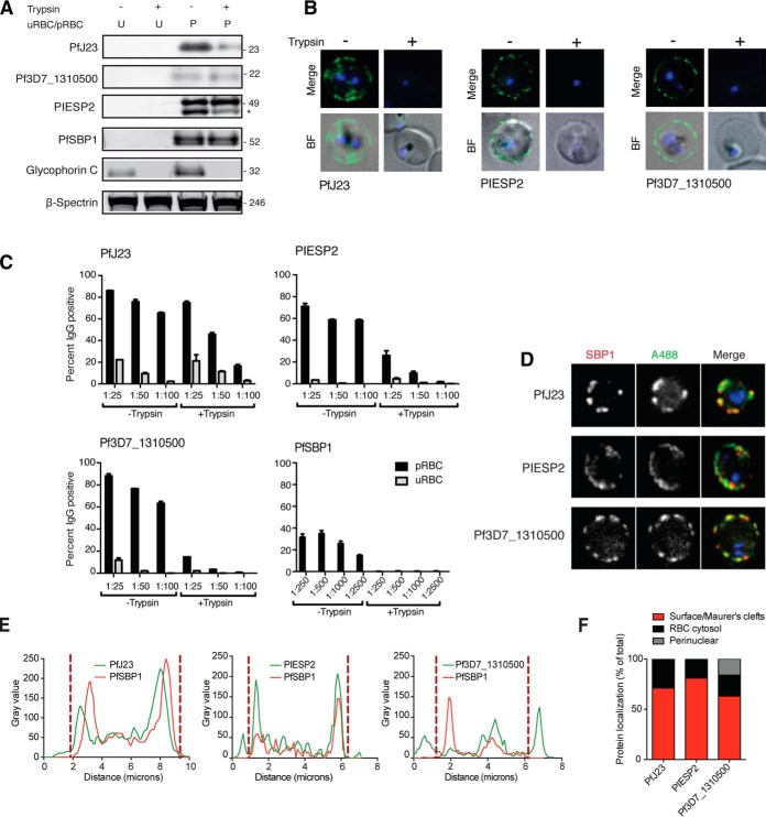
Despite recent efforts toward control and elimination, malaria remains a major public health problem worldwide. Plasmodium falciparum resistance against artemisinin, used in front line combination drugs, is on the rise, and the only approved vaccine shows limited efficacy. Combinations of novel and tailored drug and vaccine interventions are required to maintain the momentum of the current malaria elimination program. Current evidence suggests that strain-transcendent protection against malaria infection can be achieved using whole organism vaccination or with a polyvalent vaccine covering multiple antigens or epitopes. These approaches have been successfully applied to the human-infective sporozoite stage. Both systemic and tissue-specific pathology during infection with the human malaria parasite P. falciparum is caused by asexual blood stages. Tissue tropism and vascular sequestration are the result of specific binding interactions between antigens on the parasite-infected red blood cell (pRBC) surface and endothelial receptors. The major surface antigen and parasite ligand binding to endothelial receptors, PfEMP1 is encoded by about 60 variants per genome and shows high sequence diversity across strains. Apart from PfEMP1 and three additional variant surface antigen families RIFIN, STEVOR, and SURFIN, systematic analysis of the infected red blood cell surface is lacking. Here we present the most comprehensive proteomic investigation of the parasitized red blood cell surface so far. Apart from the known variant surface antigens, we identified a set of putative single copy surface antigens with low sequence diversity, several of which are validated in a series of complementary experiments. Further functional and immunological investigation is underway to test these novel P. falciparum blood stage proteins as possible vaccine candidates.


Figures

Fig. 1.
Fig. 1.
Membrane proteomics. A, Overview of the membrane proteomics process: biological replicates (A, B, and C) of purified and intact pRBCs (28–34 h post infection) were either surface-shaved with trypsin and chymotrypsin (+), or mock-treated with sucrose-supplemented PBS (−). B, Lysis optimization. Five μl of RBCs were lysed with either PBS, PBS supplemented with 5–20% sucrose, or RPMI, for 30–60 min. Lysis is presented as percentage of hemoglobin in each sample and normalized to a positive lysis control with 0.1% Triton X-100 in PBS, which was set to 100%. C, Validation of proteomics samples. Membranes were collected for Western blot analysis for each of the six samples (A±, B±, and C±). Membranes were probed with anti-Glycophorin C or anti-ATS antibodies. The anti-ATS antibody detects both full-length PfEMP1 (>300 kDa, marked with an asterisk), as well as the surface truncated PfEMP1 (70–95 kDa, box), which mainly consists of the cytoplasmic C-terminal ATS region of PfEMP1. Full-length PfEMP1 migrates very closely to cross-reactive RBC spectrins (marked with a double asterisk). Anti-Spectrin antibodies were used as a loading control. Each lane represents protein extract from 2.5 × 10⁶ pRBC. D, Membranes from the six samples (A±, B±, and C±) were analyzed by Western blot using surface reactive sera from the six mice immunized with mock-treated pRBCs. Four areas of interest (–4) where bands are present only in untreated cells or bands are truncated on treatment are marked with red boxes. U = unparasitized RBCs, p = pRBCs. E, Membranes from the six samples (A±, B±, and C±) were separated by SDS-PAGE and stained with Coomassie blue. Areas of interest were divided into multiple bands (A-L) for in-gel digestion. U = unparasitized RBCs, p = pRBCs). F, Bar charts describing distribution of transmembrane (TM, blue) and exported (green) proteins, comparing proteins significantly enriched in the untreated sample (Sign. hits) with all proteins identified (All).
Fig. 2.
Fig. 2.
Supernatant proteomics. A, Overview of supernatant proteomics process. B, Surface shaving optimization. Intact and live trophozoite stage pRBC were surface shaved with a titration of trypsin, and analyzed by Western blot. Each lane represents protein extract from 2.5 × 10⁶ pRBC. RBC refers to unparasitized RBCs. C, Validation of proteomics samples. After supernatants were collected for surface proteomics, the remaining cell pellet for each of the four samples were collected for Western blot analysis (A−, A+, B−, and B+). To validate the surface shaving method, membranes were probed with anti-Glycophorin C and anti-ATS antibodies. Anti-Spectrin antibodies were used as a loading control. Each lane represents protein extract from 2.5 × 10⁶ pRBC.
Fig. 3.
Fig. 3.
Supernatant proteomics analysis and overall comparison. A, Log2fold change scatter plot of the 2737 identified proteins from the two biological replicates. The 381 proteins with significantly different peptide abundance between surface shaved and lysis control samples are marked in red (corrected p values ≤ 0.05), B, Bar charts show distribution of transmembrane (TM) and PEXEL proteins, comparing proteins significantly enriched in surface shaving sample (Significant hits; blue/green, 100 proteins) and all proteins identified (gray, 2730). C, A comparison of total number and localization of secretory proteins (left graph) and exported proteins (right graph) across our two proteomics approaches and two previously published methods (20, 21).
Fig. 4.
Fig. 4.
In vivo expression and allelic diversity for secretory proteins from membrane proteomics (A) and supernatant proteomics (B). Both graphs show transcriptional expression of all hits (A: n = 54, B: n = 65) in three patient samples from Senegal (left) and the ratio of nonsynonymous to synonymous SNPs across all hits based on a set of 64 parasite genomes from Senegal (right). Hits proposed for further validation (Table I) are shown in bold. The subset of hits validated by specific antibodies is marked in red. Vertical dashed line indicates our diversity threshold used for selection of candidates (πΝ < 0.004).
Fig. 5.
Fig. 5.
Validation of candidates. A, Unparasitized RBC and intact and live trophozoite stage pRBC (± pre-treatment with trypsin/chymotrypsin) analyzed by Western blot, and membranes were probed with polyclonal antibodies targeting candidate. A trypsin sensitive band at the expected size was detected for pRBCs probed with PfJ23 and PIESP2, whereas detection of Pf3D7_1310500 did not appear to be trypsin dependent. PfSBP1, Glycophorin C, and β-spectrin were included as controls. Each lane represents protein extract from 2.5 × 10⁶ pRBC. Full Western blots are provided in supplemental Fig. S3. B, Surface reactivity of candidate antibodies to purified pRBCs (± pre-treatment with trypsin/chymotrypsin) was detected by live microscopy. Cells were stained with the nuclear dye Vybrant violet and the surface labeled using AlexaFluor488 goat anti-rat secondary antibody. Antibody signal is only detectable on the pRBC surface without prior trypsin treatment of the cells. C, Surface reactivity of candidate antibodies to purified pRBCs (± pre-treatment with trypsin/chymotrypsin) was detected by flow cytometry. Cells were gated for live cells and single cells, and uRBCs and pRBCs were subsequently separately gated based on Vybrant Violet fluorescence and surface reactivity measured using AlexaFluor488 goat anti-rat secondary antibody. Surface reactivity was measured as percent of cells positive for AlexaFluor488. Surface reactivity of all antibodies tested decreased with trypsin treatment, but this change was most significant for PIESP2, Pf3D7_1310500 and the lowest dilution (1:100) of PfJ23. D, Immunofluorescence analysis of the localization of PfJ23, PIESP2 and Pf3D7_1310500 (detected with specific sera) in fixed and permeabilized trophozoite pRBCs shows partial colocalization in the cytosol of pRBC with the Maurer's cleft resident protein PfSBP1, as well as pRBC surface localization. E, Fluorescence plot profile of PfJ23, PIESP2 and Pf3D7_1310500 localization confirms colocalization with PfSBP1 in addition to surface localization. Dashed lines mark the boundary between Maurer's cleft and peripheral/surface labeling based on SBP1 distribution. F, Immunofluorescence-based analysis of protein localization across multiple cells classified by fluorescence intensity percentage, shows that up to 70% of PfJ23, 90% of PIESP2, and 69% of Pf3D7_1310500, respectively, localizes to the pRBC surface or Maurer's clefts, whereas the remaining localization is cytosolic (and perinuclear in the case of Pf3D7_1310500).

References

    1. WHO. (2016) WHO Malaria Report 2016
    1. Maier A. G., Cooke B. M., Cowman A. F., and Tilley L. (2009) Malaria parasite proteins that remodel the host erythrocyte. Nature Rev. Microbiology 7, 341–354 - PubMed
    1. Su X. Z., Heatwole V. M., Wertheimer S. P., Guinet F., Herrfeldt J. A., Peterson D. S., Ravetch J. A., and Wellems T. E. (1995) The large diverse gene family var encodes proteins involved in cytoadherence and antigenic variation of Plasmodium falciparum-infected erythrocytes. Cell 82, 89–100 - PubMed
    1. Smith J. D., Chitnis C. E., Craig A. G., Roberts D. J., Hudson-Taylor D. E., Peterson D. S., Pinches R., Newbold C. I., and Miller L. H. (1995) Switches in expression of Plasmodium falciparum var genes correlate with changes in antigenic and cytoadherent phenotypes of infected erythrocytes. Cell 82, 101–110 - PMC - PubMed
    1. Baruch D. I., Pasloske B. L., Singh H. B., Bi X., Ma X. C., Feldman M., Taraschi T. F., and Howard R. J. (1995) Cloning the P. falciparum gene encoding PfEMP1, a malarial variant antigen and adherence receptor on the surface of parasitized human erythrocytes. Cell 82, 77–87 - PubMed

Publication types

LinkOut - more resources