. 2017 Nov 21;8(6):e01415-17.
doi: 10.1128/mBio.01415-17.

CD4+ T Cells Orchestrate Lethal Immune Pathology despite Fungal Clearance during Cryptococcus neoformans Meningoencephalitis

Affiliations

CD4+ T Cells Orchestrate Lethal Immune Pathology despite Fungal Clearance during Cryptococcus neoformans Meningoencephalitis

Lori M Neal et al. mBio. .

Abstract

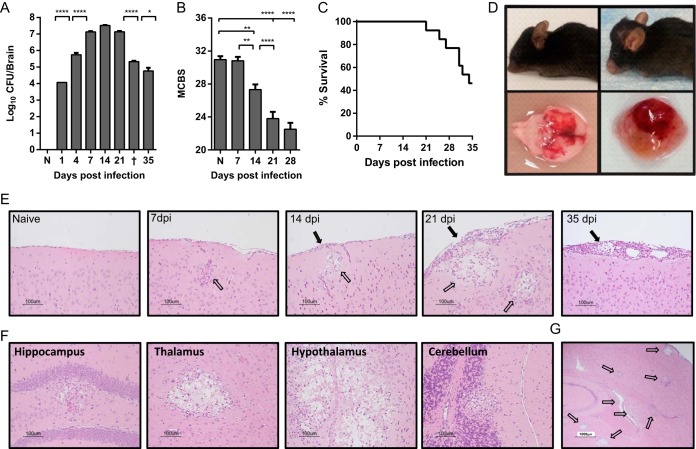
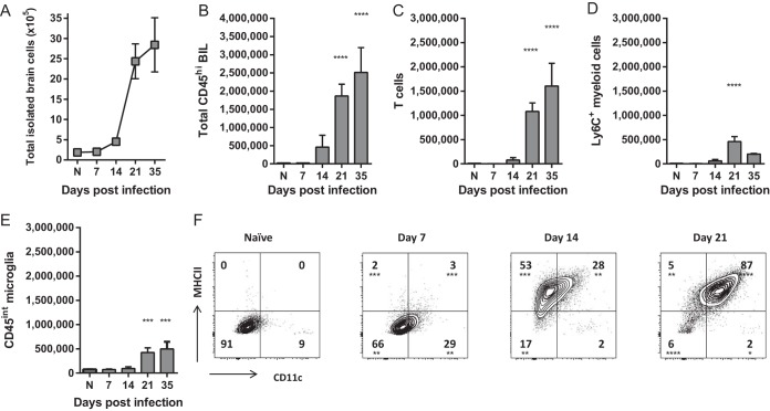
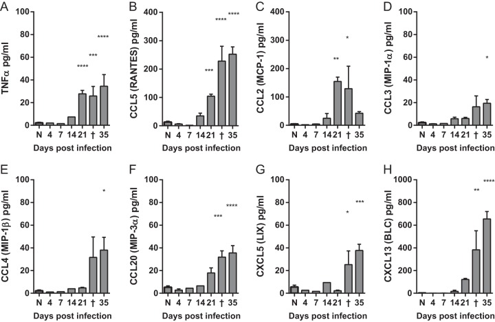
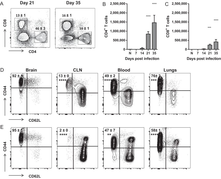
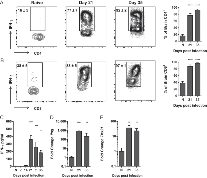
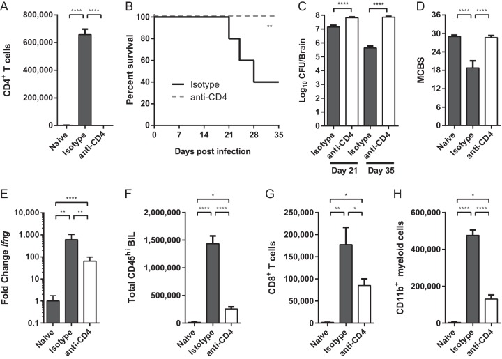
Cryptococcus neoformans is a major fungal pathogen that disseminates to the central nervous system (CNS) to cause fatal meningoencephalitis, but little is known about immune responses within this immune-privileged site. CD4+ T cells have demonstrated roles in anticryptococcal defenses, but increasing evidence suggests that they may contribute to clinical deterioration and pathology in both HIV-positive (HIV+) and non-HIV patients who develop immune reconstitution inflammatory syndrome (IRIS) and post-infectious inflammatory response syndrome (PIIRS), respectively. Here we report a novel murine model of cryptococcal meningoencephalitis and a potential damaging role of T cells in disseminated cryptococcal CNS infection. In this model, fungal burdens plateaued in the infected brain by day 7 postinfection, but activation of microglia and accumulation of CD45hi leukocytes was significantly delayed relative to fungal growth and did not peak until day 21. The inflammatory leukocyte infiltrate consisted predominantly of gamma interferon (IFN-γ)-producing CD4+ T cells, conventionally believed to promote fungal clearance and recovery. However, more than 50% of mice succumbed to infection and neurological dysfunction between days 21 and 35 despite a 100-fold reduction in fungal burdens. Depletion of CD4+ cells significantly impaired IFN-γ production, CD8+ T cell and myeloid cell accumulation, and fungal clearance from the CNS but prevented the development of clinical symptoms and mortality. These findings conclusively demonstrate that although CD4+ T cells are necessary to control fungal growth, they can also promote significant immunopathology and mortality during CNS infection. The results from this model may provide important guidance for development and use of anti-inflammatory therapies to minimize CNS injury in patients with severe cryptococcal infections.IMPORTANCE CNS infection with the fungal pathogen Cryptococcus neoformans often results in debilitating brain injury and has a high mortality rate despite antifungal treatment. Treatment is complicated by the fact that immune responses needed to eliminate infection are also thought to drive CNS damage in a subset of both HIV+ and non-HIV patients. Thus, physicians need to balance efforts to enhance patients' immune responses and promote microbiological control with anti-inflammatory therapy to protect the CNS. Here we report a novel model of cryptococcal meningoencephalitis demonstrating that fungal growth within the CNS does not immediately cause symptomatic disease. Rather, accumulation of antifungal immune cells critically mediates CNS injury and mortality. This model demonstrates that antifungal immune responses in the CNS can cause detrimental pathology and addresses the urgent need for animal models to investigate the specific cellular and molecular mechanisms underlying cryptococcal disease in order to better treat treat patients with CNS infections.

Keywords: Cryptococcus; Cryptococcus neoformans; IRIS; PIIRS; T cells; central nervous system infections; encephalitis; fungi; immunopathology; meningitis; opportunistic infections.


Figures

FIG 1
FIG 1
Infection with Cryptococcus neoformans 52D causes a highly lethal meningoencephalitis in C57BL/6 mice despite fungal clearance. C57BL/6 mice were infected with 106 CFU of C. neoformans 52D via retro-orbital intravenous inoculation. (A) Fungal burdens were measured in whole-brain homogenates at the indicated time points postinfection. Naive mice (N) and animals that succumbed to infection and were euthanized between days 31 and 33 postinfection (†) are indicated. (B) Overall health and neurological status was assessed using a modified murine coma and behavioral scale (MCBS), out of a maximum score of 36 points. Data shown in panels A and B are the means plus standard errors of the means (SEM) (error bars) from a representative experiment of 2 to 5 independent experiments with 3 to 8 mice per time point. Values that are statistically significantly different are indicated by brackets and asterisks as follows: *, P < 0.05; **, P < 0.01; ****, P < 0.0001. (C) In separate experiments, survival was monitored through day 35 postinfection. Infection was considered fatal, and animals were euthanized when they lost 20% body weight, had persistent cranial swelling, and/or developed neurological sequelae such as seizures, tremors, limb weakness or paralysis. (D) Representative images of severe cranial swelling and CNS tissue injury in infected mice. (E to G) Brains from perfused mice were paraffin embedded, coronally sectioned, and stained with hematoxylin and eosin (H&E). Note that stained C. neoformans organisms occupy a relatively small area compared to areas of displaced cerebral tissue by accumulating polysaccharide capsule material, leaving behind a characteristic “Swiss cheese” pattern. Solid black arrows indicate inflammation and cryptococcal organisms in the meninges; clear arrows indicate inflammation and cryptococcal organisms in the parenchyma. (E to G) Representative images shown are from the cortex at various time points postinfection (in days postinfection [dpi]) in panel E (20×), various anatomical structures at 21 days postinfection (20×) (F), and whole brain at 21 days postinfection (4×) (G).
FIG 2
FIG 2
Recruitment of brain-infiltrating leukocytes and activation of microglia is delayed relative to early fungal growth in the CNS. (A) The total number of cells isolated from the leukocyte-enriched fraction of brains, containing predominantly leukocytes and microglia, was quantified at various time points postinfection. (B to D) The number of CD45hi brain-infiltrating leukocytes (BIL) (B), including T cells (C) and CD11b+ LyC+ myeloid cells (D) was determined by flow cytometry. (E and F) Numbers of CD45int microglia (E) and expression of surface activation markers CD11c and MHCII (F) were also quantified. Note that there was not significant accumulation of BIL or complete activation of microglia until late on day 21 postinfection, delayed relative to peak fungal growth but coinciding with the onset of mortality (Fig. 1). Data shown are the mean ± SEM from a representative experiment of two to four independent experiments with three to eight animals per time point. Values that are statistically significantly different from the means for naive (N) animals are indicated by asterisks as follows: *, P < 0.05; **, P < 0.01; ***, P < 0.001; ****, P < 0.0001.
FIG 3
FIG 3
Production of inflammatory cytokines in the brains of mice with cryptococcal meningoencephalitis is delayed but coincides with leukocyte recruitment. (A to H) Levels of inflammatory cytokines were measured in the supernatants of whole-brain homogenates. Each brain was homogenized in 5 ml of medium. Naive mice (N) and animals that succumbed to infection and were euthanized between days 31 to 33 postinfection (†) are indicated. Note that there was not significant induction of any cytokines measured until late on day 21 postinfection. Data shown are the means ± SEM from a representative of two to four independent experiments with three to eight mice per time point. Values that are statistically significantly different from the means for naive animals are indicated by asterisks as follows: *, P < 0.05; **, P < 0.01; ***, P < 0.001; ****, P < 0.0001.
FIG 4
FIG 4
CD4+ and CD8+ T cells that accumulate in the brains of mice with cryptococcal meningoencephalitis are antigen-experienced cells. (A to C) The percentages (of total CD45hi BIL) (A) and total numbers of CD4+ and CD8+ T cells (B and C) that accumulate in the brains of C. neoformans-infected mice. (D and E) The frequencies of antigen-experienced (CD44hi CD62Llo) CD4+ (D) and CD8+ (E) T cells isolated from the brains, cervical lymph nodes (CLN), blood, and lungs of infected mice on day 35 postinfection. Similar frequencies were observed on day 21. Data shown are the mean ± SEM from a representative experiment of two to four independent experiments with three to eight mice per time point. Representative flow cytometry plots are shown. Values that are statistically significantly different from the means for naive (N) animals (B and C) or brain-infiltrating cells (D and E) are indicated by asterisks as follows: **, P < 0.01; ***, P < 0.001; ****, P < 0.0001.
FIG 5
FIG 5
CD4+ and CD8+ T cells that accumulate in the brains of mice with cryptococcal meningoencephalitis are uniformly Th1 polarized and produce IFN-γ. (A and B) Frequency of IFN-γ-producing CD4+ (A) and CD8+ (B) T cells isolated from the brains of naive (N) and infected mice on day 21 and 35 postinfection. (C to E) Levels of IFN-γ protein and expression of IFN-γ (Ifng) (D) and Tbet (Tbx21) transcript (E) were measured in whole-brain homogenates. Note that fold expression data are on a log scale. Data shown are the means ± SEM from a representative of two to four independent experiments with three to eight mice per time point. Representative flow cytometry plots are shown. Values that are statistically significantly different from the means for naive (N) animals are indicated by asterisk as follows: **, P < 0.01; ***, P < 0.001; ****, P < 0.0001.
FIG 6
FIG 6
Depletion of CD4+ T cells rescues survival despite failure to clear infection during cryptococcal meningoencephalitis. (A) Treatment with anti-CD4 depleting antibody (white bar) reduces the total number of brain-infiltrating CD4+ T cells by >99% compared to infected isotype-treated controls (dark gray bar). (B) Survival of infected CD4-depleted (broken line) and isotype-treated mice through day 35 postinfection. (C and D) Brain fungal burdens were calculated on day 21 and 35 postinfection (C), and MCBS scores were calculated on day 21 (D). (E) Expression of IFN-γ transcript was quantified in whole-brain homogenates on day 35. Note that fold expression data are on a log scale. (F to H) The total numbers of CD45hi BIL (F), CD8+ T cells (G), and CD11b+ Ly6C+ myeloid cells (H) were quantified by flow cytometry on day 35 postinfection. Similar reductions were also observed on day 21. Data shown are the means ± SEM from a representative experiment of three independent experiments with 5 to 10 mice per group. Values that are statistically significantly different are indicated by brackets and asterisks as follows: *, P < 0.05; **, P < 0.01; ****, P < 0.0001.

Comment in

References

    1. Park BJ, Wannemuehler KA, Marston BJ, Govender N, Pappas PG, Chiller TM. 2009. Estimation of the current global burden of cryptococcal meningitis among persons living with HIV/AIDS. AIDS 23:525–530. doi:10.1097/QAD.0b013e328322ffac. - DOI - PubMed
    1. Pappas PG. 2013. Cryptococcal infections in non-HIV-infected patients. Trans Am Clin Climatol Assoc 124:61–79. - PMC - PubMed
    1. Rajasingham R, Smith RM, Park BJ, Jarvis JN, Govender NP, Chiller TM, Denning DW, Loyse A, Boulware DR. 2017. Global burden of disease of HIV-associated cryptococcal meningitis: an updated analysis. Lancet Infect Dis 17:873–881. doi:10.1016/S1473-3099(17)30243-8. - DOI - PMC - PubMed
    1. Bicanic T, Harrison T, Niepieklo A, Dyakopu N, Meintjes G. 2006. Symptomatic relapse of HIV-associated cryptococcal meningitis after initial fluconazole monotherapy: the role of fluconazole resistance and immune reconstitution. Clin Infect Dis 43:1069–1073. doi:10.1086/507895. - DOI - PubMed
    1. Saag MS, Graybill RJ, Larsen RA, Pappas PG, Perfect JR, Powderly WG, Sobel JD, Dismukes WE. 2000. Practice guidelines for the management of cryptococcal disease. Infectious Diseases Society of America. Clin Infect Dis 30:710–718. doi:10.1086/313757. - DOI - PubMed

MeSH terms

Substances

LinkOut - more resources