. 2017 Nov 21;8(1):1652.
doi: 10.1038/s41467-017-01815-7.

Single-molecule visualization of conformational changes and substrate transport in the vitamin B12 ABC importer BtuCD-F

Affiliations

Single-molecule visualization of conformational changes and substrate transport in the vitamin B12 ABC importer BtuCD-F

Joris M H Goudsmits et al. Nat Commun. .

Abstract

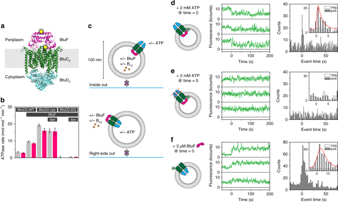
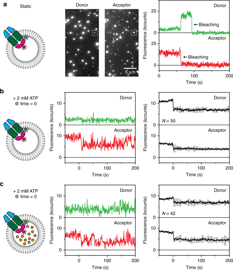
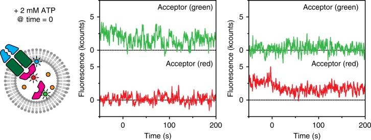
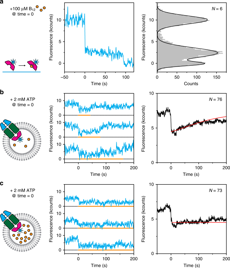
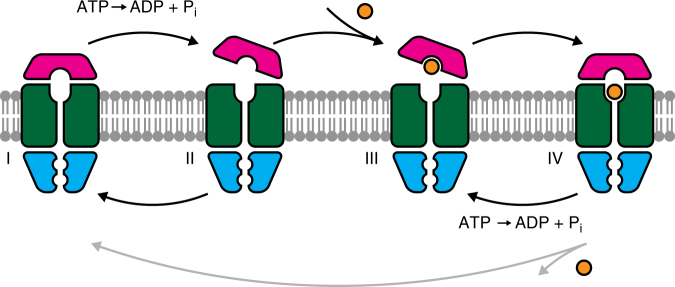
ATP-binding cassette (ABC) transporters form the largest class of active membrane transport proteins. Binding and hydrolysis of ATP by their highly conserved nucleotide-binding domains drive conformational changes of the complex that mediate transport of substrate across the membrane. The vitamin B12 importer BtuCD-F in Escherichia coli is an extensively studied model system. The periplasmic soluble binding protein BtuF binds the ligand; the transmembrane and ATPase domains BtuCD mediate translocation. Here we report the direct observation at the single-molecule level of ATP, vitamin B12 and BtuF-induced events in the transporter complex embedded in liposomes. Single-molecule fluorescence imaging techniques reveal that membrane-embedded BtuCD forms a stable complex with BtuF, regardless of the presence of ATP and vitamin B12. We observe that a vitamin B12 molecule remains bound to the complex for tens of seconds, during which several ATP hydrolysis cycles can take place, before it is being transported across the membrane.


Conflict of interest statement

The authors declare no competing financial interests.

Figures

Fig. 1
Fig. 1
Experimental setup, ATPase rate and fluorescence quenching by ATP and BtuF. a Structure of BtuF (pink) bound to BtuCD (BtuC homodimer: green, BtuD homodimer: blue) in the absence of nucleotide and substrate (Protein Data Bank (PDB) ID 2QI9). Cysteine mutations for labelling in BtuF (D141C) and BtuC (Q111C) are marked with a yellow dot. The distance between the labelling positions is ~37 Å. b ATPase rate of various BtuCD-F mutants with or without fluorescent label reconstituted in liposomes and loaded with (pink bar) or without (grey bar) vitamin B12. BtuCD WT denotes the wild-type, BtuCDcys denotes the cysteine mutant and BtuCDEQ denotes the cysteine mutant that is ATPase-impaired. For all combinations, the cysteine mutant of BtuF is used. Measured rates are not corrected for orientation of the transporter. When BtuF is present at the concentrations used, the full complex is formed. Values displayed are the mean and standard deviation of three experiments. c Experimental design (fluorescent labels are omitted). BtuCD was reconstituted in liposomes of 100-nm diameter in ratios such that on average one transporter was found per liposome. By introducing BtuF and vitamin B12 to the lumen of the vesicle and ATP on the outside or vice versa, only one particular orientation of the transporter was probed. Proteoliposomes are tethered to a glass surface via a biotin-streptavidin link and imaged using TIRF microscopy. d A complex of BtuCDcys labelled with Alexa Fluor 555 and unlabelled BtuF showed decrease in fluorescence intensity upon addition of 2 mM ATP and 10 mM Mg2+ on the outside (middle panel). The distribution of event times of the first drop of intensity is plotted in a histogram (right panel) for the positive (pos, with ATP) and negative (neg, without ATP) experiment. For a description of the data analysis, see methods. e Similar experiment as described in c, but with the ATPase impaired mutant BtuCDEQ. No events were observed. f Similar experiment as described in c, but the vesicle lumen was left empty. Upon introduction of BtuF to the outside of the liposomes an increase in fluorescence intensity was observed. For each condition in df around 1000 single-molecule fluorescence traces were analysed
Fig. 2
Fig. 2
Complex formation observed with FRET. a A stable complex of BtuCDcys (Alexa Fluor 555) and BtuF (Alexa Fluor 647) was formed when reconstituted in substrate-free liposomes. The middle panel shows a section of a field of view. On excitation of donor fluorophores (left channel), emission of acceptor fluorophores was visible (right channel), which is an indication of FRET and thus complex formation. The right panel shows the fluorescence traces of the spots marked with a square. A 2.5× higher laser power density was used to promote bleaching of the dyes, here visible at ~65 and ~100 s. b The same complex as in a was used, but now ATP and Mg2+ were introduced at time zero. The total intensity (sum of donor and acceptor fluorescence) decreased when ATP was present, and dynamics in the signal are visible (middle panel). The right panel shows the average of all traces where a drop in total intensity was observed upon introduction of ATP; the pair of traces shown in the middle panel is one of them. The grey floating bars indicate the number of times the signal exceeded a threshold (see Methods), and thus report on the extent of the fluctuations increasing at positive times. In total, over 1000 fluorescence traces were analysed. c Similar experiment as described in b, but with 100 µM vitamin B12 introduced to the lumen of the vesicles
Fig. 3
Fig. 3
Absence of exchange observed with FRET. BtuCDcys labelled with Alexa Fluor 488 was reconstituted in liposomes loaded with 50% BtuF labelled with Alexa Fluor 555 (acceptor, green), 50% BtuF labelled with Alexa Fluor 647 (acceptor, red), and 100 µM vitamin B12. One example of an initially bound ‘green’ BtuF (middle panel) and one of an initially bound ‘red’ BtuF (right panel) are displayed. While ATP and Mg2+ were present, no increase in fluorescence intensity in the opposite channel (red or green respectively) was observed
Fig. 4
Fig. 4
Transport of vitamin B12 observed with fluorescence quenching. a Dye quenching by vitamin B12. His-tagged BtuF labelled with Alexa Fluor 488 was immobilised on the glass surface via anti-His antibodies. Upon introduction of substrate, the fluorescence was quenched (middle panel) before it bleached around 100 s. Fluorescence intensities were collected in a histogram (right panel) to show that the quenching effect was ~75%. b When unlabelled BtuCD was reconstituted in liposomes loaded with 10 µM vitamin B12 (~3 molecules) and BtuF labelled with Alexa Fluor 488, a decrease in intensity was observed upon introduction of ATP and Mg2+ (middle panel). The orange line marks the periods of low fluorescence and indicates when a substrate molecule was present in the transporter. Averaging all traces (right panel) shows a gradual increase in intensity, which we attribute to depletion of substrate from the liposome. For data analysis, see methods. c Similar experiment as described in b, but with ten times higher concentration of vitamin B12. Depletion as observed in b was not visible here. For each condition, more than roughly 1000 traces were analysed
Fig. 5
Fig. 5
Transport model. BtuCD-F forms a stable complex in the ground state (I, corresponding to PDB ID 2QI9 and 4DBL). Binding and hydrolysis of ATP partially dislodges BtuF from the transporter (II, related to PDB ID 1L7V and 4R9U). In the absence of vitamin B12, the complex cycles back and forth between states I and II. When substrate is present, BtuF can catch it (III), and the complex will fully associate capturing the vitamin inside the complex (IV, corresponding to PDB ID 4FI3). From this state, the transporter can either translocate the substrate molecule and return to the ground state (I), or it can return via ATP binding and hydrolysis back to the previous state (III). The latter pathway, which does not transport any molecule, is more likely to happen

Similar articles

Cited by

References

    1. Davidson AL, Dassa E, Orelle C, Chen J. Structure, function, and evolution of bacterial ATP-binding cassette systems. Microbiol. Mol. Biol. Rev. 2008;72:317–364. doi: 10.1128/MMBR.00031-07. - DOI - PMC - PubMed
    1. Higgins CF, Linton KJ. The ATP switch model for ABC transporters. Nat. Struct. Mol. Biol. 2004;11:918–926. doi: 10.1038/nsmb836. - DOI - PubMed
    1. Wang B, et al. Membrane porters of ATP-binding cassette transport systems are polyphyletic. J. Membr. Biol. 2009;231:1–10. doi: 10.1007/s00232-009-9200-6. - DOI - PubMed
    1. Rees DC, Johnson E, Lewinson O. ABC transporters: the power to change. Nat. Rev. Mol. Cell. Biol. 2009;10:218–227. doi: 10.1038/nrm2646. - DOI - PMC - PubMed
    1. Erkens GB, et al. The structural basis of modularity in ECF-type ABC transporters. Nat. Struct. Mol. Biol. 2011;18:755–760. doi: 10.1038/nsmb.2073. - DOI - PubMed

Publication types

MeSH terms

LinkOut - more resources