. 2017 Nov 21;8(1):1654.
doi: 10.1038/s41467-017-01837-1.

Cisplatin is retained in the cochlea indefinitely following chemotherapy

Affiliations

Cisplatin is retained in the cochlea indefinitely following chemotherapy

Andrew M Breglio et al. Nat Commun. .

Abstract

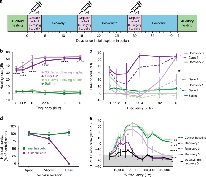
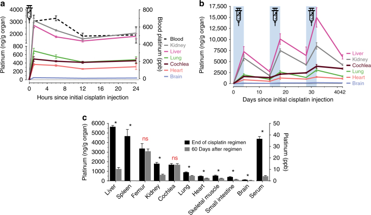
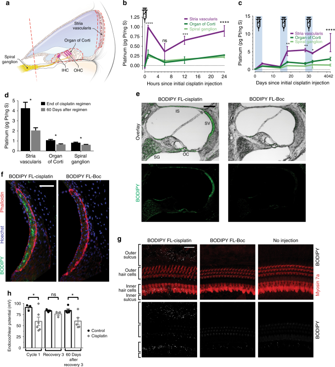
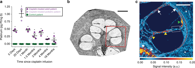
Cisplatin chemotherapy causes permanent hearing loss in 40-80% of treated patients. It is unclear whether the cochlea has unique sensitivity to cisplatin or is exposed to higher levels of the drug. Here we use inductively coupled plasma mass spectrometry (ICP-MS) to examine cisplatin pharmacokinetics in the cochleae of mice and humans. In most organs cisplatin is detected within one hour after injection, and is eliminated over the following days to weeks. In contrast, the cochlea retains cisplatin for months to years after treatment in both mice and humans. Using laser ablation coupled to ICP-MS, we map cisplatin distribution within the human cochlea. Cisplatin accumulation is consistently high in the stria vascularis, the region of the cochlea that maintains the ionic composition of endolymph. Our results demonstrate long-term retention of cisplatin in the human cochlea, and they point to the stria vascularis as an important therapeutic target for preventing cisplatin ototoxicity.


Conflict of interest statement

K.M.M. is an Applications Specialist with Electro Scientific Industries, Inc., which manufactures the laser ablation modules utilized in this study. The remaining authors declare no competing financial interests.

Figures

Fig. 1
Fig. 1
A clinically relevant mouse model of cisplatin ototoxicity shows progressive, high-frequency hearing loss. a The three-cycle cisplatin regimen administered to CBA/CaJ mice. Each cycle consisted of 4 days of once-daily i.p. injections of cisplatin followed by 10 days of recovery. b Hearing loss following the cisplatin regimen as measured by auditory brainstem response recordings. Cisplatin caused a moderate to severe hearing loss across frequencies. n = 22 mice for cisplatin and saline groups. n = 6 mice for 60 days after cisplatin and saline groups. Statistical significance as determined by two-way ANOVA followed by the Holm–Šidák multiple comparisons test is displayed for cisplatin vs. saline. c Progression of hearing loss after each cycle of cisplatin and subsequent recovery. High-frequency hearing loss becomes evident after two cycles. Robust hearing loss across all frequencies is evident after three cycles. n = 3–5 for each cisplatin treatment time point, n = 23 for saline group. Two-way ANOVA followed by the Holm–Šidák multiple comparisons test was used to determine significance. d Percentages of surviving inner and outer hair cells (as compared to control averages) following the complete cisplatin regimen. Predominantly outer hair cells in the basal portion of the cochlea are lost. n = 3 mice in each group. e Distortion product otoacoustic emission (DPOAE) amplitudes (a measure of outer hair cell function) after each cycle and recovery period. Gray shaded area depicts the testing noise floor (the underlying background noise detected by the system microphone). n = 3–5 mice at each time point. Two-way ANOVA followed by the Holm–Šidák multiple comparisons test was used to determine significance. Data are expressed as mean ± s.e.m. ns not significant, *P < 0.01, **P < 0.01, ****P < 0.0001
Fig. 2
Fig. 2
Cisplatin is readily cleared from most organs but is retained in the cochlea. a ICP-MS measured platinum concentrations in whole mouse organs isolated throughout the first 24 h following a single i.p. injection of cisplatin. 0-h data points represent mice prior to injection of cisplatin. Concentrations are normalized to organ mass (ng Pt per g of organ, left y-axis) or expressed as parts per billion (ppb, right y-axis) for whole blood. Platinum levels peak in most organs 1 h after injection. Concentrations vary significantly across organs and are highest in kidney and liver. n = 3–4 mice per time point. b Concentrations of cisplatin in whole mouse organs throughout the complete 42-day cisplatin regimen. The three, 4-day long cycles of once-daily i.p. cisplatin are indicated by blue shading. In most organs, platinum levels increase following injection and then decline during the recovery period. In contrast, platinum progressively accumulates in cochlea with no apparent elimination. n = 3–4 mice at each time point for all organs. c Concentrations of platinum in whole mouse organs at the end of the cisplatin regimen and following a subsequent 60-day recovery. During this recovery period, platinum was significantly eliminated from all organs except the cochlea and femur. n = 3–4 mice in each group. Multiple two-tailed t-tests with correction for multiple comparisons by the Holm–Šidák method were used to determine significance. Data are expressed as mean ± s.e.m. ns not significant, *P < 0.05
Fig. 3
Fig. 3
The cochlear stria vascularis accumulates high levels of cisplatin, resulting in its dysfunction. a A cross-sectional illustration of cochlear architecture. The three regions of the cochlea: stria vascularis, organ of Corti and spiral ganglion, microdissected for ICP-MS analysis are indicated by dashed red lines. Endolymph fills the blue shaded region. IHC inner hair cell, OHC outer hair cell. b Platinum concentrations in microdissected cochlear regions in the first 24 h following a single i.p. injection of cisplatin. Cisplatin readily distributes throughout these cochlear regions within the first hour. Platinum concentrations are normalized to sulfur content, a surrogate for total tissue quantity. n = 3–4 mice per time point. Statistical significance is displayed for stria vascularis values as compared to the other two cochlear regions. Two-way ANOVA followed by the Holm–Šidák multiple comparisons test was used to determine significance. c Concentrations of platinum in cochlear regions throughout the complete cisplatin regimen. Platinum progressively accumulates in all three regions, but most significantly in stria vascularis. n = 3 mice per time point. Statistical significance is displayed for stria vascularis values as compared to other two regions. Two-way ANOVA followed by the Holm–Šidák multiple comparisons test was used to determine significance. d Concentrations of platinum in cochlear regions at the end of the cisplatin regimen and following a subsequent 60-day recovery. All regions show a decrease in platinum concentrations. n = 4 mice per group. Unpaired two-tailed t-tests were used to determine significance. e Representative low-magnification confocal slices from cochlear cryosections of mice treated with a full regimen of BODIPY FL-cisplatin or the control (no cisplatin) conjugate BODIPY FL-Boc. SG spiral ganglion, OC organ of Corti, SV stria vascularis, IS interscalar septum. Scale bar = 100 µm. f Representative maximum intensity projections of thin confocal stacks of the stria vascularis only. Scale bar = 50 µm. g Representative maximum intensity projections of confocal stacks of organ of Corti whole mounts. Scale bar = 25 µm. h Endocochlear potentials (EPs) measured in the endolymph following cisplatin treatment. EPs decline at 24 h after cisplatin but recover somewhat by the end of the regimen. However, 60 days after cessation of cisplatin administration, EPs are still significantly reduced. n = 3–6 mice per group; data points represent individual mice. Unpaired two-tailed t-tests were used to determine significance. Data are expressed as mean ± s.e.m. ns not significant, *P < 0.05, **P < 0.01, ***P < 0.001, ****P < 0.0001
Fig. 4
Fig. 4
Cisplatin is retained in human cochleae long after cessation of treatment. a Platinum concentrations measured in cochlear sections of human temporal bones. Matched pairs of patients who received cisplatin chemotherapy and control (cisplatin-naive) patients are plotted by the time elapsed between the patient’s last cisplatin infusion and their death. Each data point represents an individual 20 µm cochlear section. Multiple two-tailed t-tests with correction for multiple comparisons by the Holm–Šidák method were used to determine significance. Data are expressed as mean ± s.e.m. *P < 0.05. b Representative brightfield image of a cochlear section prior to ICP-MS analysis. Similar sections were digested whole and analyzed for total cisplatin content. This section was laser ablated in the region outlined in red. Scale bar = 1 mm. c Laser ablation ICP-MS image of platinum distribution in the area in the red box in b from a patient who was treated with cisplatin. This patient died 25 days after their last cisplatin infusion. Green arrowhead marks stria vascularis, yellow arrowheads mark cochlear nerve fibers, white arrowheads mark the boundary between the cochlear nerve and the bone of the cochlear modiolus, white arrows mark the endosteum which lines the cochlear canal, asterisks mark surrounding cochlear bone, and white box frames organ of Corti. Scale bar = 500 µm. Platinum signal intensity values are in arbitrary units

References

    1. Miller KD, et al. Cancer treatment and survivorship statistics, 2016. CA Cancer J. Clin. 2016;66:271–289. doi: 10.3322/caac.21349. - DOI - PubMed
    1. Frisina RD, et al. Comprehensive audiometric analysis of hearing impairment and tinnitus after cisplatin-based chemotherapy in survivors of adult-onset cancer. J. Clin. Oncol. 2016;34:2712–2720. doi: 10.1200/JCO.2016.66.8822. - DOI - PMC - PubMed
    1. van Zeijl LG, Conijn EA, Rodenburg M, Tange RA, Brocaar MP. Analysis of hearing loss due to cis-diamminedichloroplatinum-II. Arch. Otorhinolaryngol. 1984;239:255–262. doi: 10.1007/BF00464252. - DOI - PubMed
    1. Knight KR, Kraemer DF, Neuwelt EA. Ototoxicity in children receiving platinum chemotherapy: underestimating a commonly occurring toxicity that may influence academic and social development. J. Clin. Oncol. 2005;23:8588–8596. doi: 10.1200/JCO.2004.00.5355. - DOI - PubMed
    1. Stohr W, et al. Cisplatin-induced ototoxicity in osteosarcoma patients: a report from the late effects surveillance system. Cancer Invest. 2005;23:201–207. doi: 10.1081/CNV-200055951. - DOI - PubMed

Publication types

MeSH terms