Review
. 2018 Feb;15(2):96-110.
doi: 10.1038/nrgastro.2017.150. Epub 2017 Nov 22.

Hepatitis E virus: advances and challenges

Affiliations
Review

Hepatitis E virus: advances and challenges

Ila Nimgaonkar et al. Nat Rev Gastroenterol Hepatol. 2018 Feb.

Abstract

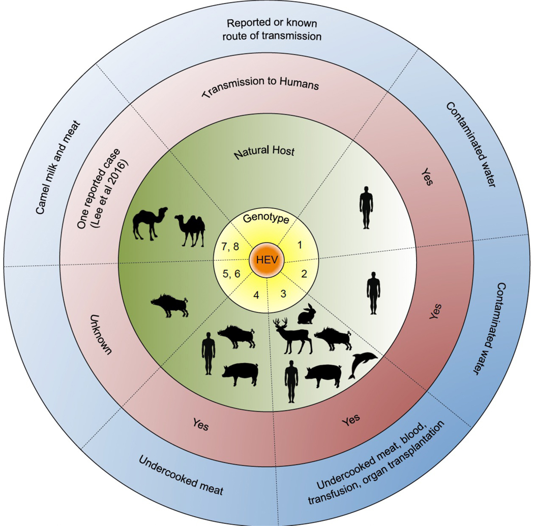
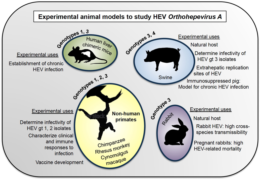
At least 20 million hepatitis E virus (HEV) infections occur annually, with >3 million symptomatic cases and ∼60,000 fatalities. Hepatitis E is generally self-limiting, with a case fatality rate of 0.5-3% in young adults. However, it can cause up to 30% mortality in pregnant women in the third trimester and can become chronic in immunocompromised individuals, such as those receiving organ transplants or chemotherapy and individuals with HIV infection. HEV is transmitted primarily via the faecal-oral route and was previously thought to be a public health concern only in developing countries. It is now also being frequently reported in industrialized countries, where it is transmitted zoonotically or through organ transplantation or blood transfusions. Although a vaccine for HEV has been developed, it is only licensed in China. Additionally, no effective, non-teratogenic and specific treatments against HEV infections are currently available. Although progress has been made in characterizing HEV biology, the scarcity of adequate experimental platforms has hampered further research. In this Review, we focus on providing an update on the HEV life cycle. We will further discuss existing cell culture and animal models and highlight platforms that have proven to be useful and/or are emerging for studying other hepatotropic (viral) pathogens.


Conflict of interest statement

Competing interests statement

A.P. and Q.D. are inventors on a patent application defining an hepatitis E virus transcomplementation system for antiviral drug screening and ORF3’s viroporin function as a antiviral drug target.

Figures

Fig 1.
Fig 1.. Host range of hepatitis E virus.
The Orthohepevirus A genus is classified into hepatitis E virus (HEV) genotype 1–8. Genotypes 1 and 2 are limited to human hosts and are transmitted via the faecal–oral route, primarily through contaminated water. Genotypes 3 and 4 have multiple hosts, and can be transmitted to humans through the consumption of undercooked meats, including pork. Genotypes 5 and 6 are known to infect wild boar; however, it is unknown whether these genotypes can be transmitted to humans (although there have been reports of wild boar genotype 3 HEV transmission to humans). Finally, genotypes 7 and 8 infect dromedary and bactrian camels, respectively. There has been one case reported of genotype 7 HEV transmission to a liver transplant patient who consumed camel meat and milk.
Fig 2.
Fig 2.. Genetic organization and translation of hepatitis E virus.
Hepatitis E virus (HEV) is a ~7.2kB, positive (+)-sense single-stranded RNA virus. The mRNA is capped at the 5’ end, polyadenylated at the 3’ end, and the junctional region (JR) between ORF1 and ORF2/3 contains a stem-loop structure that is critical for HEV replication. After viral entry and uncoating, the (+)-sense full-length viral genome is translated by host ribosomes to produce the ORF1 polyprotein, which contains the non-structural replication machinery of the virus including the methyltransferase (Met), RNA helicase (Hel), and RNA-dependent RNA polymerase (RdRp), as well as several non-enzymatic regions essential for efficient viral replication (the ‘Y’, ‘X’, and ‘hypervariable’ (HVR) regions). Additionally, ORF1 contains a putative papain-like cysteine protease (PCP) based on sequence similarity to the protease of rubella virus, though data showing protease activity for this region have been conflicting. It is unclear whether the ORF1 polyprotein undergoes processing into smaller units. HEV genotype 1 is thought to contain an additional open reading frame, ORF4, that is translated into a viral protein enhancing RdRp activity. After translation of the ORF1 polyprotein, the RdRp from ORF1 transcribes an antisense (–)-stranded intermediate RNA from the (+)-sense strand. The (–)-sense strand then serves as a template for the transcription of more (+)-sense full-length RNA for packaging into new progeny virions, as well as a shorter, ~2.2kB subgenomic RNA (sgRNA) encoding ORF2 and ORF3. These viral genes are ~2.2kB and ~360bp in length, respectively, and ORF3 entirely overlaps with ORF2 except for one leading base pair. The sgRNA, which is capped at the 5’ end and polyadenylated at the 3’ end, is then translated into the ORF2 capsid protein and the ORF3 viroporin based on a leaky scanning mechanism by host ribosomes. Regulation of transcription of the sgRNA is poorly understood.
Fig 3.
Fig 3.. Life cycle of hepatitis E virus.
(1) Viral entry: hepatitis E virus (HEV) is a quasi-enveloped virus, meaning it can exist in the non-enveloped state (HEV) or can be coated in a lipid-derived membrane (eHEV). HEV and eHEV have distinct entry mechanisms. Little is known about entry mechanisms for HEV. For eHEV, the virus enters the cell through clathrin-dependent and dynamin-dependent, receptor-mediated endocytosis. A specific cell surface receptor mediating eHEV entry remains to be identified, but the GTPases Rab5 and Rab7 are known to have a role in eHEV entry. Upon entering the cell, the envelope of eHEV undergoes lysosome-mediated lipid degradation, and uncoats in a poorly understood process to expose the viral mRNA. (2) ORF1 polyprotein (pORF1) containing the RdRp is translated from the (+)-strand, and the RdRp then transcribes full-length (–)-sense RNA. The (–)-sense RNA serves as a template for transcribing more full-length (+)-sense RNA to be packaged into progeny virions, as well as a shorter subgenomic RNA (sgRNA) which encodes ORF2 and ORF3. The ORF2 capsid protein (pORF2), and the ORF3 protein (pORF3), a viroporin essential for viral release, are translated from the sgRNA. (3) pORF3 binds to TSG101, a member of the endosomal sorting complexes required for transport (ESCRT) pathway that is used by several other RNA viruses to bud from cell membranes. The interaction of pORF3 with TSG101 probably promotes budding of progeny virions into multivesicular bodies (MVBs), which then fuse with the plasma membrane to release virions from the cell. The lipid envelope of eHEV is thought to be derived from the trans-Golgi network, and viral particles contained in eHEV have been shown to be associated with pORF3. pORF3 has additionally been shown to exhibit viroporin activity, and it is possible that pORF3 exists in multiple forms to perform distinct functions. (4) eHEV released from the apical membrane enters the bile duct, where the lipid envelope is thought to be degraded by detergents and proteases in the bile. This feature would explain why HEV in the faeces is non-enveloped. On the other hand, eHEV released from the basal membrane of hepatocytes enters the serum in its quasi-enveloped form, where it is protected from neutralizing antibodies against pORF2 and pORF3, but is less efficient at infecting cells.
Fig 4.
Fig 4.. Towards more physiologically relevant 2D and 3D cell culture models for studying HEV.
The species permissive to HEV infection include (but are not limited to) humans, rabbits, swine, deer, wild boar, and camel. In order to better study HEV infection in physiologically relevant in vitro models, it will be desirable to generate co-cultures that recapitulate the complexity of the liver including endothelial, stellate, cholangiocytic, Kupffer, and hepatic cells in the appropriate ratios. These cells can be harvested from primary tissue or differentiated from stem cells, and could be derived from the aforementioned species to explore viral host tropism. Primary cultures have the disadvantage of limited durability; this issue can be overcome by differentiating the various cell types from indefinitely self-renewing stem cells. The latter, however, is technically challenging and requires advances in current hepatic differentiation protocols. Incorporating primary and stem-cell derived tissues into a 3D architecture will also be important to more closely mimic the physiological hepatic environment and preserve cell morphology. The architecture of the liver leads to heterogeneous environmental cues (e.g. nutrients, oxygen, inflammatory factors, etc) reaching individual cells, and 3D cultures can better capture this phenomenon. Furthermore it was previously shown that primary human hepatocyte dedifferentiation can be delayed or prevented in collagen sandwich cultures, by aggregation in spheroids, or in co-culture with non-parenchymal cells.
Fig 5.
Fig 5.. Experimental animal models to study HEV Orthohepevirus A.
Experimental animal models that have been used to study HEV include non-human primates, swine, rabbits, and human liver chimeric mice. Chimpanzees, rhesus monkeys, and Cynomolgus macaques were the earliest animal models in HEV research, and have been used to study HEV pathogenesis and vaccine efficacy. Swine, which are naturally infected with gts 3 and 4 of HEV and can transmit these strains to humans, have been used to determine the infectivity of gt 3 isolates, and to show extrahepatic replication sites of HEV. Recently, an iatrogenically immunosuppressed swine model was shown to support chronic infection with gt 3, and similarly, human liver chimeric mice can support chronic infection with gt1 and gt3 HEV. Gt, genotype.

References

    1. Khuroo MS, Khuroo MS & Khuroo NS Hepatitis E: Discovery, global impact, control and cure. World J Gastroenterol 22, 7030–7045, doi: 10.3748/wjg.v22.i31.7030 (2016). - DOI - PMC - PubMed
    1. Khuroo MS Study of an epidemic of non-A, non-B hepatitis. Possibility of another human hepatitis virus distinct from post-transfusion non-A, non-B type. Am J Med 68, 818–824 (1980). - PubMed
    1. Wong DC, Purcell RH, Sreenivasan MA, Prasad SR & Pavri KM Epidemic and endemic hepatitis in India: evidence for a non-A, non-B hepatitis virus aetiology. Lancet 2, 876–879 (1980). - PubMed
    1. Balayan MS et al. Evidence for a virus in non-A, non-B hepatitis transmitted via the fecal-oral route. Intervirology 20, 23–31 (1983).

      A volunteer ingested pooled stool extracts from patients infected with HEV to show for the first time that the virus is fecal-orally transmitted.

    1. Reyes GR et al. Isolation of a cDNA from the virus responsible for enterically transmitted nonA, non-B hepatitis. Science 247, 1335–1339 (1990).

      The first partial cDNA of ET-NANBH was cloned, and the virus renamed ‘hepatitis E virus’.

Publication types

LinkOut - more resources