

A Complex Interplay of Vitamin B1 and B6 Metabolism with Cognition, Brain Structure, and Functional Connectivity in Older Adults
- PMID: 29163003
- PMCID: PMC5663975
- DOI: 10.3389/fnins.2017.00596
A Complex Interplay of Vitamin B1 and B6 Metabolism with Cognition, Brain Structure, and Functional Connectivity in Older Adults
Abstract
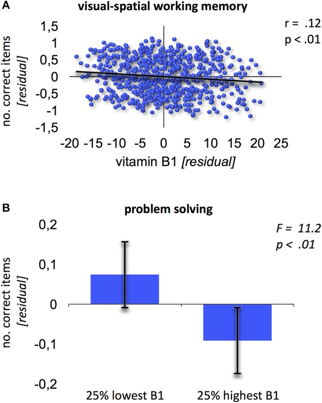
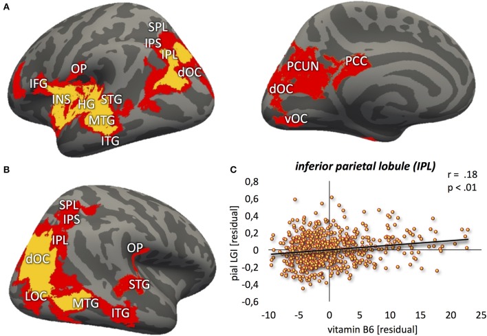
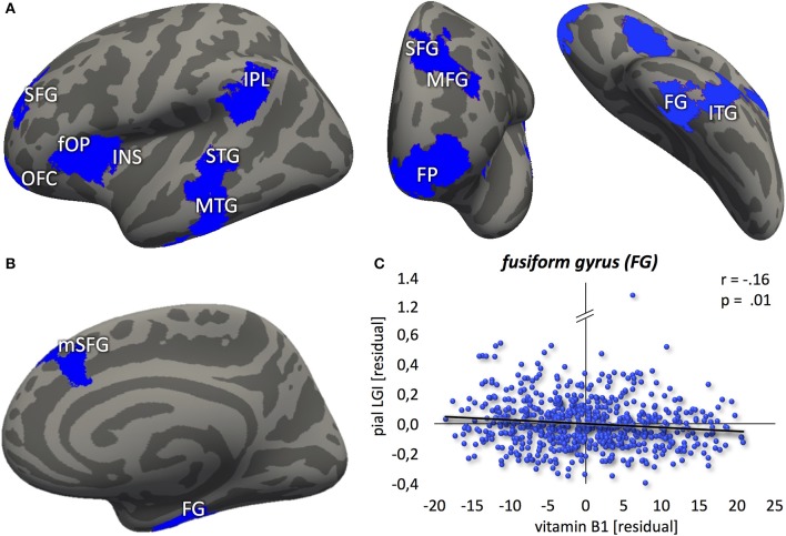
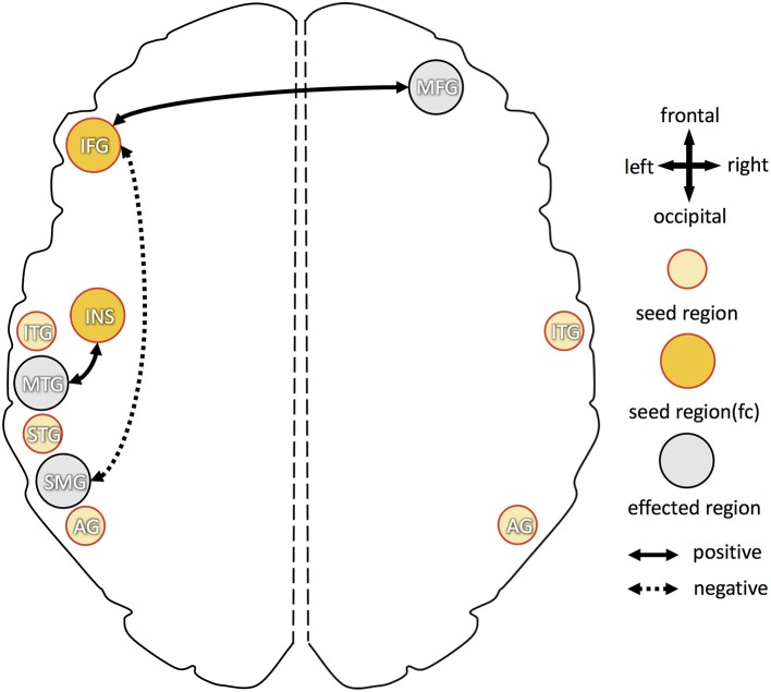
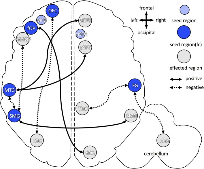
Aging is associated with brain atrophy, functional brain network reorganization and decline of cognitive performance, albeit characterized by high interindividual variability. Among environmental influencing factors accounting for this variability, nutrition and particularly vitamin supply is thought to play an important role. While evidence exists that supplementation of vitamins B6 and B1 might be beneficial for cognition and brain structure, at least in deficient states and neurodegenerative diseases, little is known about this relation during healthy aging and in relation to reorganization of functional brain networks. We thus assessed the relation between blood levels of vitamins B1 and B6 and cognitive performance, cortical folding, and functional resting-state connectivity in a large sample of older adults (N > 600; age: 55-85 years), drawn from the population-based 1000BRAINS study. In addition to blood sampling, subjects underwent structural and functional resting-state neuroimaging as well as extensive neuropsychological testing in the domains of executive functions, (working) memory, attention, and language. Brain regions showing changes in the local gyrification index as calculated using FreeSurfer in relation to vitamin levels were used for subsequent seed-based resting-state functional connectivity analysis. For B6, a positive correlation with local cortical folding was found throughout the brain, while only slight changes in functional connectivity were observed. Contrarily, for B1, a negative correlation with cortical folding as well as problem solving and visuo-spatial working memory performance was found, which was accompanied by pronounced increases of interhemispheric and decreases of intrahemispheric functional connectivity. While the effects for B6 expand previous knowledge on beneficial effects of B6 supplementation on brain structure, they also showed that additional effects on cognition might not be recognizable in healthy older subjects with normal B6 blood levels. The cortical atrophy and pronounced functional reorganization associated with B1, contrarily, was more in line with the theory of a disturbed B1 metabolism in older adults, leading to B1 utilization deficits, and thus, an effective B1 deficiency in the brain, despite normal to high-normal blood levels.
Keywords: B vitamins; FreeSurfer; aging; cognitive performance; gyrification index; interindividual variability; resting state.
Figures
Similar articles
-
Influence of age and cognitive performance on resting-state brain networks of older adults in a population-based cohort.Cortex. 2017 Apr;89:28-44. doi: 10.1016/j.cortex.2017.01.008. Epub 2017 Jan 23. Cortex. 2017. PMID: 28192723
-
Age- and function-related regional changes in cortical folding of the default mode network in older adults.Brain Struct Funct. 2017 Jan;222(1):83-99. doi: 10.1007/s00429-016-1202-4. Epub 2016 Mar 4. Brain Struct Funct. 2017. PMID: 26943919
-
Resting-State Network Patterns Underlying Cognitive Function in Bipolar Disorder: A Graph Theoretical Analysis.Brain Connect. 2020 Sep;10(7):355-367. doi: 10.1089/brain.2019.0709. Epub 2020 Jul 21. Brain Connect. 2020. PMID: 32458698 Free PMC article.
-
Effects of nutrients (in food) on the structure and function of the nervous system: update on dietary requirements for brain. Part 1: micronutrients.J Nutr Health Aging. 2006 Sep-Oct;10(5):377-85. J Nutr Health Aging. 2006. PMID: 17066209 Review.
-
Healthy aging by staying selectively connected: a mini-review.Gerontology. 2014;60(1):3-9. doi: 10.1159/000354376. Epub 2013 Sep 28. Gerontology. 2014. PMID: 24080587 Review.
Cited by
-
Association between Serum Essential Metal Elements and the Risk of Schizophrenia in China.Sci Rep. 2020 Jul 3;10(1):10875. doi: 10.1038/s41598-020-66496-7. Sci Rep. 2020. PMID: 32620780 Free PMC article.
-
Revisiting the Role of Vitamins and Minerals in Alzheimer's Disease.Antioxidants (Basel). 2023 Feb 8;12(2):415. doi: 10.3390/antiox12020415. Antioxidants (Basel). 2023. PMID: 36829974 Free PMC article. Review.
-
Sustainable Valorization of Sambucus nigra L. Berries: From Crop Biodiversity to Nutritional Value of Juice and Pomace.Foods. 2021 Dec 31;11(1):104. doi: 10.3390/foods11010104. Foods. 2021. PMID: 35010230 Free PMC article.
-
Gut Microbiota and its Metabolites: Bridge of Dietary Nutrients and Alzheimer's Disease.Adv Nutr. 2023 Jul;14(4):819-839. doi: 10.1016/j.advnut.2023.04.005. Epub 2023 Apr 17. Adv Nutr. 2023. PMID: 37075947 Free PMC article. Review.
-
Cognitive Improvement in Healthy Older Adults Can Parallel That of Younger Adults Following Lifestyle Modification: Support for Cognitive Reserve During Aging.J Alzheimers Dis Rep. 2018 Nov 2;2(1):201-205. doi: 10.3233/ADR-180056. J Alzheimers Dis Rep. 2018. PMID: 30480262 Free PMC article.
References
-
- Amunts K., Weiss P. H., Mohlberg H., Pieperhoff P., Eickhoff S., Gurd J. M., et al. . (2004). Analysis of neural mechanisms underlying verbal fluency in cytoarchitectonically defined stereotaxic space—The roles of Brodmann areas 44 and 45. Neuroimage 22, 42–56. 10.1016/j.neuroimage.2003.12.031 - DOI - PubMed
-
- Arendt T., Bruckner M. K., Bigl V., Marcova L. (1995). Dendritic reorganisation in the basal forebrain under degenerative conditions and its defects in Alzheimer's disease. III. The basal forebrain compared with other subcortical areas. J. Comp. Neurol. 351, 223–246. 10.1002/cne.903510204 - DOI - PubMed
LinkOut - more resources
Full Text Sources
Other Literature Sources