Review
. 2017 Nov 1:8:887.
doi: 10.3389/fphys.2017.00887. eCollection 2017.

An Update on Sec61 Channel Functions, Mechanisms, and Related Diseases

Affiliations
Review

An Update on Sec61 Channel Functions, Mechanisms, and Related Diseases

Sven Lang et al. Front Physiol. .

Abstract

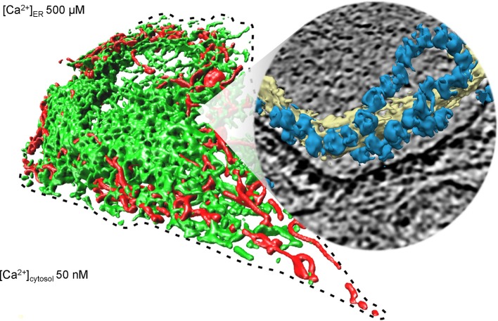
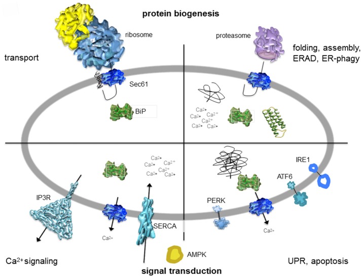
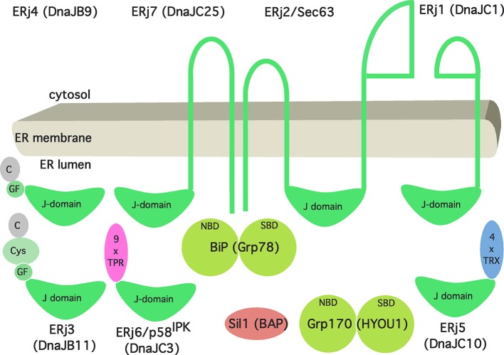
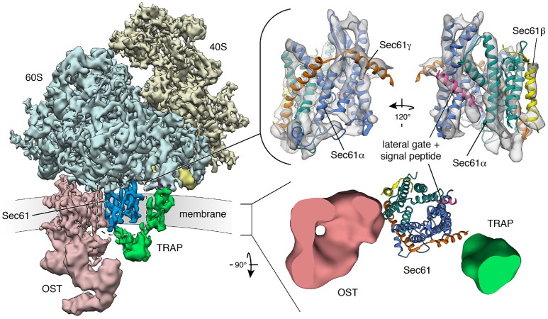
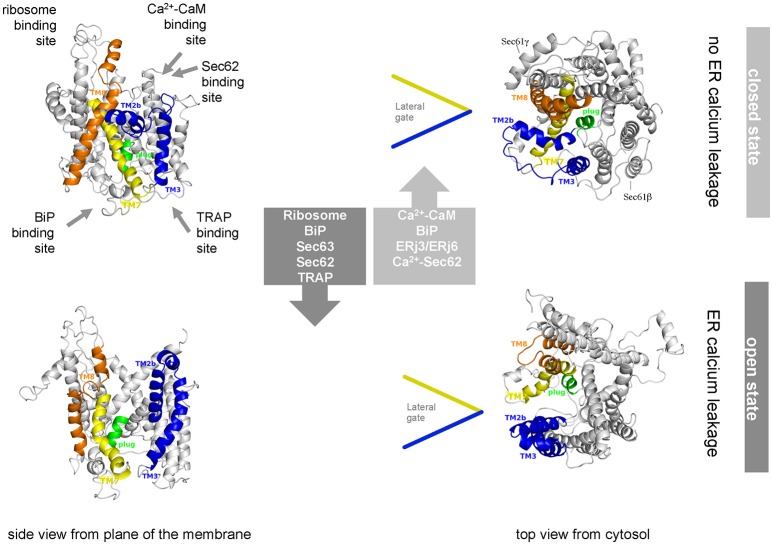
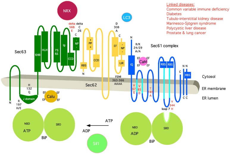
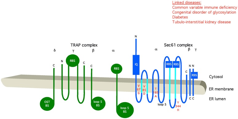
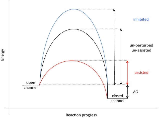
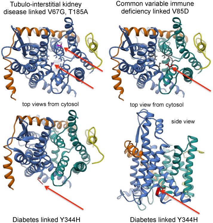
The membrane of the endoplasmic reticulum (ER) of nucleated human cells harbors the protein translocon, which facilitates membrane integration or translocation of almost every newly synthesized polypeptide targeted to organelles of the endo- and exocytotic pathway. The translocon comprises the polypeptide-conducting Sec61 channel and several additional proteins and complexes that are permanently or transiently associated with the heterotrimeric Sec61 complex. This ensemble of proteins facilitates ER targeting of precursor polypeptides, modification of precursor polypeptides in transit through the Sec61 complex, and Sec61 channel gating, i.e., dynamic regulation of the pore forming subunit to mediate precursor transport and calcium efflux. Recently, cryoelectron tomography of translocons in native ER membrane vesicles, derived from human cell lines or patient fibroblasts, and even intact cells has given unprecedented insights into the architecture and dynamics of the native translocon and the Sec61 channel. These structural data are discussed in light of different Sec61 channel activities including ribosome receptor function, membrane insertion, and translocation of newly synthesized polypeptides as well as the putative physiological roles of the Sec61 channel as a passive ER calcium leak channel. Furthermore, the structural insights into the Sec61 channel are incorporated into an overview and update on Sec61 channel-related diseases-the Sec61 channelopathies-and novel therapeutic concepts for their treatment.

Keywords: ATP import; BiP; Sec61 complex; calcium leakage; endoplasmic reticulum; protein biogenesis.


Figures

Figure 1
Figure 1
Collage of 3D reconstructions of mammalian mitochondria and ER, respectively. The left part of the figure represents a 3D reconstruction after live cell fluorescence imaging, following import of a green fluorescent protein into the ER and of a red fluorescent protein into the mitochondria. The plasma membrane is indicated by a dashed line; the position of the round nucleus can be estimated in the upper part of the cell void of ER and mitochondria. Typical concentrations of free calcium are given for cytosol and ER of a resting cell. The right part represents a 3D reconstruction of cellular ER after CET, on top of a slice through the respective tomogram. ER membranes are shown in yellow; 80S ribosomes are shown in blue. The collage is based on Zimmermann (2016).
Figure 2
Figure 2
Artist's depiction of cross-section through the mammalian ER with a focus on signal transduction and protein biogenesis. The non-annotated structures refer to a not yet-folded polypeptide, a natively folded protein, and an aggregate of non-native polypeptides, respectively. AMPK, AMP-activated protein kinase; IP3R, IP3-receptor; SERCA, sarcoplasmic/endoplasmic reticulum Ca2+ ATPase. The cartoon is based on Zimmermann (2016). See text for details.
Figure 3
Figure 3
Artist's view of the Hsp70/Hsp40 chaperone network of the human ER. See text for details. The following binding characteristics (KD) were observed for BiP binding in the presence of ATP (in μM): ERj1, 0.12; Sec63, 5; ERj3, 3.5; ERj4, 6.07; ERj5, 0.45; ERj6, 0.59; ERj7, 1.1. The cartoon and affinities are based on Schorr et al. (2015).
Figure 4
Figure 4
Structure and architecture of the native mammalian translocon visualized using CET. (Left) Overall structure of the native ribosome-translocon complex (EMD 3069) with the ribosomal subunits (40S: yellow; 60S: light blue) and the translocon components Sec61 (dark blue), TRAP (green) and OST (red) depicted. Within the 60S subunit, eL38 (purple) and the short expansion segment (bright yellow), which are contacted by the cytosolic domain of TRAPγ, are highlighted. Right, upper panel: Isolated density for the Sec61 complex with an atomic model of the laterally opened Sec61 complex (PDB 3jc2) superposed. The Sec61α (N-terminal: green; C-terminal half: blue), Sec61β (yellow) and Sec61γ (orange) subunits are indicated. A signal peptide (magenta) is intercalated at the lateral gate. Right, lower panel: Transmembrane region of the translocon with down-filtered densities for membrane-embedded segments of TRAP (green) and OST (red) depicted. Sec61 is represented by an atomic model. The ER membrane resides in the paper plane.
Figure 5
Figure 5
Artist's view of the dynamic equilibrium and gating mechanisms of the human Sec61 complex. Allosteric effectors of the dynamic equilibrium of the Sec61 channel and their binding sites are indicated. The cartoon is based on Dudek et al. (2015). See text for details.
Figure 6
Figure 6
Artist's depiction of the domain organization of Sec61 complex and its auxiliary components BiP, Sec62, and Sec63. Additional interaction partners of BiP (Sil1), Sec61 (Calmodulin, CaM), Sec62 (LC3), and Sec63 (Nucleoredoxin, NRX; Calumenin, Calu) are shown. Furthermore, relevant motifs (such as IQ and LIR) and domains are indicated, as well as point mutations that disturb the respective interaction or are linked to disease (in red). CCD, coiled-coil domain; EF, EF hand; NBD, nucleotide-binding domain; NP, negatively charged patch; PP, positively charged patch; RBS, ribosome-binding site; SBD, substrate-binding domain. The following binding characteristics were observed: BiP/Sec61α Kd 500 μM, ATP-dependent; BiP/Sec63 KD 5 μM; CaM/Sec61α KD 115 nM, Ca2+-dependent, TFP-sensitive; Sec62/Sec61α Ca2+-sensitive; Sec62/LC3 KD 20 μM; Sec63/NRX H2O2-dependent; Sec63/Sec62 KD 5 nM. C, carboxy-terminus; N, amino-terminus. See text for details.
Figure 7
Figure 7
Artist's depiction of the organization of Sec61 complex and its auxiliary component TRAP. Relevant motifs (IQ) and domains are indicated, as well as point mutations that disturb the respective interaction or are linked to disease (in red). BS, binding site; OST, oligosaccharyltransferase; RBS, ribosome-binding site. C, carboxy-terminus; N, amino-terminus. See text for details.
Figure 8
Figure 8
Energetics of Sec61 channel gating. See text for details.
Figure 9
Figure 9
Position of disease-linked mutations in 3D reconstructions of the Sec61 complex. See text for details.

References

    1. Alberti S., Esser C., Höhfeld J. (2003). BAG-1—a nucleotide exchange factor of Hsc70 with multiple cellular functions. Cell Stress Chaperones 8, 225–231. - PMC - PubMed
    1. Ampofo E., Welker S., Jung M., Müller L., Greiner M., Zimmermann R., et al. . (2013). CK2 phosphorylation of human Sec63 regulates its interaction with Sec62. Biochim. Biophys. Acta 1830, 2938–2945. 10.1016/j.bbagen.2012.12.020 - DOI - PubMed
    1. Ast T., Cohen G., Schuldiner M. (2013). A network of cytosolic factors targets SRP-independent proteins to the endoplasmic reticulum. Cell 152, 1134–1145. 10.1016/j.cell.2013.02.003 - DOI - PubMed
    1. Ast T., Michaelis S., Schuldiner M. (2016). The protease Ste24 clears clogged translocons. Cell 164, 103–114. 10.1016/j.cell.2015.11.053 - DOI - PMC - PubMed
    1. Aviram N., Ast T., Costa E. A., Arakel E., Chuartzman S. G., Jan C. H., et al. . (2016). The SND proteins constitute an alternative targeting route to the endoplasmic reticulum. Nature 540, 134–138. 10.1038/nature20169 - DOI - PMC - PubMed

LinkOut - more resources