. 2017 Nov 1;8(11):7306-7311.
doi: 10.1039/c7sc03023a. Epub 2017 Sep 21.

Displacement and hybridization reactions in aptamer-functionalized hydrogels for biomimetic protein release and signal transduction

Affiliations

Displacement and hybridization reactions in aptamer-functionalized hydrogels for biomimetic protein release and signal transduction

Jinping Lai et al. Chem Sci. .

Abstract

A variety of hydrogels have been synthesized for controlling the release of signaling molecules in applications such as drug delivery and regenerative medicine. However, it remains challenging to synthesize hydrogels with the ability to control the release of signaling molecules sequentially or periodically under physiological conditions as living cells do in response to the variation of metabolism. The purpose of this work was to study a novel biomimetic hydrogel system with the ability of recapitulating the procedure of cellular signal transduction and controlling the sequential release of signaling molecules under physiological conditions. In the presence of a small chemical, the signaling molecule is regulated to change from a DNA-bound state to a free state and the freed signaling molecule is able to regulate intracellular signal transduction and cell migration. Moreover, periodic exposure of the hydrogel system to the small chemical leads to sequential protein release. Since signaling molecules are important for every activity of the cell, this hydrogel system holds potential as a metabolism-responsive platform for controlled release of signaling molecules and cell regulation in various applications.


Figures

Fig. 1
Fig. 1. (a) Schematic illustration of regulating the DNA-bound and free states of protein via sequential DNA displacement and hybridization reactions. TM: triggering small molecule; AA: aptamer sequence binding to TM (e.g., adenosine used herein as a model chemical); TS: triggering DNA sequence; AP: aptamer sequence binding to a target protein (e.g., PDGF-BB). (b) Secondary structures of aptamers of adenosine (left) and PDGF-BB (right).
Fig. 2
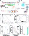
Fig. 2. Examination of adenosine-triggered strand displacement. (a) Rational design of AA and TS sequences for the displacement reaction. The functional regions I and II in AA were designed to hybridize with the 5′-end of TS. The hybridization with 14 base pairs is shown here for clear legibility. (b) Structure of 2-aminopurine and schematic illustration of its fluorescence on/off status. (c) Fluorescence emission spectra of AA and AA–TS duplex in solution. Ex = 307 nm. (d) Effects of hybridization length on the fluorescence intensity and melting temperature of the AA–TS duplex (Em = 370 nm). (e) Relationship between fluorescence recovery and hybridization length of the AA–TS duplex in the presence of adenosine (5 mM). (f) Fluorescence emission spectra of the AA–TS solutions in the presence of adenosine. The number of base pairs was 14. (g) Fluorescence recovery of the AA–TS solution in the presence of nucleosides (1 mM). Error bars represent s. e. m (n = 3).
Fig. 3
Fig. 3. Evaluation of displacement and hybridization reactions in the hydrogels. (a) Fluorescence imaging of the core compartment to verify the displacement reaction. Scale bar: 2 mm (b) effect of displacement time on TS release in the presence of adenosine (5 mM). (c) Effect of adenosine concentration on displacement reaction. (d) Fluorescence imaging of the shell hydrogel stained with TYE665-CS. Scale bar: 5 mm. (e) Examination of TS-mediated protein release from the shell hydrogel. Error bars represent s. e. m (n = 3).
Fig. 4
Fig. 4. Displacement and hybridization reactions for PDGF-BB output from the hydrogel. (a) Fluorescence imaging of the core–shell hydrogel with (+)/without (–) adenosine treatment. The core was stained with FAM-TS (green, Ex/Em = 488 nm/540 nm) and the shell was stained with TYE665-CS (red, Ex/Em = 590 nm/650 nm). Fluorescence profiles along dotted lines drew in the merged images were also shown for clear comparison. Scale bar is 2 mm. (b) Effect of the concentration of adenosine on PDGF-BB output. (c) Hourly PDGF-BB output from the hydrogels treated with (+) or without (–) adenosine. In the (+) group, the hydrogel was treated with 1 mM of adenosine at two time points. Each time, the treatment last for 1 h.
Fig. 5
Fig. 5. Signal output from the hydrogel for regulating intracellular signal transduction and cell migration. (a) Ratiometric fluorescence analysis of intracellular Ca2+ response using fura-2. Blank: without hydrogel or adenosine; Group I: hydrogel with adenosine (without PDGF-BB); Group II: PDGF-BB-loaded hydrogel without adenosine treatment; Group III: PDGF-BB-loaded hydrogel with adenosine treatment. 1st and 2nd: SMCs were treated twice following the pattern of no or periodic PDGF-BB release. The time point of exposing the cells to the release medium was set at 0. (b) Fluorescence images of SMCs stained with Calcium Green™ before and after stimulation. The colors represent the intensity of fluorescence emission at 540 nm. Ex/Em = 488/540 nm. Scale bar: 50 μm. (c) PDGF-BB output for regulating pAkt expression in SMCs examined by western blot. Error bars represent s. e. m (n = 10), ***, p < 0.001. (d) PDGF-BB output for regulation of cell migration. See group information in (a). Error bars represent s. e. m (n = 3). ns = not significant; **, p < 0.01.

References

    1. Hoffman A. S. Adv. Drug Delivery Rev. 2002;54:3–12. - PubMed
    2. Lutolf M. P., Hubbell J. A. Nat. Biotechnol. 2005;23:47–55. - PubMed
    3. Yu L., Ding J. D. Chem. Soc. Rev. 2008;37:1473–1481. - PubMed
    4. Li J., Mo L. T., Lu C. H., Fu T., Yang H. H., Tan W. H. Chem. Soc. Rev. 2016;45:1410–1431. - PMC - PubMed
    5. Lin C. C. RSC Adv. 2015;5:39844–39853. - PMC - PubMed
    1. Yang H. H., Liu H. P., Kang H. Z., Tan W. H. J. Am. Chem. Soc. 2008;130:6320–6321. - PMC - PubMed
    1. Huang F. J., Liao W. C., Sohn Y. S., Nechushtai R., Lu C. H., Willner I. J. Am. Chem. Soc. 2016;138:8936–8945. - PubMed
    2. Hu Y. W., Guo W. W., Kahn J. S., Aleman-Garcia M. A., Willner I. Angew. Chem., Int. Ed. 2016;55:4210–4214. - PubMed
    3. Cheng E. J., Xing Y. Z., Chen P., Yang Y., Sun Y. W., Zhou D. J., Xu L. J., Fan Q. H., Liu D. S. Angew. Chem., Int. Ed. 2009;48:7660–7663. - PubMed
    1. Zhang Z. Y., Chen N. C., Li S. H., Battig M. R., Wang Y. J. Am. Chem. Soc. 2012;134:15716–15719. - PubMed
    1. Tomatsu I., Peng K., Kros A. Adv. Drug Delivery Rev. 2011;63:1257–1266. - PubMed
    2. Fomina N., Sankaranarayanan J., Almutairi A. Adv. Drug Delivery Rev. 2012;64:1005–1020. - PMC - PubMed
    3. Yan B., Boyer J. C., Habault D., Branda N. R., Zhao Y. J. Am. Chem. Soc. 2012;134:16558–16561. - PubMed
    4. Pasparakis G., Manouras T., Vamvakaki M., Argitis P. Nat. Commun. 2014;5:3623. - PMC - PubMed
    5. Liu J. W. Soft Matter. 2011;7:6757–6767.

LinkOut - more resources