Review
. 2017 Nov 1;8(11):7330-7338.
doi: 10.1039/c7sc01462g. Epub 2017 Aug 9.

Magnetic control of cellular processes using biofunctional nanoparticles

Affiliations
Review

Magnetic control of cellular processes using biofunctional nanoparticles

Cornelia Monzel et al. Chem Sci. .

Erratum in

Abstract

Remote control of cellular functions is a key challenge in biomedical research. Only a few tools are currently capable of manipulating cellular events at distance, at spatial and temporal scales matching their naturally active range. A promising approach, often referred to as 'magnetogenetics', is based on the use of magnetic fields, in conjunction with targeted biofunctional magnetic nanoparticles. By triggering molecular stimuli via mechanical, thermal or biochemical perturbations, magnetic actuation constitutes a highly versatile tool with numerous applications in fundamental research as well as exciting prospects in nano- and regenerative medicine. Here, we highlight recent studies, comment on the advancement of magnetic manipulation, and discuss remaining challenges.


Figures
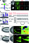
Fig. 1
Fig. 1. Magneto-mechanical stimulation. (a) Left: Mechanoreceptor activation via pulling. A magnetic tip in close proximity of an MNP bound receptor mechanically loads the MNP–receptor complex and activates intracellular signaling. Right: Immunofluorescence staining for MNPs, E-cadherin and the signal activation reporters, actin and vinculin. 9 pN forces result in enhanced molecular colocalization and recruitment compared to 1 pN forces. Scale bar, 2 μm. Taken from Seo et al. with permission from Elsevier. (b) Left: Gating ion channels of the inner ear via mechanical deflection. A magnetic field gradient tilts stereocilia with attached MNPs. Bundle displacement opens the tip link at the channel top (yellow) resulting in ion influx. Right: Traces of pulsed magnetic stimulation, the corresponding hair bundle displacement, the fluorescence signal of a Ca2+-sensitive dye inside the bundle, and the control experiment, where channel opening was prevented. Reprinted with permission from Lee et al. Copyright 2014 American Chemical Society. (c) Left: TWIST expression correlates with mechanical cell compression in a Drosophila embryo. To probe this, the natural compression of endodermal cells is blocked and mimicked instead by magnetically pulling on cells containing a ferrofluid. These magnetized cells are adjacent to endodermal cells and compress the tissue upon magnetic field application. Right: (i) cell compression induced by magnetic manipulation (yellow arrow). (ii) Particle imaging velocimetry indicates compression changes – lowest in black and highest in red. (iii) Inhibition of TWIST expression in the uncompressed cells in the ablated embryo (between the red arrows). (iv) Recovery of TWIST expression in the cells of the ablated embryo by inducing physiological compression with magnetic fields. Taken from Desprat et al. with permission from Elsevier.
Fig. 2
Fig. 2. Magneto-thermal stimulation. (a) Controlling membrane ion channels via MNP heating. An MNP in close proximity to the temperature-sensitive ion channel TRPV1 is locally heated to >42 °C with a radio-frequency (RF) magnetic field. Subsequently, TRPV1 opens, enabling Ca2+ influx. The ion concentration increase is used to activate selected signaling processes. (b) Magneto-thermal TRPV1 stimulation for the controlled activation of hippocampal neurons. The ion channel gating evokes correlated and repeated trains of action potentials in TRPV1 expressing neurons. Top: 10 fluorescence traces (orange) with an average overlay (black) before, during, and after magnetic stimulation (blue bar). Bottom: Raster plots of 100 randomly selected neurons exhibiting repetitive calcium spikes. Shaded blue bars represent alternating magnetic field pulses. Taken from Chen et al. with permission from AAAS. (c) Remote regulation of glucose homeostasis in mice by TRPV1 activation. During channel activation in a RF magnetic field (pink area) a calcium-dependent transgene expression of insulin is initiated. Enhanced insulin expression followed by reduced blood glucose levels in mice are predominantly observed for ferritin–MNPs directly coupled to TRPV1 via the GFP–antiGFP nanobody interaction (αGFP–TRPV1/GFP–ferritin), as well as for ferritin–MNPs associated to the cell plasma membrane (TRPV1/myrferritin). *P < 0.05. Data are mean ± s.e.m. Taken from Stanley et al. with permission from AAAS.
Fig. 3
Fig. 3. Magneto-molecular stimulation. (a) and (b) In vivo control of receptor signal transduction via functionalized MNP accumulation. (a) Left: magnetic field induced aggregation of MNPs bound to plasma membrane receptors. Right: (top) scanning electron microscopy images of antibody–MNP-treated cells before and after magnetic field application. Antibody–MNPs shown in yellow are individually distributed before and clustered after magnetic field treatment. Taken from Cho et al. with permission from NPG. (bottom) Oligomerization of FcεRI–DNP receptor–ligand complexes triggers calcium signaling. Graph depicting successively enhanced intracellular calcium levels (Δ[Ca2+]) for cells subjected to five electromagnetic pulses (arrows). Taken from Mannix et al. with permission from NPG. (b) Left: remote control of Rac-GTPase signaling via cytosolic accumulation of functionalized MNPs. Right: Guanine nucleotide exchange factors TIAM1 coupled to MNP (orange) are attracted to distinct subcellular sites in a magnetic field gradient. TIAM1–MNPs localize into an inactive area of the cell border and activate Rac1, which in turn triggers actin branched polymerization and protrusion formation. An actin comet is observed at 64 min. Scale bar, 1 μm. Taken from Etoc et al. with permission from NPG. (c) Left: in vitro formation of microtubule asters from cellular extract via magnetic accumulation of MNPs conjugated with Ran, a molecular switch regulating microtubule self-assembly during mitosis. Middle: (top) schematic representation and (bottom) fluorescence image of microtubule nucleation and assembly at the point of Ran-NP accumulation (pink dotted circle). Scale bar, 10 μm. Right: quantification of microtubule-based structures, with and without magnetically accumulated Ran-NP. Taken from Hoffmann et al. with permission from NPG.

References

    1. Gautier M., Dhennin-Duthille I., Ay A. S., Rybarczyk P., Korichneva I., Ouadid-Ahidouch H. Br. J. Pharmacol. 2014;171:2582–2592. - PMC - PubMed
    1. Wu Y. I., Frey D., Lungu O. I., Jaehrig A., Schlichting I., Kuhlman B., Hahn K. M. Nature. 2009;461:104–108. - PMC - PubMed
    1. Liu Z., Liu Y., Chang Y., Seyf H. R., Henry A., Mattheyses A. L., Yehl K., Zhang Y., Huang Z., Salaita K. Nat. Methods. 2015;13:143–146. - PMC - PubMed
    1. van Bergeijk P., Adrian M., Hoogenraad C. C., Kapitein L. C. Nature. 2015;518:111–114. - PMC - PubMed
    1. Seo D., Southard K. M., Kim J., Lee H. J., Farlow J., Lee J., Litt D. B., Haas T., Alivisatos A. P., Cheon J., Gartner Z. J., Jun Y. Cell. 2016;165:1507–1518. - PMC - PubMed

LinkOut - more resources