. 2017 Nov 1;8(11):7464-7475.
doi: 10.1039/c7sc01599b. Epub 2017 Sep 4.

Design, crystal structure and atomic force microscopy study of thioether ligated d,l-cyclic antimicrobial peptides against multidrug resistant Pseudomonas aeruginosa

Affiliations

Design, crystal structure and atomic force microscopy study of thioether ligated d,l-cyclic antimicrobial peptides against multidrug resistant Pseudomonas aeruginosa

Runze He et al. Chem Sci. .

Abstract

Here we report a new family of cyclic antimicrobial peptides (CAMPs) targeting MDR strains of Pseudomonas aeruginosa. These CAMPs are cyclized via a xylene double thioether bridge connecting two cysteines placed at the ends of a linear amphiphilic alternating d,l-sequence composed of lysines and tryptophans. Investigations by transmission electron microscopy (TEM), dynamic light scattering and atomic force microscopy (AFM) suggest that these peptide macrocycles interact with the membrane to form lipid-peptide aggregates. Amphiphilic conformations compatible with membrane disruption are observed in high resolution X-ray crystal structures of fucosylated derivatives in complex with lectin LecB. The potential for optimization is highlighted by N-methylation of backbone amides leading to derivatives with similar antimicrobial activity but lower hemolysis.


Figures

Scheme 1
Scheme 1. Synthesis of RH11. Conditions: (a) SPPS: Fmoc deprotection: piperidine/NMP 1 : 4 (v/v), 20 min; amino acid coupling: 3 eq. Fmoc-aa-OH, 3 eq. PyBOP, 5 eq. DIEA in NMP, 2–4 hours; (b) cleavage: TFA/TIS/H2O (95/4/1/); (c) cyclization: KI (20 eq.) DIPEA (5 eq.) α,α′-dichloro-p-xylene (3 eq.) in CH3CN/H2O (1 : 1, v/v), RT, 1–2 hours under Argon.
Fig. 1
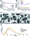
Fig. 1. (A) d,l-Peptides RH6 (red), RH11 (blue) and all l-peptide RH17 (black) (at c = 685 μg mL–1) were incubated in human serum over 24 hours and remaining peptide was quantified by LC/MS. (B) PAO1 membrane integrity was measured by monitoring DNA release using the SYTOX dye and fluorescence spectroscopy. PAO1 cells in minimal salts medium were treated with SYTOX and each of the antibiotics/AMPs (added at time = 0) at a concentration of 4 × MIC. The fluorescence change is shown during 60 min. (C) TEM images showing morphology of PAO1 cells incubated for 60 min without compound (left), or with 10 × MIC polymyxin B (center) and 10 × MIC RH11 (right). (D) Particle size distribution observed by DLS in solutions of 100 μg mL–1 E. coli LPS without or with antibiotic compound at 100 μg mL–1. Each sample was measured in triplicate and the experiments were repeated three times.
Fig. 2
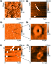
Fig. 2. AFM image of B. subtilis cells on mica surface with or without treatment with CAMP RH11 at 4 × MIC (see methods for details). (A) Control cells without treatment; (B) cells after treatment with RH11, the AFM image shows doughnut-like structures around the bacteria. (C) Focus on doughnut-like structures observed with B. subtilis treated with RH11. (D) zoom image of (C). (E) AFM image of phosphatidylglycerol lipid vesicles treated with RH11. (F) zoom image of phosphatidylglycerol lipid vesicles with RH11.
Fig. 3
Fig. 3. X-ray crystal structure of fucosylated analogs of RH11 in complex with lectin LecB. (a) Overview of the FdRH11o–LecB complex showing 2 symmetric copies of the LecB tetramer. The protein is shown as ribbons and the cyclic peptides in stick model, each symmetry non-equivalent LecB monomer and its bound ligand are shown in the same color. The orange (FdRH11o-1), green (FdRH11o-2), and blue (FdRH11o-3) monomers reveal a fully resolved bound ligand. (b) Detail of the FdRH11o-1 shown with blue dashed cloud density. (c) Cyclic peptide portion of FdRH11o-1 in stick model with cationic lysine side chains in blue and hydrophobic residues (Trp and the xylene bridge) in ruby, showing one water-bridged hydrogen bond and four backbone H-bonds forming an improper short β-sheet like structure. The chosen cut-off distance for hydrogen bridges is 3.2 Å. (d) Cyclic peptide portion of FdRH11o-2 in stick model with two backbone H-bonds and two water-bridged backbone H-bonds. (e) Cyclic peptide portion of FdRH11o-3 in stick model, with backbone H-bonds connected via four crystallographic water molecules, one backbone H-bond involving the C-terminus, and a backbone-to-lysine side chain H-bond. (f) Cyclic peptide portion of FRH11o in stick model as observed in its LecB complex, with missing side-chains modeled in, showing two backbone H-bonds. (g) Overlay of α-carbon backbone observed in the three occupied sites of the FdRH11o–LecB complex (orange, green and blue) and the single occupied site of FRH11o–LecB complex (magenta). (h) Cyclic peptide in stick model as observed in the FdRH11m–LecB complex, with missing side-chains modeled in, showing one water bridged backbone H-bond and one H-bond between an indole NH and a backbone carbonyl. (i) Ramachandran plot for all l-residue dihedrals in the cyclic peptides. (j) Ramachandran plot for all d-residue dihedrals in the cyclic peptides. In both plots the allowed regions are shown in red and yellow. See also Table S2 and Fig. S6.

References

    1. O'Connell K. M. G., Hodgkinson J. T., Sore H. F., Welch M., Salmond G. P. C., Spring D. R. Angew. Chem., Int. Ed. Engl. 2013;52:10706–10733. - PubMed
    1. Chatterjee M., Anju C. P., Biswas L., Anil Kumar V., Gopi Mohan C., Biswas R. Int. J. Med. Microbiol. 2016;306:48–58. - PubMed
    1. Hill T. A., Shepherd N. E., Diness F., Fairlie D. P. Angew. Chem., Int. Ed. Engl. 2014;53:13020–13041. - PubMed
    1. Yudin A. K. Chem. Sci. 2015;6:30–49. - PMC - PubMed
    1. Valeur E., Guéret S. M., Adihou H., Gopalakrishnan R., Lemurell M., Waldmann H., Grossmann T. N., Plowright A. T. Angew. Chem., Int. Ed. Engl. 2017;56:10294–10323. - PubMed