. 2017 Nov 2:7:252.
doi: 10.3389/fonc.2017.00252. eCollection 2017.

Development of Novel Patient-Derived Xenografts from Breast Cancer Brain Metastases

Affiliations

Development of Novel Patient-Derived Xenografts from Breast Cancer Brain Metastases

María J Contreras-Zárate et al. Front Oncol. .

Abstract

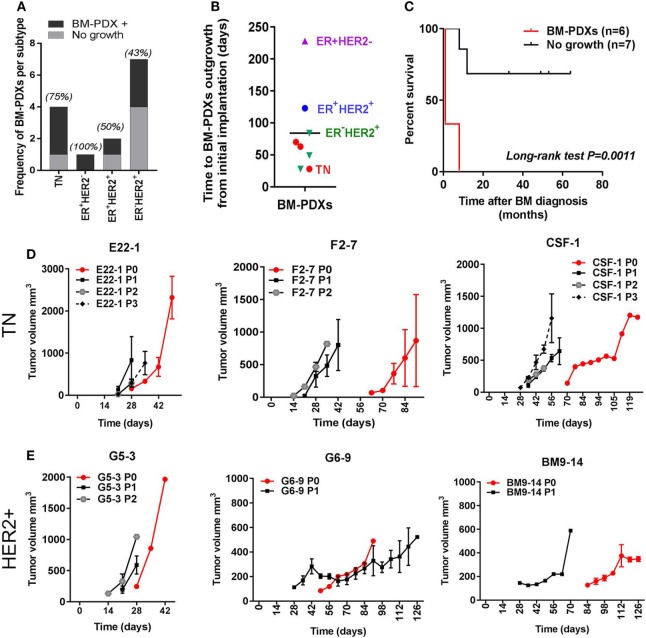
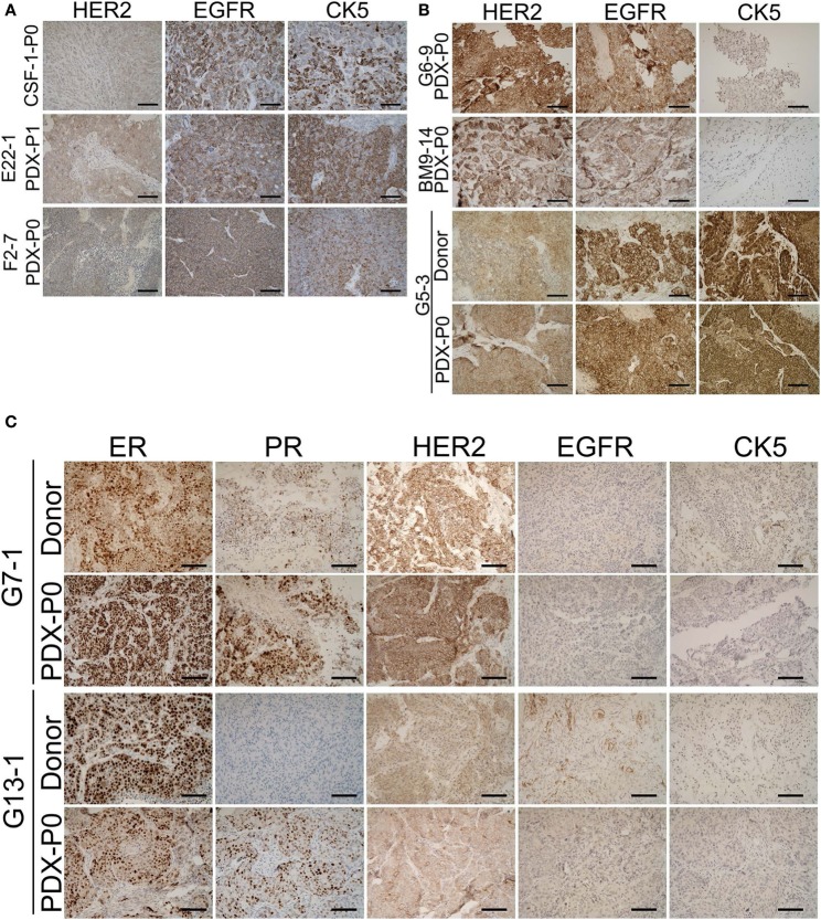
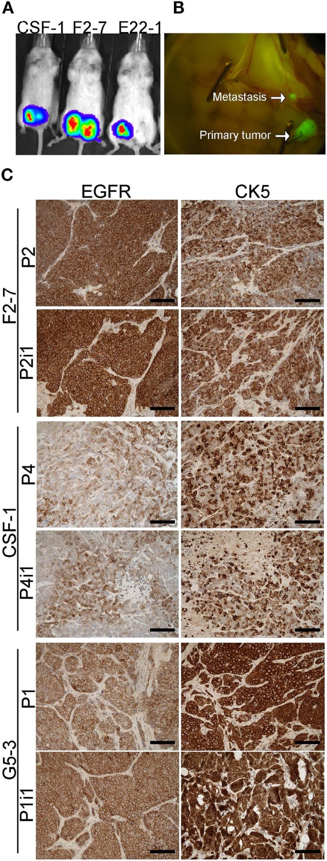
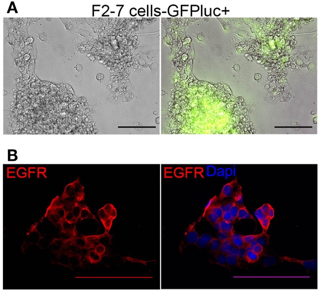
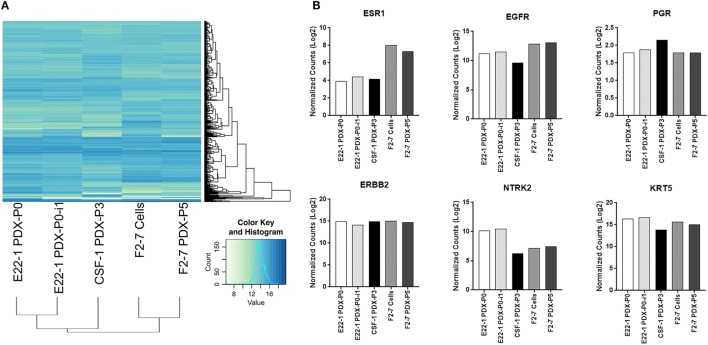
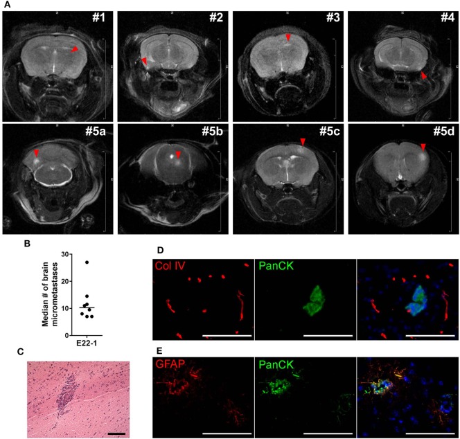
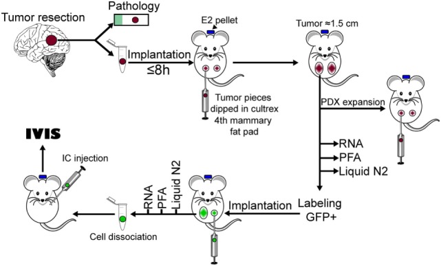
Brain metastases are an increasing burden among breast cancer patients, particularly for those with HER2+ and triple negative (TN) subtypes. Mechanistic insight into the pathophysiology of brain metastases and preclinical validation of therapies has relied almost exclusively on intracardiac injection of brain-homing cells derived from highly aggressive TN MDA-MB-231 and HER2+ BT474 breast cancer cell lines. Yet, these well characterized models are far from representing the tumor heterogeneity observed clinically and, due to their fast progression in vivo, their suitability to validate therapies for established brain metastasis remains limited. The goal of this study was to develop and characterize novel human brain metastasis breast cancer patient-derived xenografts (BM-PDXs) to study the biology of brain metastasis and to serve as tools for testing novel therapeutic approaches. We obtained freshly resected brain metastases from consenting donors with breast cancer. Tissue was immediately implanted in the mammary fat pad of female immunocompromised mice and expanded as BM-PDXs. Brain metastases from 3/4 (75%) TN, 1/1 (100%) estrogen receptor positive (ER+), and 5/9 (55.5%) HER2+ clinical subtypes were established as transplantable BM-PDXs. To facilitate tracking of metastatic dissemination using BM-PDXs, we labeled PDX-dissociated cells with EGFP-luciferase followed by reimplantation in mice, and generated a BM-derived cell line (F2-7). Immunohistologic analyses demonstrated that parental and labeled BM-PDXs retained expression of critical clinical markers such as ER, progesterone receptor, epidermal growth factor receptor, HER2, and the basal cell marker cytokeratin 5. Similarly, RNA sequencing analysis showed clustering of parental, labeled BM-PDXs and their corresponding cell line derivative. Intracardiac injection of dissociated cells from BM-E22-1, resulted in magnetic resonance imaging-detectable macrometastases in 4/8 (50%) and micrometastases (8/8) (100%) mice, suggesting that BM-PDXs remain capable of colonizing the brain at high frequencies. Brain metastases developed 8-12 weeks after ic injection, located to the brain parenchyma, grew around blood vessels, and elicited astroglia activation characteristic of breast cancer brain metastasis. These novel BM-PDXs represent heterogeneous and clinically relevant models to study mechanisms of brain metastatic colonization, with the added benefit of a slower progression rate that makes them suitable for preclinical testing of drugs in therapeutic settings.

Keywords: HER2+; brain colonization; brain metastases models; breast cancer; patient-derived xenograft; triple negative.


Figures

Figure 1
Figure 1
Establishment of brain-metastases-patient-derived xenografts (BM-PDXs) from breast cancer. PDXs were established in NOD/SCID/ILIIrg−/− mice, and subsequently propagated via direct transplantation of solid tumor pieces into new recipient mice. Tumors were grown under continuous estrogen supplementation. (A) BM-PDXs established as a function of breast cancer subtypes. Graph depicts the number (bars) and percentage of BM-PDXs established (BM-PDXs+) per tumor subtype compared to tumors that did not grow after 8 months of implantation. (B) Time in days from initial implantation to outgrowth as measurable tumors (~62.5 mm3) for all PDXs, colors indicate breast cancer subtypes. (C) Survival (months) after brain metastases diagnosis of patients with breast cancer whose surgical samples had in vivo tumorigenic potential (n = 6), or not (n = 7). Log-rank (Mantel-Cox) test P value is shown. (D,E) Tumor growth after implantation (P0, red lines), and subsequent passaging (P1, P2, P3, black/gray lines) in (D) TN and (E) ERHER2+ BM-PDXs. For P0, 1–2 tumors were implanted in a single recipient. For P1-P3, data shows average tumor volume from two tumors in 1–2 recipient mice.
Figure 2
Figure 2
Retention of estrogen receptor (ER), progesterone receptor (PR), HER2, epidermal growth factor receptor (EGFR), and cytokeratin (CK) expression in brain-metastases-patient-derived xenografts (BM-PDXs). Sections of BM-PDXs were stained by IHC for ER, PR, HER2, EGFR, and CK5 at first passage (P0) and compared to donor tumor (when available). (A) Expression of EGFR, HER2, and CK5 in triple negative (TN) BM-PDXs (ER, PR, negative, not shown). (B) Expression of HER2, EGFR, and CK5 in HER2+BM-PDXs in donor and BM-PDXs P0. (C) Expression of ER, PR, HER2, EGFR, and CK5 in ER+HER2+BM-PDXs and their donor counterparts. Scale bars, 100 μm.
Figure 3
Figure 3
GFP-Luciferase-labeled brain-metastases-patient-derived xenografts (BM-PDXs) retain expression of epidermal growth factor receptor (EGFR), cytokeratin 5 (CK5), and HER2. (A) Luciferase imaging (IVIS) of NSG mice carrying GFP-luciferase BM-PDXs F2-7, CSF-1, and G5-3 in the mammary fat pad. (B) Spontaneous metastases to lymph node in G5-3 GFP-luc carrying mice. (C) Expression of EGFR and CK5 in sections of F2-7, CSF-1, and G5-3 BM-PDXs before (P1, P2) and after GFP-luciferase labeling (P2-I1, P1-I1). Scale bars, 100 μm.
Figure 4
Figure 4
Characteristics of F2-7 BM-cell line in vitro. (A) F2-7 cells were labeled in vitro with GFP-luciferase and cultured in collagen-I coated plates. Left: Brightfield, right: GFP expression of live cells. (B) Immunofluorescence staining shows epidermal growth factor receptor (EGFR) (red) expression in F2-7 cells grown in coverslips. 4′,6-diamidino-2-phenylindole (DAPI) labels nuclei. Scale bars are 100 μm.
Figure 5
Figure 5
RNA expression profiles of F2-7 cell line and brain-metastases-patient-derived xenografts (BM-PDXs) before and after labeling. RNA sequencing of BM-PDX E22-1 prior to (E22-1-PDX-P0) and after GFP-luciferase labeling (E22-1 PDX-P0-I1), BM-PDX CSF-1 P3, and F2-7 cell-line compared to F2-7 BM-PDX-P5. (A) Unsupervised hierarchical clustering of samples using the 500 most variable genes across all samples. (B) Normalized expression (log 2) plots of clinically relevant genes (ESR1, EGFR, PGR, ERBB2, NTRK2, KRT5). Differential expression was calculated using cufflinks (cuffdiff), and hierarchical clustering was performed in R.
Figure 6
Figure 6
Experimental metastases using brain-metastases-patient-derived xenografts (BM-PDXs). (A) Dissociated cells (250,000/100 μl PBS) from BM-PDX E22-1 were injected intracardially in female NSG mice (n = 8). Representative T2-weigthed rapid acquisition with relaxation enhancement (RARE) magnetic resonance imaging (MRI) image shows large metastases in 4/10 mice, 8 weeks after injection (top panel). Lower panel shows large and multiple metastases brain metastases in one mouse at 13 weeks postinjection. The summed diameter of all metastases in this mouse was 3.7 mm. (B) Median number of micrometastases per mouse, counted in six brain sections, 300 μm apart. (C) H&E staining shows micrometastasis in brain section from mice injected with E22-1 BM-PDX. (D) Double immunofluorescence staining shows metastatic E22-1 cells (Pan-cytokeratin, PanCK, green) surrounded by reactive astrocytes (GFAP+, red) (20×). (E) Double immunofluorescence staining shows PanCK+E22-1 cells outside of blood vessels (Col-IV) (20×).
Figure 7
Figure 7
Diagram of procedures used to develop brain-metastases-patient-derived xenografts (BM-PDXs) in this study.

Similar articles

Cited by

References

    1. Kaal ECA, Niël CGJH, Vecht CJ. Therapeutic management of brain metastasis. Lancet Neurol (2005) 4(5):289–98.10.1016/S1474-4422(05)70072-7 - DOI - PubMed
    1. Lin NU, Bellon JR, Winer EP. CNS metastases in breast cancer. JCO (2004) 22(17):3608–17.10.1200/JCO.2004.01.175 - DOI - PubMed
    1. Anders C, Carey LA. Understanding and treating triple-negative breast cancer. Oncology (Williston Park) (2008) 22(11):1233–9; discussion 9-40, 43. - PMC - PubMed
    1. Evans AJ, James JJ, Cornford EJ, Chan SY, Burrell HC, Pinder SE, et al. Brain metastases from breast cancer: identification of a high-risk group. Clin Oncol (R Coll Radiol) (2004) 16(5):345–9.10.1016/j.clon.2004.03.012 - DOI - PubMed
    1. Tham Y-L, Sexton K, Kramer R, Hilsenbeck S, Elledge R. Primary breast cancer phenotypes associated with propensity for central nervous system metastases. Cancer (2006) 107(4):696–704.10.1002/cncr.22041 - DOI - PubMed