. 2017 Nov 3:5:232.
doi: 10.3389/fped.2017.00232. eCollection 2017.

Pretreatment with Human Chorionic Gonadotropin Protects the Neonatal Brain against the Effects of Hypoxic-Ischemic Injury

Affiliations

Pretreatment with Human Chorionic Gonadotropin Protects the Neonatal Brain against the Effects of Hypoxic-Ischemic Injury

Tammy Z Movsas et al. Front Pediatr. .

Abstract

Introduction: Though the human fetus is exposed to placentally derived human chorionic gonadotropin (hCG) throughout gestation, the role of hCG on the fetal brain is unknown. Review of the available literature appears to indicate that groups of women with higher mean levels of hCG during pregnancy tend to have offspring with lower cerebral palsy (CP) risk. Given that newborn cerebral injury often precedes the development of CP, we aimed to determine whether hCG may protect against the neurodegenerative effects of neonatal brain injury.

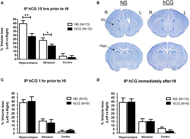
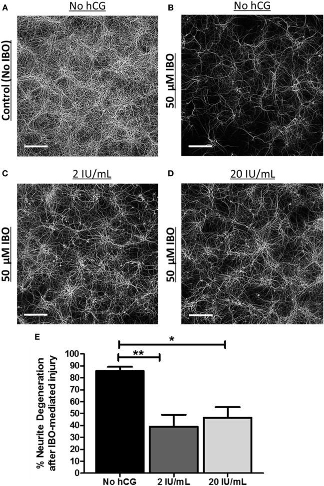
Methods: We utilized the Rice-Vannucci model of neonatal cerebral hypoxia-ischemia (HI) in postnatal day 7 mice to examine whether intraperitoneal administration of hCG 15-18 h prior, 1 h after or immediately following HI decrease brain tissue loss 7 days after injury. We next studied whether hCG has pro-survival and trophic properties in neurons by exposing immature cortical and hippocampal neurons to hCG in vitro and examining neurite sprouting and neuronal survival prior and after glutamate receptor-mediated excitotoxic injury.

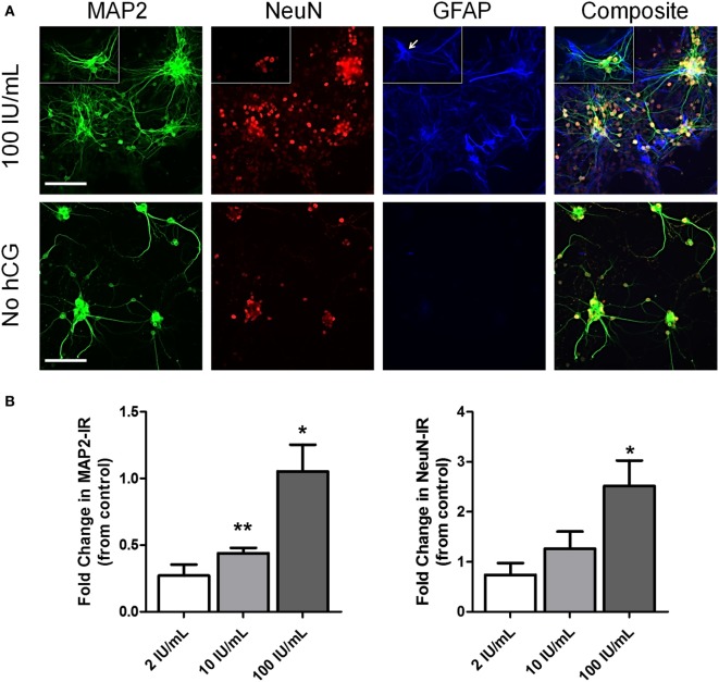
Results: We found that intraperitoneal injection of hCG 15 h prior to HI, but not at or 1 h after HI induction, resulted in a significant decrease in hippocampal and striatal tissue loss 7 days following brain injury. Furthermore, hCG reduced N-methyl-d-aspartate (NMDA)-mediated neuronal excitotoxicity in vitro when neurons were continuously exposed to this hormone for 10 days or when given at the time and following neuronal injury. In addition, continuous in vitro administration of hCG for 6-9 days increased neurite sprouting and basal neuronal survival as assessed by at least a 1-fold increase in MAP2 immunoreactivity and a 2.5-fold increase in NeuN + immunoreactivity.

Conclusion: Our findings suggest that hCG can decrease HI-associated immature neural degeneration. The mechanism of action for this neuroprotective effect may partly involve inhibition of NMDA-dependent excitotoxic injury. This study supports the hypothesis that hCG during pregnancy has the potential for protecting the developing brain against HI, an important CP risk factor.

Keywords: brain injury; cerebral palsy; chorionic gonadotropin; excitotoxicity; fetal brain; human chorionic gonadotropin; ischemia; neuroprotection.


Figures

Figure 1
Figure 1
In vivo hCG IP administration protects the brain from the neurodegenerative effects of HI in a time-dependent manner. (A) The relative tissue losses [calculated by the percent difference between left (L) ipsilateral, ischemic, and right (R) contralateral, non-ischemic, hemisphere] in the hippocampus, striatum and cerebral cortex 7 days after carotid ligation followed by 45 min of hypoxia from NS-injected vs. hCG-injected (1,500 IU/kg) P7 mouse pups treated 15 h prior to HI. hCG treatment decreased hippocampal (NS 44.5 ± 3.9% vs. hCG 28.5 ± 3.8%) and striatal (NS 23.5 ± 3.1% vs. hCG 16.7 ± 2.3%) tissue loss following HI. Actual area values (average ± SEM mm2) in left vs. right hippocampus are 1.8 ± 0.12 vs. 0.97 ± 0.10 mm2 for NS and 1.8 ± 0.14 vs. 1.3 ± 0.10 mm2 for hCG. Average area values in left vs. right striatum are 3.3 ± 0.19 vs. 2.4 ± 0.12 mm2 for NS and 3.0 ± 0.16 vs. 2.5 ± 0.12 mm2 for hCG. (B) Representative coronal digital micrographs taken from injured NS- and hCG-injected animals treated 15 h prior to HI. Note the ex-vacuo dilatation of the left, ischemic, cerebral ventricle, and relative atrophy of the left hippocampal formation in the NS group compared with the contralateral brain. (C,D) IP injection of hCG (1,500 IU/kg) 1 h (C) or immediately after (D) neonatal injury in P7 neonatal pups did not protect against the neurodegenerative effects of HI. Error bars show SE; *p < 0.05 and **p < 0.01. hCG, human chorionic gonadotropin; HI, hypoxia-ischemia; Hipp, hippocampus; IP, intraperitoneal; NS, normal saline; Str, striatum.
Figure 2
Figure 2
IP administration of hCG (1,500 IU/kg) does not alter neonatal pup body temperature. (A,B) Body temperature as assessed by rectal temperature in neonatal P7 mouse pups did not change 1 h or 15–18 h following hCG injection when examined during pup huddling or during a constant physiological ambient temperature of 36.5°C. (C) Removal of individual neonatal P7 pups from physiological (36.5°C) to room temperature (23°C) produced a gradual decline in body temperature. hCG administration did not alter the rate of temperature decline upon ambient temperature exposure when examined 15–18 h after drug injection. Statistical comparisons were performed by repeated measures ANOVA followed by Bonferroni post hoc test. (D) Second-degree polynomial curve fit calculated from data in (C), demonstrating nearly identical temperature drops upon change in ambient temperature from 36.5 to 23°C in NS control and hCG-treated neonatal P7 pups. hCG, human chorionic gonadotropin; IP, intraperitoneal.
Figure 3
Figure 3
Serum hCG concentrations obtained from neonatal P7 mouse pups 1 and 15–18 h following IP 1,500 IU/kg hCG injection. Serum hCG levels (calculated in ng/mL) were obtained utilizing a commercially available hCG ELISA assay (ab100533, Abcam, Cambridge, MA, USA). Conversion to IU/mL was done using this assay and a known concentration of the hCG source employed in the animal and cell experiments (CG10, Sigma, St. Louis, MO, USA). No hCG IR was observed in control, NS-injected, mouse serum indicating no mouse protein cross-reactivity. The serum hCG concentration did not appear to differ significantly between 1 h (31.4 ± 3 ng/mL; 0.41 ± 0.04 IU/mL) and 15–18 h (29.8 ± 4 ng/mL; 0.39 ± 0.06 IU/mL) post-IP hormone injection. These serum concentrations are similar to those found in human term amniotic fluid (43, 48). hCG, human chorionic gonadotropin; IP, intraperitoneal, IR, immunoreactivity; NS, normal saline.
Figure 4
Figure 4
In vitro chronic, 10-day, hCG exposure to immature cortical and hippocampal neurons is not toxic and protects them from NMDA-dependent excitotoxic injury. (A,B) MAP2 fluorescence following immunocytochemical staining of dissociated cortical and hippocampal neurons exposed to control vs. 2 IU/mL of hCG for 9 days prior and 24 h following IBO (50 µM; IBO) exposure. Prolonged hCG did not appear to affect neuronal survival. Note also the relative preservation of neurites in hCG-exposed neurons after IBO exposure (most noticeable in cortical neurons) compared with control (CSS; controlled salt solution). (C) Quantitative bar graph analysis of the effects of hCG on neuronal survival as a measure of LDH activity in the neuronal media or MAP2 IR in control and hCG-treated neuronal cultures 24 h after injury with IBO. Cortical and hippocampal neurons exposed to hCG demonstrated a reduction in IBO-mediated increases in LDH activity and a relative preservation of neurite staining compared with non-hCG-treated cells. Scale bars = 165 µm. Error bars show SE; *p < 0.05 and **p < 0.01. hCG, human chorionic gonadotropin; IBO, ibotenic acid; IR, immunoreactivity; LDH, lactase dehydrogenase.
Figure 5
Figure 5
Acute neuronal hCG exposure in vitro protects neurons against NMDA-mediated excitotoxic neurodegeneration. (A) Representative low-power digital micrograph of MAP2 IR in DIV12 cultured, non-injured/non-hCG-treated, cortical neurons. (B–D) Representative digital micrographs of DIV12 cultured MAP2-stained neuronal fibers 24 h following exposure to the NMDA-receptor agonist, IBO in the presence or absence of 2 or 20 IU/mL of hCG when hCG is administered during and following IBO exposure. (E) Quantitative summary of the neuroprotective effect of acute hCG administration 24 h post-IBO administration. Percent neurite degeneration was obtained as a function of the percent change in MAP2-IR from non-IBO control neurons. Scale bar = 220 µm. N = 4/condition. Error bars show SE; *p < 0.05 and **p < 0.01. hCG, human chorionic gonadotropin; IBO, ibotenic acid; IR, immunoreactivity.
Figure 6
Figure 6
Early (DIV3) high-dose hCG (100 IU/mL) exposure in vitro increases neuronal cell count and neurite sprouting. (A) Representative low-power micrographs of DIV9 cortical neurons exposed to high-dose (10 and 100 IU/mL) hCG for 6 days and triple-stained with the neurite marker, MAP2, the neuron-specific marker, NeuN, and the astrocyte-specific marker, GFAP. Prolonged high concentrations of hCG resulted in large grouping of neurons with more complex neurite processes surrounded by GFAP + astrocytes. In contrast, GFAP + astrocytes are nearly absent in non-hCG-treated cultures at DIV9. Small rectangular inserts demonstrate a morphologically appearing GFAP + astrocyte (arrow) nearby a small group of NeuN + neurons. (B) A significant increase in MAP2 IR and visible increases in neuronal sprouting were observed with greater than 2 IU/mL concentrations of hCG. High-dose hCG (100 IU/mL) appeared to qualitatively and quantitatively increased NeuN + IR. Scale bar = 80 µm. N = 3/condition. Error bars show SE; *p < 0.05 and **p < 0.01. hCG, human chorionic gonadotropin; IR, immunoreactivity.

References

    1. Cole LA. New discoveries on the biology and detection of human chorionic gonadotropin. Reprod Biol Endocrinol (2009) 7:8.10.1186/1477-7827-7-8 - DOI - PMC - PubMed
    1. Goldsmith PC, McGregor WG, Raymoure WJ, Kuhn RW, Jaffe RB. Cellular localization of chorionic gonadotropin in human fetal kidney and liver. J Clin Endocrinol Metab (1983) 57(3):654–61.10.1210/jcem-57-3-654 - DOI - PubMed
    1. Abdallah MA, Lei ZM, Li X, Greenwold N, Nakajima ST, Jauniaux E, et al. Human fetal nongonadal tissues contain human chorionic gonadotropin/luteinizing hormone receptors. J Clin Endocrinol Metab (2004) 89(2):952–6.10.1210/jc.2003-030917 - DOI - PubMed
    1. Lei ZM, Rao CV, Kornyei JL, Licht P, Hiatt ES. Novel expression of human chorionic gonadotropin/luteinizing hormone receptor gene in brain. Endocrinology (1993) 132(5):2262–70.10.1210/endo.132.5.8477671 - DOI - PubMed
    1. AL-Hader AA, Tao YX, Lei ZM, Rao CV. Fetal rat brains contain luteinizing hormone/human chorionic gonadotropin receptors. Early Pregnancy (1997) 3(4):323–9. - PubMed

LinkOut - more resources