. 2017 Nov 1;58(13):5887-5896.
doi: 10.1167/iovs.17-22989.

Mast Cell-Derived Tryptase in Geographic Atrophy

Affiliations

Mast Cell-Derived Tryptase in Geographic Atrophy

D Scott McLeod et al. Invest Ophthalmol Vis Sci. .

Abstract

Purpose: Our previous study demonstrated significantly more degranulating mast cells (MCs) in choroids from subjects with age-related macular degeneration compared to aged controls. This study examined the immunolocalization of tryptase, the most abundant MC secretory granule-derived serine protease, in aged control eyes and eyes with geographic atrophy (GA).

Methods: Postmortem human eyes with and without GA were obtained from the National Disease Research Interchange. Tissue was fixed, cryopreserved, sectioned, and immunostained with a monoclonal antibody against tryptase. Sections were imaged on a Zeiss 710 Confocal Microscope.

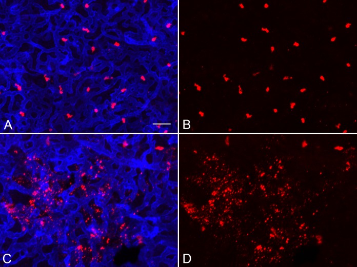
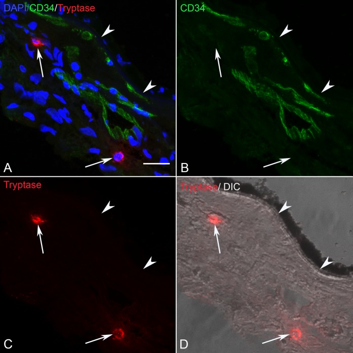
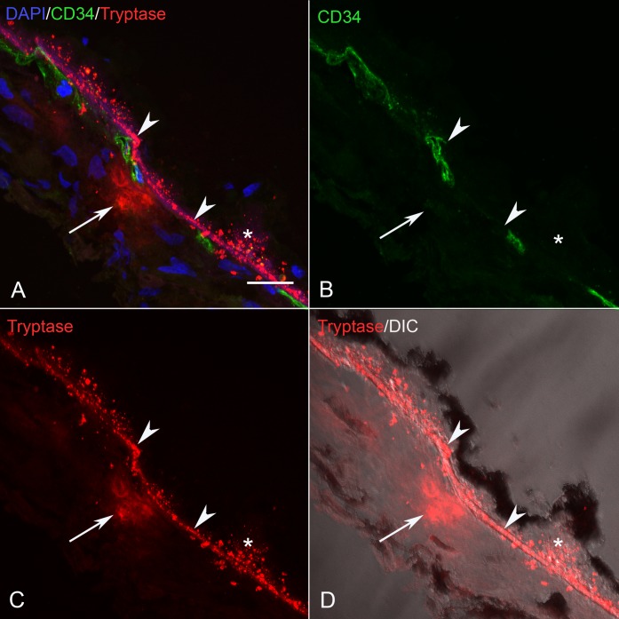
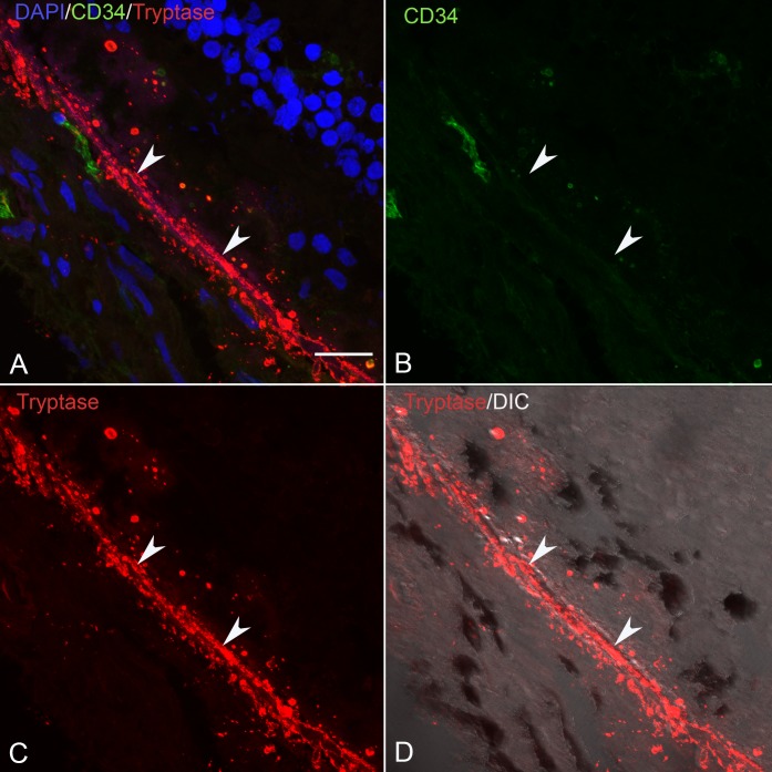
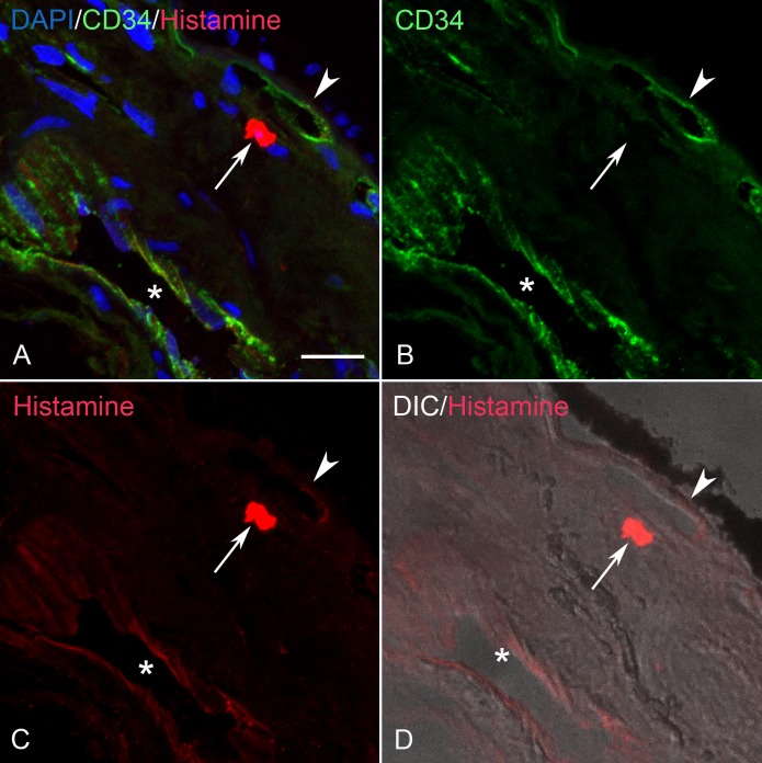
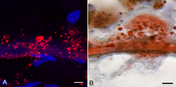
Results: In the posterior pole of all aged control eyes, tryptase was confined to choroidal MCs, which were located primarily in Sattler's layer. In eyes with GA, many MCs were located in the inner choroid near choriocapillaris and Bruch's membrane (BM). Tryptase was found not only in MCs but also diffusely around them in stroma, suggesting they had degranulated. In contrast with aged control eyes, eyes with GA also had strong tryptase staining in BM. Tryptase was observed within BM in regions of RPE atrophy, at the border of atrophy, and extending well into the nonatrophic region.

Conclusions: Our results demonstrate that tryptase, released during choroidal MC degranulation, binds to BM in GA in advance of RPE atrophy. Tryptase activates MMPs that can degrade extracellular matrix (ECM) and basement membrane components found in BM. ECM modifications are likely to have a profound effect on the function and health of RPE and choroidal thinning in GA.


Figures

Figure 1
Figure 1
Aged control whole mount choroid immunolabeled for (A) UEA lectin (blue), (B) tryptase (red), and (C) chymase (green). The vast majority of human choroidal MCs are of the MCT phenotype (tryptase+/chymase). Only a small percentage of MCs are MCtc (tryptase+/chymase+) (arrows). Scale bar: 30 μm.
Figure 2
Figure 2
Aged control (A, B) and GA (C, D) whole mount choroids immunolabeled with UEA lectin (blue) and anti-tryptase (red). MC degranulation near the border region of RPE atrophy demonstrates the association between MC degranulation and choriocapillaris atrophy. The free tryptase+ granules were at the level of the inner aspect of the choriocapillaris as determined in z stacks. Scale bar: 30 μm.
Figure 3
Figure 3
(A–D) Section from an 85-year-old aged control subject showing (C) tryptase+ MCs (arrows), (B) CD34+ blood vessels (green) and BM (arrowhead). Most MCs in aged control eyes were located in Sattler's and Haller's layer where tryptase+ granules were confined to MC cytoplasm. In (A) the colors are merged and (D) is differential interference contrast (DIC) illumination so RPE can be distinguished. Scale bar: 20 μm.
Figure 4
Figure 4
(A–D) Section from an 89-year-old GA subject showing (C) a degranulated tryptase+ MC (arrow), (B) CD34+ blood vessels and BM (arrowhead) in a nonatrophic region of RPE (*). (D) DIC illumination and (A) has all colors merged. MCs in GA eyes often were located near the choriocapillaris where diffuse tryptase immunoreactivity was observed in the surrounding choroidal stroma. Granular tryptase reaction product was localized in BM (arrowhead), sub RPE deposits and drusen. Scale bar: 20 μm.
Figure 5
Figure 5
(A–D) Section from the same 89-year-old GA subject in shown in Figure 4 near the border of RPE atrophy demonstrates granular tryptase immunoreactivity in BM (arrowhead). (B) The density of CD34+ choriocapillaris is reduced in this border region and (C) RPE are multilayered with granular tryptase immunoreactivity around them and within BM. (D) DIC illumination and (A) has all colors merged. Scale bar: 20 μm.
Figure 6
Figure 6
(A–D) Section from the same 89-year-old GA subject shown in Figure 5 in a region with RPE atrophy showing, (B) reduced CD34+ choriocapillaris density and diffuse tryptase staining (C) surrounding a degranulating MC (arrow) and granular tryptase immunoreactivity in BM (arrowhead). (D) DIC and (A) has all colors merged. Scale bar: 20 μm.
Figure 7
Figure 7
(A–D) Section from an 86-year-old aged control subject showing (C) a histamine+ MC (arrow), (B) CD34+ blood vessels and BM (arrowhead). (D) DIC and (A) has all colors merged. The endothelium of an artery is weakly histamine+ (*). BM (arrowhead) is unlabeled D. Scale bar: 20 μm.
Figure 8
Figure 8
(A–D) Section from an 89-year-old GA subject near the border of RPE atrophy showing (C) a histamine+ MC (arrow), (B) CD34+ blood vessels, and BM (arrowhead). (D) DIC illumination shows a drusen+ (*), and (A) has all colors merged. BM (arrowhead), drusen (*) and RPE that are unlabeled. Scale bar: 20 μm.
Figure 9
Figure 9
(A–D) Section from the same 89-year-old GA subject shown in Figure 8 in a region of RPE atrophy showing (C) a histamine+ MC (arrow), (B) CD34+ blood vessel attenuation, and unstained BM (arrowhead). (D) DIC illumination and (A) has all colors merged. Scale bar: 20 μm.
Figure 10
Figure 10
Sections from an 81-year-old aged control subject (A) and an 89-year-old GA subject (B–D) showing oil red O staining. In aged controls, weak diffuse staining was observed in BM. In the GA eyes, intense staining was observed in BM and drusen. The staining was granular in appearance and was remarkably similar to tryptase immunoreaction product in GA. Scale bar: 10 μm.
Figure 11
Figure 11
Semiserial sections of a 90-year-old GA choroid comparing tryptase localization (A) with oil red O staining (B) in BM and drusen. Note the rounded particulate bodies are labeled in both and reside in the same tissue compartment. Scale bar: 5 μm.

References

    1. Theoharides TC, Alysandratos KD, Angelidou A,et al. Mast cells and inflammation. Biochim Biophys Acta. 2012; 1822: 21– 33. - PMC - PubMed
    1. Wernersson S, Pejler G. . Mast cell secretory granules: armed for battle. Nat Rev Immunol. 2014; 14: 478– 494. - PubMed
    1. Kritikou E, Kuiper J, Kovanen PT, Bot I. . The impact of mast cells on cardiovascular diseases. Eur J Pharmacol. 2016; 778: 103– 115. - PubMed
    1. Krystel-Whittemore M, Dileepan KN, Wood JG. . Mast cell: a multi-functional master cell. Front Immunol. 2015; 6: 620. - PMC - PubMed
    1. Schwartz LB, Irani AM, Roller K, Castells MC, Schechter NM. . Quantitation of histamine, tryptase, and chymase in dispersed human T and TC mast cells. J Immunol. 1987; 138: 2611– 2615. - PubMed

Publication types

LinkOut - more resources