

Microcystic Calcifying Epithelial Odontogenic Tumor
- PMID: 29164474
- PMCID: PMC6232214
- DOI: 10.1007/s12105-017-0868-0
Microcystic Calcifying Epithelial Odontogenic Tumor
Abstract
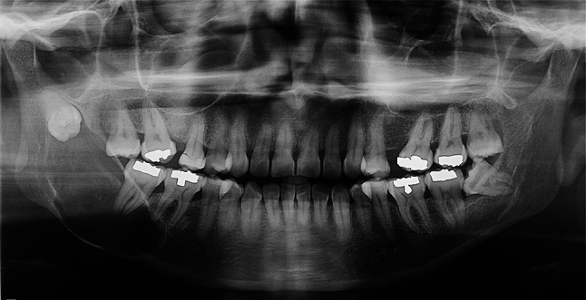
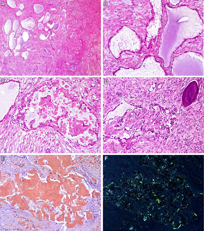
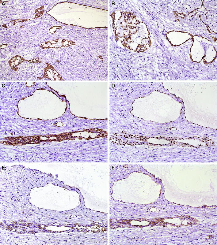
Microcystic variant of calcifying epithelial odontogenic tumor is rare. We herein describe an additional well-documented case of microcystic CEOT. The affected patient is a Guatemalan 42-year-old female with an expansile well-defined mixed radiolucent-radiopaque lesion located in the right posterior mandible. The lesion was associated to an unerupted third molar. Histopathologic examination revealed nests and cords of moderately pleomorphic, eosinophilic polyhedral epithelial cells surrounded by a fibromyxoid stroma. The neoplastic cells showed microcystic pattern made of pseudo-glandular spaces with variable diameter. Occasional amyloid deposits and calcified acellular material were observed. Tumor cells were positive for AE1/AE3, CK14, CK19, p63, CD138, and beta-catenin. Conservative surgical resection was performed with an uneventful immediate post-surgical follow-up. After 1 year follow-up there is no evidence of recurrence. Pathologists should be aware of this unusual microcystic presentation of CEOT, which may pose a diagnostic challenge and potential diagnostic dilemma.
Keywords: Calcifying epithelial odontogenic tumor; Immunohistochemistry; Mandible; Microcystic.
Conflict of interest statement
All of authors have indicated they have no potential conflicts of interest and no financial relationships relevant to this article to disclose.
Figures
References
-
- Wright JM, Devilliers P. Calcifying epithelial odontogenic tumour. In: El-Naggar AK, Chan JKC, Grandis JR, Takata T, Slootweg PJ, editors. World Health Organization classification of head and neck tumours. Lyon: IARC; 2017. pp. 220–221.
Publication types
MeSH terms
Supplementary concepts
LinkOut - more resources
Full Text Sources
Other Literature Sources
Medical
Miscellaneous
