. 2018 Jan;7(1):87-97.
doi: 10.1002/sctm.17-0066. Epub 2017 Nov 21.

A Universal Approach to Correct Various HBB Gene Mutations in Human Stem Cells for Gene Therapy of Beta-Thalassemia and Sickle Cell Disease

Affiliations

A Universal Approach to Correct Various HBB Gene Mutations in Human Stem Cells for Gene Therapy of Beta-Thalassemia and Sickle Cell Disease

Liuhong Cai et al. Stem Cells Transl Med. 2018 Jan.

Abstract

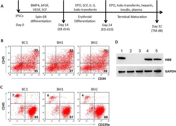
Beta-thalassemia is one of the most common recessive genetic diseases, caused by mutations in the HBB gene. Over 200 different types of mutations in the HBB gene containing three exons have been identified in patients with β-thalassemia (β-thal) whereas a homozygous mutation in exon 1 causes sickle cell disease (SCD). Novel therapeutic strategies to permanently correct the HBB mutation in stem cells that are able to expand and differentiate into erythrocytes producing corrected HBB proteins are highly desirable. Genome editing aided by CRISPR/Cas9 and other site-specific engineered nucleases offers promise to precisely correct a genetic mutation in the native genome without alterations in other parts of the human genome. Although making a sequence-specific nuclease to enhance correction of a specific HBB mutation by homology-directed repair (HDR) is becoming straightforward, targeting various HBB mutations of β-thal is still challenging because individual guide RNA as well as a donor DNA template for HDR of each type of HBB gene mutation have to be selected and validated. Using human induced pluripotent stem cells (iPSCs) from two β-thal patients with different HBB gene mutations, we devised and tested a universal strategy to achieve targeted insertion of the HBB cDNA in exon 1 of HBB gene using Cas9 and two validated guide RNAs. We observed that HBB protein production was restored in erythrocytes derived from iPSCs of two patients. This strategy of restoring functional HBB gene expression will be able to correct most types of HBB gene mutations in β-thal and SCD. Stem Cells Translational Medicine 2018;7:87-97.

Keywords: Beta-thalassemia; CRISPR/Cas9; Gene editing; Gene therapy; Hemoglobinopathies; Stem cells.


Figures

Figure 1
Figure 1
Characterization of BH1 and BH2 induced pluripotent stem cells (iPSCs) generated from beta‐thalassemia major patients. (A): Live cell staining of iPSCs at reprogramming day 14 under feeder‐free and xeno‐free culture conditions. The iPSC colonies were incubated with anti‐human TRA‐1‐60‐PE in a cell culture incubator for 1 hour. The representative images of live staining were obtained using a fluorescence microscope. The colony morphologies were observed using light phase. Bar size = 100 μm. (B): Positive iPS cell colonies of alive TRA‐1–60 staining were picked up and pooled to establish BH1 and BH2 human iPS cell lines. The iPSCs were expanded on vitronectin‐coated plate in E8 media. The pluripotent stem cell markers, TRA‐1‐60 and SSEA‐4, were measured by flow cytometry and shown in histograms. Black lines represented isotype‐matched antibody controls. (C): Real‐time quantitative PCR analyses of OCT4 and NANOG gene expression in BH1 and BH2 iPSCs. The relative gene expression levels were normalized by housekeeping gene GAPDH. The BC1 iPSCs were used as controls. (D): The HBB gene mutations in BH1 and BH2 iPSCs were confirmed by Sanger DNA sequencing. β17 and β‐thal654 mutations were identified in BH1 iPSCs, while β17 and β41‐42 mutations were identified in BH2 iPSCs. Abbreviation: GAPDH, glyceraldehyde‐3‐phosphate dehydrogenase.
Figure 2
Figure 2
Erythrocyte differentiation of BH1 and BH2 iPSCs. (A): Diagrammatic sketch of differentiation procedure. To generate red blood cells, the iPSCs underwent three stages of spin‐EB differentiation, erythroid differentiation, and erythrocyte terminal maturation (TM) in feeder‐free and xeno‐free culture conditions. (B): Flow cytometric analyses of hematopoietic stem/progenitor cells (CD34+CD45+) cells at EB day 14. (C): Flow cytometric analyses of erythrocytes (CD235a+CD45) at TM day 8. The differentiation derivatives from BC1 iPSCs were used as controls. (D): Western blot to detect HBB proteins of erythrocytes from various iPSCs after terminal differentiation. GAPDH was used as loading control. Lane 1: BC1; Lane 2: BH1; Lane 3: BH2; Lanes 4, 5: two other control iPSC lines. Abbreviations: EB, embryoid bodies; GAPDH, glyceraldehyde‐3‐phosphate dehydrogenase; iPSCs, induced pluripotent stem cells.
Figure 3
Figure 3
CRISPR/Cas9 mediated genome editing of the human HBB gene by its cDNA. (A): A diagram of a strategy to replace the HBB genomic DNA by its CDS linked by a P2A self‐cleaving peptide with the GFP reporter gene. The two guide RNAs, gR‐HBB‐a and gR‐HBB‐UTR, were designed to target on exon 1 and 3′ UTR of HBB gene, respectively. The donor vector containing HBB CDS, a L‐HA and a R‐HA was transfected into BH1, BH2, or BC1 induced pluripotent stem cells (iPSCs) along with guide‐RNA vectors and Cas9 vector. The positive colonies with homology‐directed repair (HDR) were identified by genomic PCR using indicated primers in Supporting Information Figure S3. Next, the edited iPSCs were transfected with plasmid pCAG‐Cre‐IRES2‐GFP to excise the loxP‐flanked PGK‐puromycin expression cassette. The positive colonies with anticipated excision were identified by genomic PCR using primer L2‐F and V4193‐R. (B): Genomic PCR screening for HDR positive (either homozygous or heterozygous) colonies using primer gDNA‐75‐F and gDNA363‐R. The wildtype HBB locus gave rise to a 438 bp PCR product (WT) whereas the corrected edited locus would be a 306 bp PCR product due to the lack of the intron (132 bp) in the donor cDNA. (C): Genomic PCR screening for excision of the PGK‐Puro cassette using primer L2‐F and V4193‐R. The targeted colonies without excision (such as BC1, #6) gave rise to a 952 bp PCR product whereas the positive colonies with excision did not give rise to specific PCR product. (D): Sanger sequencing to confirm HBB gene corrections in BH1 and BH2 iPSCs. The synonymous change of β14 (Leucine) from CTG to TTA, in addition to the corrected β17 (AAG), was observed in selected clones of BH1 and BH2 iPSCs. Abbreviations: bp, basepair; CDS, coding sequence; L‐HA, left homology arm; R‐HA, right homology arm; WT, wild type.
Figure 4
Figure 4
The GFP expression under the control of the HBB locus from induced pluripotent stem cell (iPSC)‐derived erythrocytes after genome editing. (A): Histogram of flow cytometric analyses at terminally differentiated erythrocytes from genome edited BC1 iPSCs. BC1 cells with a heterozygous integration of the HBB‐GFP allele (#6C2) or homozygous integration (#8C8) were used, together with parental BC1 iPSCs. (B): Real‐time quantitative PCR analyses of HBB and HBG gene expression. GFP positive and negative cells were isolated by fluorescence‐activated cell sorting at day 8 after terminal differentiation. The relative gene expression levels were normalized by housekeeping gene GAPDH. (C): Flow cytometric analyses of other marker expression in the GFP positive versus negative cells at day 8 of terminal differentiation of #6C2 iPSCs. CD235a and CD45 were used to characterize erythrocytes. A DNA‐staining fluorescent dye DRAQ5 was used to evaluate enucleation of erythrocytes lacking nuclear DNA. Abbreviation: GAPDH, glyceraldehyde‐3‐phosphate dehydrogenase.
Figure 5
Figure 5
Expression of HBB protein in erythrocytes from the HBB‐corrected BH1 and BH2 iPSCs. (A): Histogram of flow cytometric analyses of erythrocytes from HBB gene targeted BH1, BH2, and BC1 iPSCs at TM day 8. Efficient erythrocyte generation was demonstrated by a high percentage of cells expressing CD235a and a low percentage of cells expressing CD45. (B): Western blot of HBB expression in erythrocytes at TM day 8. The defective beta hemoglobin expression in erythrocytes from beta thalassemia patient iPSCs was rescued by mono allelic HBB gene targeting (heterozygous). The undifferentiated iPSCs of each line were used as negative controls. (C): Quantification (mean ± SEM) of HBB protein expression based on Western blot data of multiple experiments (n = 3). Abbreviations: GAPDH, glyceraldehyde‐3‐phosphate dehydrogenase; iPSC, induced pluripotent stem cells; TM, terminal maturation.

Similar articles

Cited by

References

    1. Weatherall DJ. Phenotype‐genotype relationships in monogenic disease: Lessons from the thalassaemias. Nat Rev Genet 2001;2:245–255. - PubMed
    1. Wu C, Dunbar CE. Stem cell gene therapy: The risks of insertional mutagenesis and approaches to minimize genotoxicity. Front Med 2011;5:356–371. - PMC - PubMed
    1. Naldini L. Ex vivo gene transfer and correction for cell‐based therapies. Nat Rev Genet 2011;12:301–315. - PubMed
    1. Rivière I, Dunbar CE, Sadelain M. Hematopoietic stem cell engineering at a crossroads. Blood 2012;119:1107–1116. - PMC - PubMed
    1. Goodman MA, Malik P. The potential of gene therapy approaches for the treatment of hemoglobinopathies: Achievements and challenges. Ther Adv Hematol 2016;7:302–315. - PMC - PubMed

Publication types