Review
. 2017 Nov;7(2):10.1128/ecosalplus.ESP-0002-2017.
doi: 10.1128/ecosalplus.ESP-0002-2017.

The Sec System: Protein Export in Escherichia coli

Affiliations
Review

The Sec System: Protein Export in Escherichia coli

Jennine M Crane et al. EcoSal Plus. 2017 Nov.

Abstract

In Escherichia coli, proteins found in the periplasm or the outer membrane are exported from the cytoplasm by the general secretory, Sec, system before they acquire stably folded structure. This dynamic process involves intricate interactions among cytoplasmic and membrane proteins, both peripheral and integral, as well as lipids. In vivo, both ATP hydrolysis and proton motive force are required. Here, we review the Sec system from the inception of the field through early 2016, including biochemical, genetic, and structural data.


Figures

Figure 1
Figure 1
Composite of the structures of the proteins of the Sec system. The structures are shown in ribbon representation of SecYEG in complex with SecA from T. maritima PDB 3DIN; SecDF from T. thermophilus PDB 3AQO; YidC from E. coli PDB 1B12; signal peptide peptidase soluble domain PDB 3BF0; SecA dimer from B. subtilis PDB 1M6N; SecA monomer from B. subtilis 1TF5; SecB from E. coli PDB 1QYN.
Figure 2
Figure 2
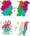
Structure of SecB, a dimer of dimers. SecB is a tetramer organized as a dimer of dimers. (a) and (c) show the two protomers that make eight-stranded β sheets on the flat sides of SecB. (b) and (d) are related to (a) and (c) by 90° rotation to show the interface of the dimer of dimers. Each protomer is shown as a different color. PDB 1QYN.
Figure 3
Figure 3
Possible pathways of ligand binding. Site-directed spin labeling and electron paramagnetic resonance spectroscopy were used to map the contact sites between SecB and polypeptide ligands. The sites of contact are shown in green. Residues that showed no contact are shown in gray, and residues not tested are shown in yellow. (a) Flat eight-stranded β-sheet on the side of the tetramer. (b) is related to (a) by a 90° rotation around the vertical axis to show the channel at the interface between the dimers. (c) The end view of the tetramer shows the depth of the channel. The structure was generated by threading the E. coli sequence through the H. influenzae structure (PDB 1FX3) which has more C-terminal residues resolved than does the E. coli structure. Reprinted from reference , with permission.
Figure 4
Figure 4
Structures of SecA monomers. (a) The sequence is E. coli SecA with the domains colored as in the structures. See text for domain abbreviations. (b) SecA from E. coli in CPK representation, PDB 2FSF with the PBD modeled based on B. subtilis SecA, PDB ITF5, by A. Economou. (c) Ribbon representation of E. coli SecA shown in (b). (d) to (g) Ribbon representation of SecA from the following species: (d) T. thermophilus, PDB 2IPC; (e) M. tuberculosis, PDB 1NL3; (f) B. subtilis, PDB 1M6N; and (g) T. maritima, PDB 3JUX.
Figure 5
Figure 5
Comparison of the open and closed structures of SecA. The closed conformation of B. subtilis SecA (PDB 1M6N) is shown as the gray ribbon. The open conformation of B. subtilis SecA (PDB 1TF5) is shown in ribbon representation with the domains colored as in Fig. 4. The Protein Binding Domain (pink) is the only domain that has moved.
Figure 6
Figure 6
Dimeric forms of SecA. Dimeric structures of SecA with the domains colored as in Fig. 4. The SecA species are from (a) B. subtilis PDB 1M6N, (b) T. thermophilus PDB 2IPC, (c) M. tuberculosis PDB 1NL3, (d) E. coli PDB 2FSF, (e) B. subtilis PDB 2IB; the three-stranded β-sheet that forms the interface is circled and enlarged in (f).
Figure 7
Figure 7
Structure of SecYE. The structure of SecYEβ (PDB 1RHZ) is shown as an example of the common structure of the SecYE core. SecY is shown as the orange ribbon, SecE as the green ribbon, and Secβ (SecG in E. coli) as purple. The view in (a) is in the plane of the membrane with the cytoplasmic face at the top and the periplasmic face at the bottom. The view in (b) results from a 90° rotation toward the viewer to show the channel in the translocon from the cytoplasmic face. The plug can be seen in the middle of the channel at the periplasmic side.
Figure 8
Figure 8
Movement of the protein binding domain. Ribbon representation of SecA from T. maritima with the PBD in different positions. The Linker Helix is shown in green and the NBD2 in brown to serve as references for movement of the PBD, shown in magenta. The reminder of the SecA is represented in gray. The SecY loop between TM6 and TM7 which inserts into SecA is shown in cyan in (c). (a) SecA in solution, PDB 3JUX; (b) SecA in solution with ADP bound, PDB 4YS0; and (c) SecA with ADP and BeFx bound in complex with SecY, PDB 3DIN.
Figure 9
Figure 9
Inversion of SecG in the cytoplasmic membrane. SecG has two transmembrane domains represented by orange and a segment connecting the two membrane domains represented by blue. The N and C termini lie on the same side of the membrane. The left-hand image represents SecG in the idle state. During protein translocation SecG inverts as shown on the right-hand side. The open arrows indicate sites of protease cleavage. The region at the C terminus recognized by anti-SecG antibody is indicated.
Figure 10
Figure 10
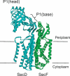
Structure of SecDF. The structure of T. thermophilus SecDF, which is encoded as a single polypeptide chain, is colored to represent the individual SecD and SecF polypeptides found in E. coli. Transmembrane helices 1 to 6 (blue) represent E. coli SecD. The periplasmic P1 domain between TM1 and TM2 is shown at the top of the figure. The head and the base subdomains are indicated. Transmembrane helices 7 and 8 represent E. coli SecF.
Figure 11
Figure 11
Two conformations of SecDF. The P1 domain of SecDF, shown extending into the periplasm comprises two subdomains: a P1 head (orange) and P1 base (blue). The protein was crystallized in the F form (left-hand side) with the P1 domain positioned so that the head is bent toward the membrane. The I form shows the head directly above the base. This structure is a model built from superimposing the base subdomain of the isolated P1 structure onto that of the full-length SecDF. Reprinted from reference , with permission.

References

    1. Redman CM, Siekevitz P, Palade GE. 1966. Synthesis and transfer of amylase in pigeon pancreatic micromosomes. J Biol Chem 241:1150–1158. [PubMed] - PubMed
    1. Andrews TM, Tata JR. 1971. Protein synthesis by membrane-bound and free ribosomes of secretory and non-secretory tissues. Biochem J 121:683–694. [PubMed] - PMC - PubMed
    1. Blobel G, Dobberstein B. 1975. Transfer of proteins across membranes. I. Presence of proteolytically processed and unprocessed nascent immunoglobulin light chains on membrane-bound ribosomes of murine myeloma. J Cell Biol 67:835–851. [PubMed] - PMC - PubMed
    1. Schlessinger D. 1963. Protein synthesis by polyribosomes on protoplast membranes of B. megaterium. J Mol Biol 7:569–582. [PubMed] - PubMed
    1. Hendler RW, Tani J. 1964. On the cytological unit for protein synthesis in vivo in E. coli. II. Studies with intact cells of type B. Biochim Biophys Acta 80:294–306. [PubMed] - PubMed

MeSH terms

Substances

LinkOut - more resources