

doi: 10.1038/ijos.2017.40.
Meeting report: a hard look at the state of enamel research
Affiliations
- PMID: 29165423
- PMCID: PMC5775332
- DOI: 10.1038/ijos.2017.40
Item in Clipboard
Meeting report: a hard look at the state of enamel research
Int J Oral Sci.
.
Abstract
The Encouraging Novel Amelogenesis Models and Ex vivo cell Lines (ENAMEL) Development workshop was held on 23 June 2017 at the Bethesda headquarters of the National Institute of Dental and Craniofacial Research (NIDCR). Discussion topics included model organisms, stem cells/cell lines, and tissues/3D cell culture/organoids. Scientists from a number of disciplines, representing institutions from across the United States, gathered to discuss advances in our understanding of enamel, as well as future directions for the field.
Figures
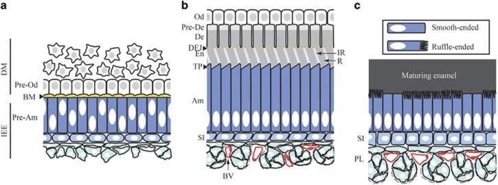
Complex cellular interaction and differentiation processes involved in enamel development. The pre-secretion (a), secretion (b) and maturation (c) stages of amelogenesis are represented in the context of the tissues surrounding the developing enamel. During tooth development, the formation of enamel and dentin, the two major mineralized constituents of the tooth, is initiated at the interface between the dental mesenchyme (DM) and the inner enamel epithelium (IEE), which are separated by a basement membrane (BM). The cells from the dental mesenchyme at this interface will differentiate into odontoblasts (Od) that produce predentin (Pre-De) and drive its progression into mineralized dentin (De). In the crown, cells from the inner enamel epithelium will differentiate into enamel-producing ameloblasts (Am). Prior to enamel and dentin deposition (pre-secretion stage), interactions between pre-odontoblasts (Pre-Od) and pre-ameloblasts (Pre-Am) play a crucial role in the specification of both compartment. Pre-dentin is secreted first and is comprised mainly of type I collagen, which starts to mineralize. Pre-ameloblasts secrete enamel matrix proteins and initiate enamel mineral ribbon deposition at the dentin-enamel junction (DEJ). Ameloblasts then go through a secretory stage where they deposit enamel matrix proteins into highly structured enamel rods (R) and interrods (IR). During this stage ameloblasts are elongated and develop a specialized structure at the secretion front called the Tomes’ process (TP). The secretion phase is followed by a maturation phase during which enamel matrix proteins are degraded by proteases to leave space for the full expansion of the hydroxyapatite crystals. During this stage, ameloblasts are shorter and cycle between ruffle-ended and smooth-ended phases. The epithelial cells underlying the ameloblasts progressively develop into a stratum intermedium (SI), directly in contact with the ameloblasts, and a papillary layer (PL) populated by blood vessels (BV). Although these layers certainly play an important role in enamel development, their function remains poorly understood.
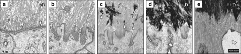
Focused ion beam scanning electron microscopy (FIB-SEM) images of early dentin and enamel mineralization in the mouse mandibular incisor. (a) The tips of unmineralized type I collagen predentin (pD) matrix deposited by odontoblasts (not shown) pass through the basement membrane (downward arrowheads) and associate with the ameloblast (Am) plasma membrane. (b) Fenestration of the basement membrane and the extension of ameloblast processes (*) into the unmineralized collagen matrix. (c) The onset of dentin mineralization as discrete mineral foci (upward arrowheads); (d) expansion of the dentin (d) mineral into a continuous layer; and (e) deposition of enamel mineral ribbons (e) on mineralized dentin. The ameloblast has already formed a Tomes process (Tp) that organizes the ribbons into rod and interrod enamel. FIB-SEM technology is allowing scientists to obtain ultrastructural information of enamel formation in wild-type and knockout mice.
References
-
- Qu Q, Haitina T, Zhu M et al. New genomic and fossil data illuminate the origin of enamel. Nature 2015; 526 (7571): 108–111. - PubMed
-
- Sire JY. Light and TEM study of nonregenerated and experimentally regenerated scales of Lepisosteus oculatus (Holostei) with particular attention to ganoine formation. Anat Rec 1994; 240 (2): 189–207. - PubMed
-
- Sire JY. Ganoine formation in the scales of primitive actinopterygian fishes, lepisosteids and polypterids. Connect Tissue Res 1995; 33 (1/2/3): 213–222. - PubMed
-
- Satchell PG, Shuler CF, Diekwisch TG. True enamel covering in teeth of the Australian lungfish Neoceratodus forsteri. Cell Tissue Res 2000; 299 (1): 27–37. - PubMed
Publication types
MeSH terms
Grants and funding
LinkOut - more resources
Full Text Sources
Other Literature Sources
