Review
. 2018 Mar;19(3):158-174.
doi: 10.1038/nrm.2017.103. Epub 2017 Nov 22.

Functional 5' UTR mRNA structures in eukaryotic translation regulation and how to find them

Affiliations
Review

Functional 5' UTR mRNA structures in eukaryotic translation regulation and how to find them

Kathrin Leppek et al. Nat Rev Mol Cell Biol. 2018 Mar.

Erratum in

Abstract

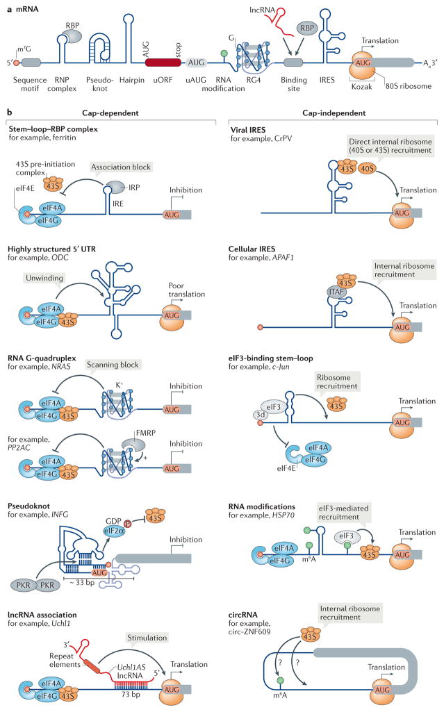
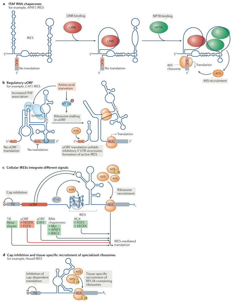
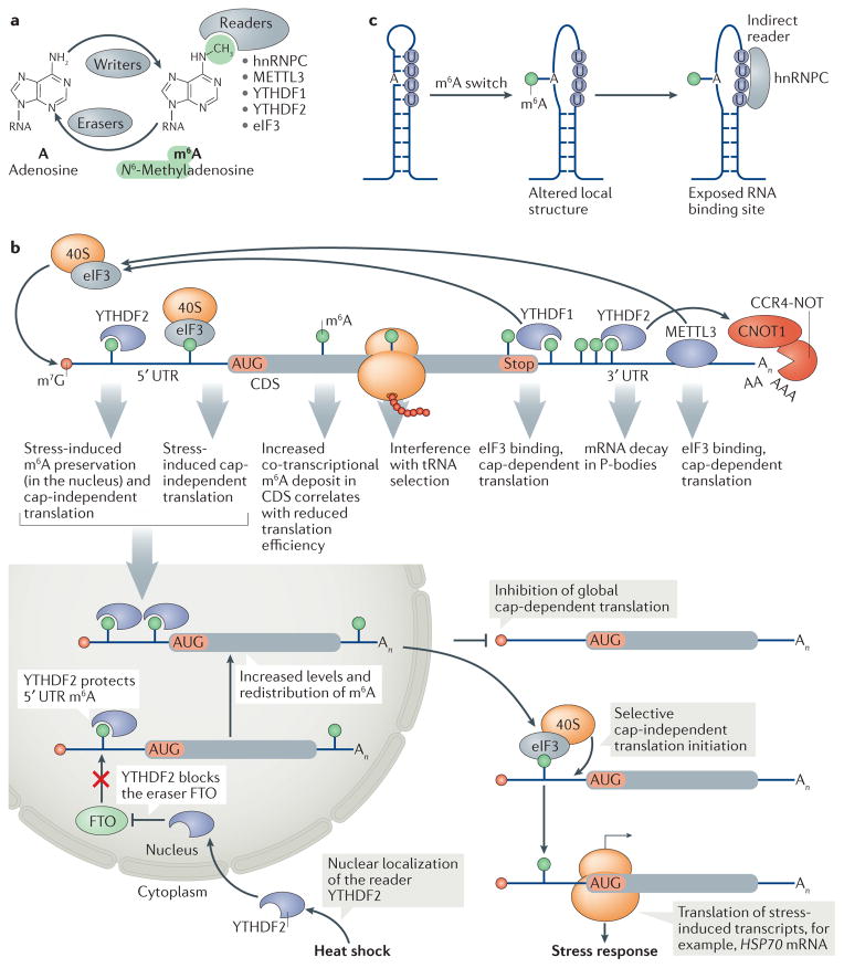
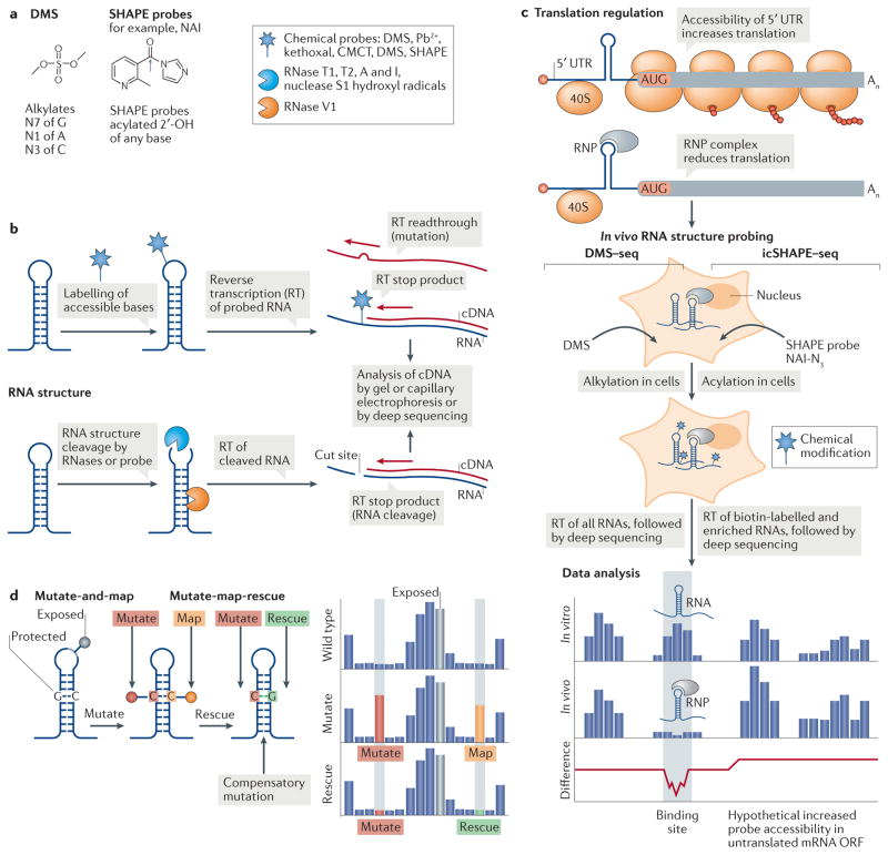
RNA molecules can fold into intricate shapes that can provide an additional layer of control of gene expression beyond that of their sequence. In this Review, we discuss the current mechanistic understanding of structures in 5' untranslated regions (UTRs) of eukaryotic mRNAs and the emerging methodologies used to explore them. These structures may regulate cap-dependent translation initiation through helicase-mediated remodelling of RNA structures and higher-order RNA interactions, as well as cap-independent translation initiation through internal ribosome entry sites (IRESs), mRNA modifications and other specialized translation pathways. We discuss known 5' UTR RNA structures and how new structure probing technologies coupled with prospective validation, particularly compensatory mutagenesis, are likely to identify classes of structured RNA elements that shape post-transcriptional control of gene expression and the development of multicellular organisms.


Conflict of interest statement

Competing interests statement

The authors declare no competing interests.

Figures

Figure 1
Figure 1. Evolutionary expansion of eukaryotic 5′ UTR lengths
The length of 5′ untranslated regions (UTRs) has increased in eukaryotes during evolution, with median lengths ranging between 53–218 nucleotides (nt). We compared RefSeq-annotated 5′ UTR lengths of reviewed and validated transcripts (n) between species for which at least 100 5′ UTRs are annotated. For yeast, we used the 5′ UTR lengths as annotated in REFS ,. The violin plots depict the distribution of 5′ UTR lengths for 15 species sorted according to decreasing median 5′ UTR length, including human (Homo sapiens), fruit fly (Drosophila melanogaster), thale cress (Arabidopsis thaliana), mouse (Mus musculus), maize (Zea mays), zebrafish (Danio rerio), rat (Rattus norvegicus), wasp (Nasonia vitripennis), western clawed frog (Xenopus tropicalis), cow (Bos taurus), wild boar (Sus scrofa), tomato (Solanum lycopersicum), chicken (Gallus gallus), dog (Canis lupus familiaris) and the budding yeast Saccharomyces cerevisiae. The data range for each species was trimmed to a maximum of the third quartile plus three times the interquartile range (Q3 + 3 × IQR).
Figure 2
Figure 2. Cis-acting regulatory RNA elements and structures in eukaryotic 5′ UTRs influence mRNA translation
a | The 7 methylguanosine (m7G) 5′ cap structure (circle) at the 5′ end of the mRNA and the poly(A) tail (An) at the 3′ end stabilize the mRNA and stimulate translation. The 5′ untranslated region (UTR) contains secondary and tertiary structures and other sequence elements. RNA structures such as pseudoknots, hairpins and RNA G-quadruplexes (RG4s), as well as upstream open reading frames (uORFs) and upstream start codons (uAUGs), mainly inhibit translation. Internal ribosomal entry sites (IRESs) mediate translation initiation independently of the cap. RNA modifications, or RNA binding proteins (RBPs) and long non coding RNAs (lncRNAs) that interact with RNA binding sites or form ribonucleoprotein (RNP) complexes, as well as the Kozak sequence around the start codon, can additionally regulate translation initiation. b | Regulatory 5′ UTR RNA structures can influence protein synthesis by promoting or inhibiting either cap-dependent (left) or cap-independent (right) translation. Whereas the structures that regulate cap-dependent initiation — RG4s, stem–loop structures and pseudoknots (but not lncRNAs) — tend to repress initiation, cap-independent regulatory RNA structures, including IRESs, eukaryotic initiation factor 3 (eIF3)-binding stem–loop structures, RNA modifications and circular RNAs (circRNAs), can internally assemble the translation machinery onto the mRNA and generally stimulate translation. 3d, eIF3d; APAF1, apoptotic peptidase activating factor 1; circ-ZNF609, circular-zinc-finger protein 609; CrPV, cricket paralysis virus; FMRP, fragile X mental retardation protein 1; HSP70, heat shock protein 70; IFNG, interferon gamma; IRE, iron responsive element; IRP, iron regulatory protein; ITAF, IRES trans-acting factor; K+, potassium; m6A, N6-methyladenosine; NRAS, NRAS proto oncogene, GTPase; ODC, ornithine decarboxylase; P, phosphorylation; PKR, protein kinase RNA activated; PP2AC, protein phosphatase 2 catalytic subunit alpha (also known asPPP2CA); Uchl1, ubiquitin carboxyl terminal hydrolase L1.
Figure 3
Figure 3. Cellular IRES structures employ different mechanisms for ribosome recruitment
Cellular structured internal ribosome entry sites (IRESs) use diverse modes to recruit ribosomes, and their activity is often induced following a change in cellular condition. a | Many IRESs rely on binding by RNA-binding proteins known as IRES trans-acting factors (ITAFs) for ribosome recruitment. Such RNA chaperones remodel the IRES structure and thereby prepare a landing platform for the ribosome. In the IRES of apoptotic peptidase activating factor 1 (APAF1) for example, binding of NRAS upstream gene protein (UNR) to a purine-rich region in a stem–loop opens two stem–loop structures and allows the binding of neural polypyrimidine tract binding protein (NPTB), which creates a ribosome- accessible site for translation. b | Cellular IRESs can also use upstream open reading frames (uORFs) to regulate IRES activity. In the 5′ untranslated region (UTR) of the arginine–lysine transporter amino acid transporter, cationic 1 (CAT1) mRNA, translation of a uORF within the IRES is induced upon amino acid stress. This stalls ribosomes in the uORF and causes a structural switch in the IRES to an active conformation, which enables the translation of the main ORF,. In addition, the association of the ITAFs PTB and heterogeneous nuclear ribonucleoprotein L (hnRNPL) with the IRES increases and is required for translation during starvation. c | Cellular IRESs can integrate signals in cis and trans to modulate internal ribosome recruitment. Whereas a translation inhibitory element (TIE) blocks cap-dependent initiation, uORF translation, ITAFs or RNA G-quadruplexes (RG4s) can all increase (green) IRES- mediated translation in a transcript-specific manner; uORF translation can also inhibit (red) IRES activity. d | In a subset of homeobox a (Hoxa) mRNAs in the mouse embryo, an IRES recruits ribosomes in a tissue-specific manner. Several of these Hoxa IRESs additionally depend on the ribosomal protein RPL38 (L38) for their activity and a TIE at the cap that blocks cap-dependent initiation. BAG1, BCL2 associated athanogene 1; eIF2α, eukaryotic initiation factor 2α; FGF, fibroblast growth factor; P, phosphorylation; VEGFA, vascular endothelial growth factor A.
Figure 4
Figure 4. The effects of N6-methyladenosine on mRNA translation and decay
a | ‘Writer’ proteins establish N6-methyladenosine (m6A) at internal RNA sites, ‘eraser’ proteins remove them, and ‘reader’ proteins directly bind the N6-methyl group of m6A (reviewed in REF. 148). The listed readers affect the translation and stability of m6A-modified mRNAs. b | m6A modifications in mRNAs occur with a preference for the last exon and 3′ untranslated region (UTR) and are increased during stress. According to its position in the mRNA, m6A is bound by readers that can induce cap-dependent translation or internal ribosome recruitment. In the 5′ UTR, eukaryotic initiation factor 3 (eIF3) can directly bind m6A and facilitate internal translation initiation. Stress-responsive 5′ UTR N6-adenosine methylation, for example during heat shock, is preserved by YTH domain-containing family protein 2 (YTHDF2), which blocks binding of the eraser fat mass and obesity-associated protein (FTO), thereby promoting cap-independent translation initiation of stress response mRNAs. m6A in the coding sequence (CDS) is linked to tRNA selection, and at the 3′ UTR it is linked to increased translation owing to YTHDF1 binding to m6A and eIF3 recruitment for cap-dependent translation. The writer methyltransferase like 3 protein (METTL3) can also directly bind to eIF3 to increase translation of m6A-containing mRNAs independently of its m6A writer activity. By contrast, YTHDF2 promotes degradation of m6A-modified mRNAs by recruiting the deadenylase complex CCR4–NOT. Together, increased translation efficiency and activated decay of m6A-modified mRNAs allow dynamic regulation of protein synthesis. c | m6A mRNA modifications are associated with unfolded RNA structures. N6 adenosine methylation in a stem disrupts based paired regions (‘m6A switch’), which allows binding of the ‘indirect reader’ heterogeneous nuclear ribonucleoprotein C (hnRNPC) to exposed U rich motifs in the nucleus. CNOT1, CCR4–NOT transcription complex subunit 1; HSP70, heat shock protein 70; P body, processing body. Part c is adapted from REF. , Macmillan Publishers Limited.
Figure 5
Figure 5. Global RNA structure probing to assess translation regulation
Global RNA structure probing inside cells can assess the transcriptome structure in the presence of proteins. a | Chemical schematics of the RNA structure probes dimethyl sulfate (DMS) and the selective 2′-hydroxyl acylation analysed by primer extension (SHAPE) reagent 2 methylnicotinic acid imidazolide (NAI) and their reactivity. The grey arrow indicates the site of 2′-OH attack of the RNA by the probe. Different probes induce single-strand-specific chemical labelling (star) or cleavage by enzymes or probes (blue Pac Man shape, single strand specific; orange Pac Man, double strand specific). b | RNA probing either labels RNA through a covalent reaction of a chemical probe with accessible nucleotides (top) or cleaves the RNA backbone with RNases (bottom). A pool of modified or cleaved RNAs is transcribed into cDNA by reverse transcription (RT), and modified or cleaved sites are identified by their effect on RT. c | A 5′ untranslated region (UTR) ribonucleoprotein (RNP) complex that inhibits ribosome scanning reduces the accessibility of the mRNA for the probe in cells. In in vivo RNA structure probing, shown here for DMS treatment followed by deep sequencing (DMS–seq) and in vivo click SHAPE followed by deep sequencing (icSHAPE–seq), RNA structures areprobedincells bychemicalmodification. Data analysis of in vitro-probed and in vivo-probed RNA can indicate the presence of RNA structures as protein binding sites, owing to masked probe accessibility or to remodelling of the structure by protein interaction. Where a 5′ UTR RNP complex inhibits the translation of an open reading frame (ORF), the accessibility of the probe to poorly translated coding sequences might increase in the absence of ribosomes. d | In multidimensional mutate and map chemical probing, a mutation (red) that eliminates base pairing exposes the mutated nucleotide and its partner nucleotide (orange) to chemical modification (pins). In mutate-map-rescue probing, a mutation is rescued from modification by a compensatory mutation of the partner nucleotide (green). The reactivity profile reflects changes in probe accessibility upon mutation (red), which allows mapping (orange) of the base-paired nucleotide, while rescue (green) confirms base pairing. Nucleotides in loops are exposed and accessible to the probe. Pb2+, lead; CMCT, 1 cyclohexyl (2-morpholinoethyl)carbodiimide metho-p-toluene sulfonate.

References

    1. Breaker RR. Riboswitches and the RNA world. Cold Spring Harb Perspect Biol. 2012;4:1–15. - PMC - PubMed
    1. Bowman JC, Hud NV, Williams LD. The ribosome challenge to the RNA world. J Mol Evol. 2015;80:143–161. - PubMed
    1. Nahvi A, et al. Genetic control by a metabolite binding mRNA. Chem Biol. 2002;9:1043–1049. - PubMed
    1. Wachter A. Gene regulation by structured mRNA elements. Trends Genet. 2014;30:172–181. - PubMed
    1. Martin W, Koonin EV. Introns and the origin of nucleus-cytosol compartmentalization. Nature. 2006;440:41–45. - PubMed

Publication types