. 2018 Jan 25;46(2):661-676.
doi: 10.1093/nar/gkx1142.

The regulatory G4 motif of the Kirsten ras (KRAS) gene is sensitive to guanine oxidation: implications on transcription

Affiliations

The regulatory G4 motif of the Kirsten ras (KRAS) gene is sensitive to guanine oxidation: implications on transcription

Susanna Cogoi et al. Nucleic Acids Res. .

Abstract

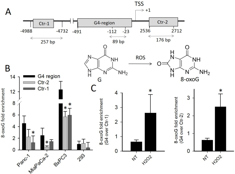
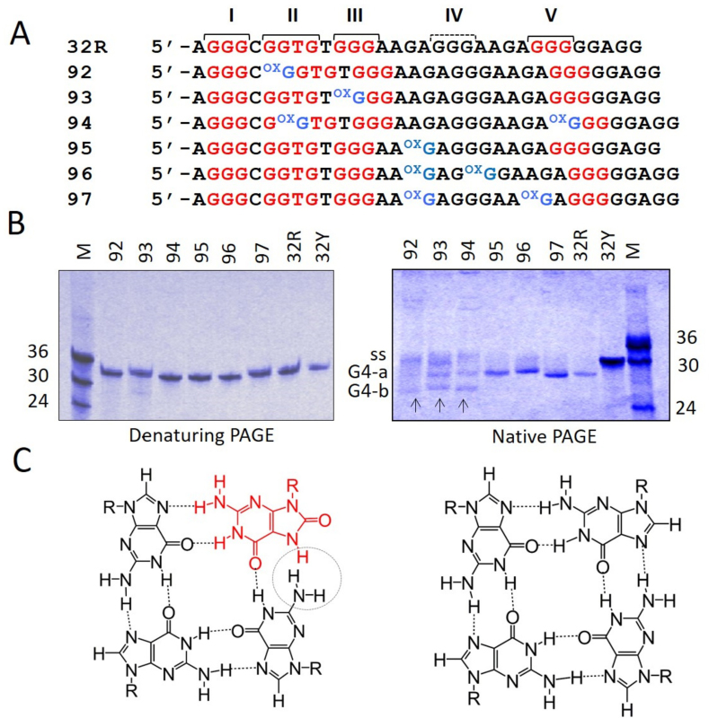
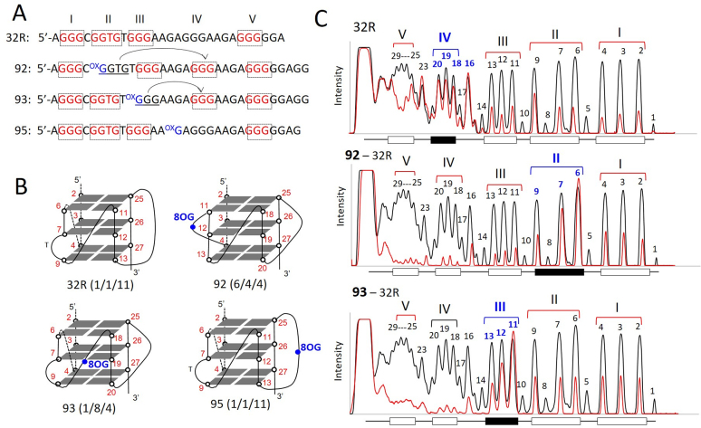
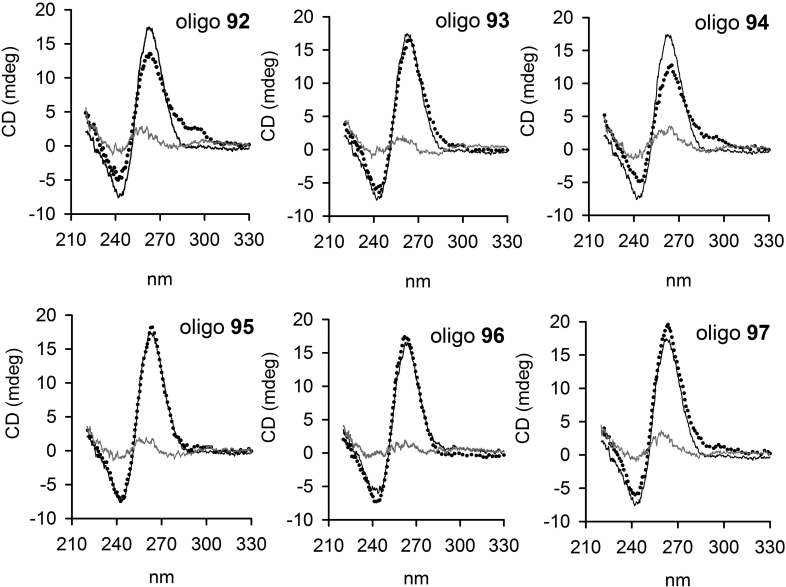
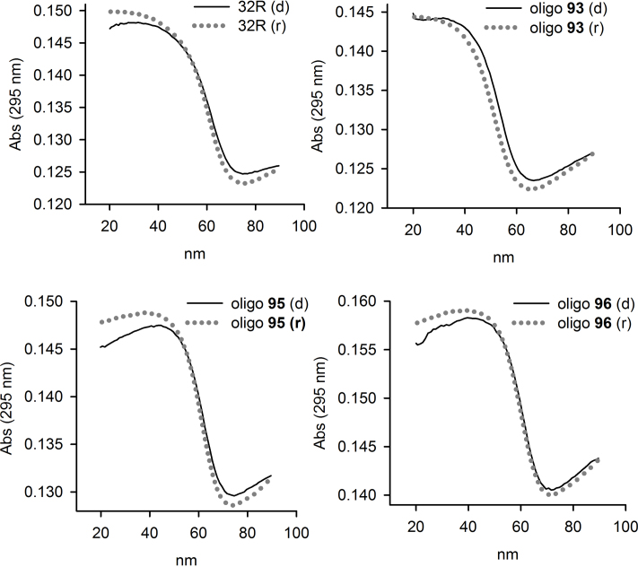
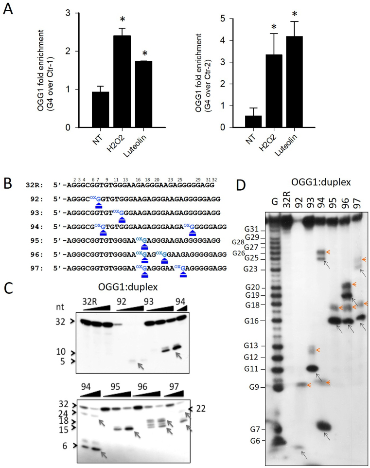
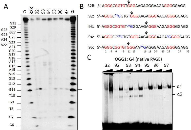
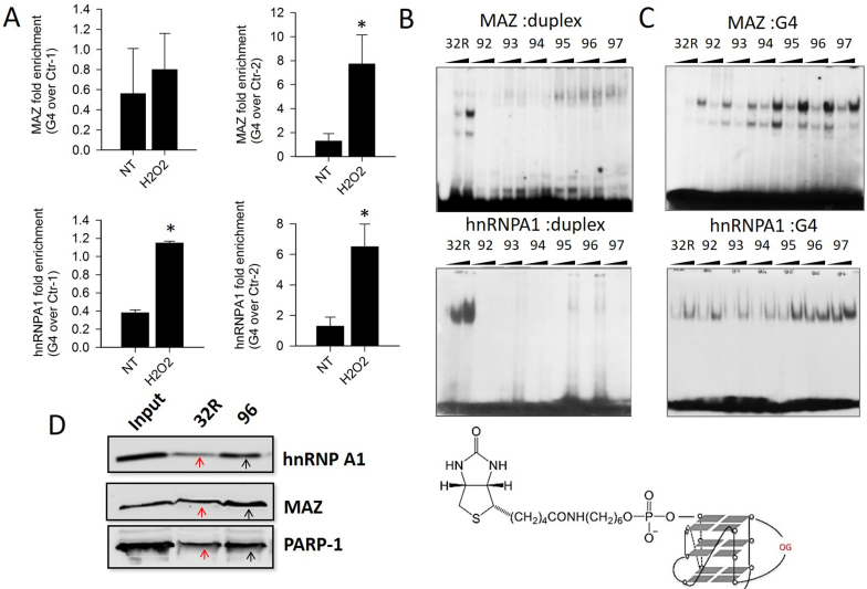
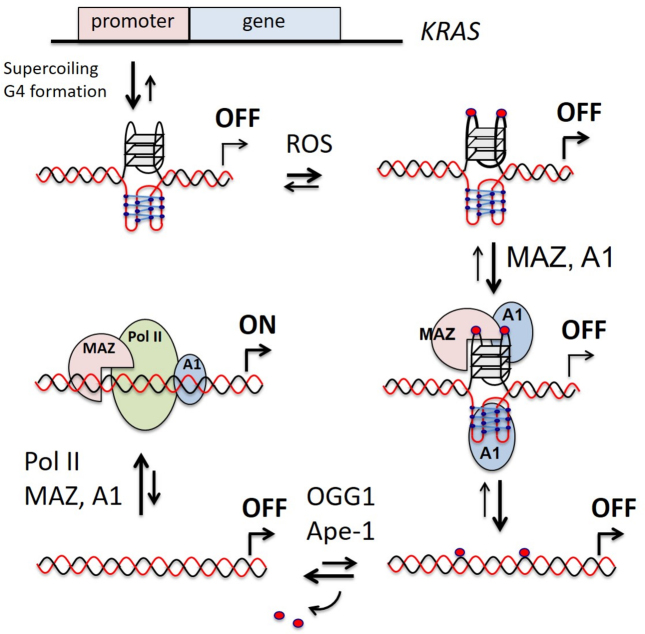
KRAS is one of the most mutated genes in human cancer. It is controlled by a G4 motif located upstream of the transcription start site. In this paper, we demonstrate that 8-oxoguanine (8-oxoG), being more abundant in G4 than in non-G4 regions, is a new player in the regulation of this oncogene. We designed oligonucleotides mimicking the KRAS G4-motif and found that 8-oxoG impacts folding and stability of the G-quadruplex. Dimethylsulphate-footprinting showed that the G-run carrying 8-oxoG is excluded from the G-tetrads and replaced by a redundant G-run in the KRAS G4-motif. Chromatin immunoprecipitation revealed that the base-excision repair protein OGG1 is recruited to the KRAS promoter when the level of 8-oxoG in the G4 region is raised by H2O2. Polyacrylamide gel electrophoresis evidenced that OGG1 removes 8-oxoG from the G4-motif in duplex, but when folded it binds to the G-quadruplex in a non-productive way. We also found that 8-oxoG enhances the recruitment to the KRAS promoter of MAZ and hnRNP A1, two nuclear factors essential for transcription. All this suggests that 8-oxoG in the promoter G4 region could have an epigenetic potential for the control of gene expression.


Figures

Figure 1.
Figure 1.
(A) Relative distance from TSS of G4 and non-G4 sequences used in quantitative real-time ChIP experiments. The length of each amplified DNA fragment is indicated. The structures of guanine and 8-oxo-7,8-dihydroguanine (8-oxoG) are shown; (B) ChIP qPCR showing the basal level of 8-oxoG in G4 and in non-G4 regions Ctr-1 and Ctr-2 in pancreatic cancer cells harboring mutated KRAS (Panc-1 and MIAPaCa-2) or wild-type KRAS (BxPC3) and in non-cancer HEK-293 cells. The histogram shows the fold enrichment of 8-oxoG in pancreatic cancer cells compared to 8-oxoG in G4 region of 293 cells; (C) ChIP qPCR showing the relative level of 8-oxoG in G4 compared to non-G4 regions (Ctr-1 and Ctr-2). The ordinate reports the ratio between the level of 8-oxoG in G4 and in non-G4 region, in Panc-1 cells treated with 1 mM H2O2. The asterisk (*) means P < 0.05 (n = 4), a Student’s t-test was performed.
Figure 2.
Figure 2.
(A) Sequences of 32R and of the designed oligonucleotides with one or two 8-oxoG either in the major 11-nt loop of KRAS G4 (92, 93 and 94) or in G-tetrads (95, 96 and 97). The G-runs I–V are pointed out; (B) 20% PAGE of 32R and 8-oxoG-substituted oligonucleotides in denaturing (left) and native (right) gels. As reference oligonucleotides of 24, 30 and 36 nt have been loaded. G4-a and G4-b are due to G4 structures, ss indicates unstructured oligonucleotides. Gels were repeated three times; (C) Structure of a G-tetrad with 8-oxoG (left) and of a canonical G-tetrad (right).
Figure 3.
Figure 3.
(A) Sequences of 32R, 92, 93 and 95 showing the G-runs involved in G4 formation (indicated in red). Compared to 32R, 92 and 93 undergo a different folding in order to exclude 8-oxoG from a G-tetrad; (B) Structure of the putative G-quadruplexes with 8-oxoG; (C) Comparison of the DMS-footprinting of unstructured 32R with those of structured 32R and 92, 93 analogs. Note that the fifth G-run of 32R replaces the G-run with 8-oxoG through an alternative folding. Experiment was repeated three times.
Figure 4.
Figure 4.
CD spectra of 32R at 20°C (black) and of 8-oxoG-substituted oligonucleotides at 20°C (dotted spectra) and 90°C (gray spectra), in 50 mM Tris–HCl, pH 7.4, 100 mM KCl. Each panel compare the CD spectrum of 32R with that of an oligonucleotide with 8-oxoG. The ordinate reports the ellipticity signal expressed in mdeg.
Figure 5.
Figure 5.
Denaturing and annealing UV-melting curves of 32R and 8-oxoG substituted oligonucleotides in 50 mM cacodylate pH 7.4, 100 mM KCl. The curves have been obtained by measuring the absorbance at 295 nm as a function of temperature, at a heating/cooling rates of 0.5°C/min. Denaturing, black filled curves; renaturing, dotted gray curves.
Figure 6.
Figure 6.
(A) ChIP qPCR showing that after treatment of Panc-1 cells with 1 mM H2O2 or 10 μM luteolin, the recruitment of OGG1 to the KRAS promoter increases more at G4 region than non-G4 regions (Ctr-1 and Ctr-2). Asterisk (*) indicate P < 0.05 (n = 4), a Student’s t-test was performed; (B) Primary sequences of 32R and designed 8-oxoG-substituted oligonucleotides where the positions of 8-oxoG are indicated; (C) The panel shows in a denaturing PAGE that OGG1 excises 8-oxoG in the duplexes formed by the designed 8-oxoG oligonucleotides and the complementary strand; (D) Sequencing 18% PAGE showing that OGG1 (1 μM) cleaves the radiolabeled duplexes (2 nM) exactly at the positions where there is 8-oxoG. The experiments in (C) and (D) were repeated three times.
Figure 7.
Figure 7.
(A) Sequencing 18% PAGE showing the effect of OGG1 (1 μM) on the G-quadruplexes. A very weak cleavage activity is detected at specific guanines: G11 in the G4s formed by 32R, 95, 96 and 97; G16/A17 in the G4s formed by 93, G4-(1/8/4) and 94, G4-(6/4/5). This cleavage activity is not correlated with 8-oxoG; (B) Sequences of the G4 motifs 32R, 92, 93, 94 and 95 showing the positions where the G4s are cleaved by OGG1 (96 and 97 behave as 95); (C) Native PAGE showing that OGG1 binds to radiolabeled 32R and 8-oxoG oligonucleotides (20 nM) in the G-quadruplex form. Note that 92, 93 and 94 form two complexes (c1 and c2) as they form in solution two quadruplexes. OGG1 (0.3 and 0.6 μg) and oligonucleotides (20 nM) have been incubated 45 min at 37°C prior to PAGE. The experiment in A was repeated three times, that in C two times.
Figure 8.
Figure 8.
(A) ChIP qPCR showing that the recruitment of MAZ and hnRNP A1 to the promoter G4 region is higher than to non-G4 regions (Ctr-1 and Ctr-2), following cell treatment with 1 mM H2O2. Asterisk (*) indicates P < 0.05 (n = 4), a Student’s t-test was performed; (B) The panels show the binding of MAZ and hnRNP A1 (2.5 and 5 μg) to 20 nM radiolabeled 32R and 8-oxoG oligonucleotides in duplex. The binding of MAZ and hnRNP A1 to the duplexes bearing 8-oxoG is strongly inhibited; (C) The panels show the binding of MAZ and hnRNP A1 to G-quadruplexes 32R and analogs bearing 8-oxoG. The proteins bind to the G4s, even though they harbor 8-oxoG. The G4s with 8-oxoG in the 11-nt loop (95, 96 and 97) bind MAZ much more than wild-type G4. Before EMSA, the G4s or duplexes have been incubated with MAZ for 1 h at 37°C and with hnRNP A1 for 30 min at 25°C; (D) Streptavidin-biotin pull-down assay with nuclear Panc-1 extract and biotinylated 32R and 96 used as DNA baits (the structure is shown). Cellular proteins bound to G4 were pulled down with streptavidin magnetic beads and analyzed by western blot. The experiments in (B) and (C) were repeated three times.
Figure 9.
Figure 9.
A model for KRAS transcription regulation involving 8-oxoG. A1 and Pol II stand for hnRNP A1 and RNA Pol II.

References

    1. Liou G.-Y., Storz P.. Reactive oxygen species in cancer. Free Radic. Res. 2010; 44:1–31. - PMC - PubMed
    1. Sosa V., Moliné T., Somoza S., Paciucci R., Kondoh H.. Oxidative stress and cancer: an overview. Ageing Res. Rev. 2012; 12:376–390. - PubMed
    1. Murphy M.P. How mitochondria produce reactive oxygen species. Biochem. J. 2009; 417:1–13. - PMC - PubMed
    1. Giorgio M., Trinei M., Migliaccio E., Pelicci P.G.. Hydrogen peroxide: a metabolic by-product or a common mediator of ageing signals. Nat. Rev. Mol. Cell Biol. 2007; 8:722–728. - PubMed
    1. St-Pierre J., Buckingham J., Roebuck S.J., Brand M.D.. Topology of superoxide production from different sites in mitochondrial electron transport chain. J. Biol. Chem. 2002; 277:44784–44790. - PubMed

Publication types

MeSH terms