

Initial clinical observations of intra- and interfractional motion variation in MR-guided lung SBRT
- PMID: 29166129
- PMCID: PMC5965474
- DOI: 10.1259/bjr.20170522
Initial clinical observations of intra- and interfractional motion variation in MR-guided lung SBRT
Abstract
Objective: To evaluate variations in intra- and interfractional tumour motion, and the effect on internal target volume (ITV) contour accuracy, using deformable image registration of real-time two-dimensional-sagittal cine-mode MRI acquired during lung stereotactic body radiation therapy (SBRT) treatments.
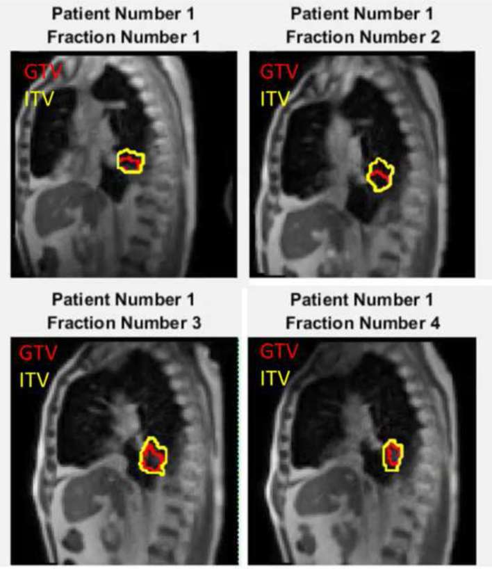
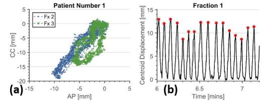
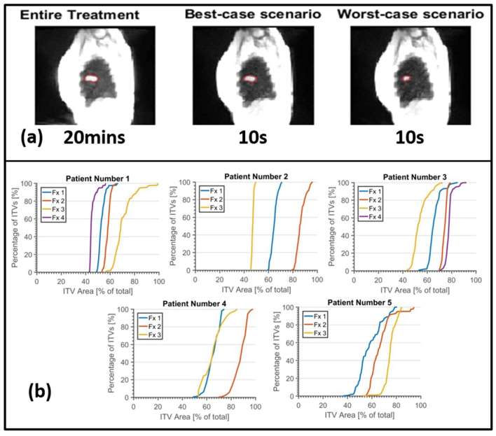
Methods: Five lung tumour patients underwent free-breathing SBRT treatments on the ViewRay system, with dose prescribed to a planning target volume (defined as a 3-6 mm expansion of the 4DCT-ITV). Sagittal slice cine-MR images (3.5 × 3.5 mm2 pixels) were acquired through the centre of the tumour at 4 frames per second throughout the treatments (3-4 fractions of 21-32 min). Tumour gross tumour volumes (GTVs) were contoured on the first frame of the MR cine and tracked for the first 20 min of each treatment using offline optical-flow based deformable registration implemented on a GPU cluster. A ground truth ITV (MR-ITV20 min) was formed by taking the union of tracked GTV contours. Pseudo-ITVs were generated from unions of the GTV contours tracked over 10 s segments of image data (MR-ITV10 s).
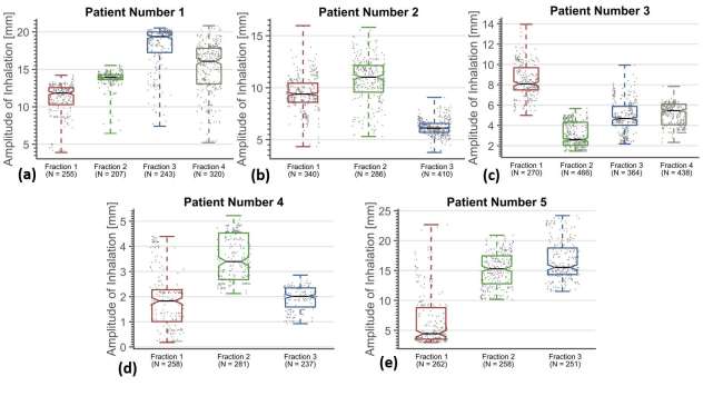
Results: Differences were observed in the magnitude of median tumour displacement between days of treatments. MR-ITV10 s areas were as small as 46% of the MR-ITV20 min.
Conclusion: An ITV offers a "snapshot" of breathing motion for the brief period of time the tumour is imaged on a specific day. Real-time MRI over prolonged periods of time and over multiple treatment fractions shows that ITV size varies. Further work is required to investigate the dosimetric effect of these results. Advances in knowledge: Five lung tumour patients underwent free-breathing MRI-guided SBRT treatments, and their tumours tracked using deformable registration of cine-mode MRI. The results indicate that variability of both intra- and interfractional breathing amplitude should be taken into account during planning of lung radiotherapy.
Figures
References
-
- Zamora DA, Riegel AC, Sun X, Balter P, Starkschall G, Mawlawi O, et al. Thoracic target volume delineation using various maximum-intensity projection computed tomography image sets for radiotherapy treatment planning. Med Phys 2010; 37: 5811–20. doi: https://doi.org/10.1118/1.3504605 - DOI - PMC - PubMed
-
- Underberg RW, Lagerwaard FJ, Cuijpers JP, Slotman BJ, van Sörnsen de Koste JR, Senan S. Four-dimensional CT scans for treatment planning in stereotactic radiotherapy for stage I lung cancer. Int J Radiat Oncol Biol Phys 2004; 60: 1283–90. doi: https://doi.org/10.1016/j.ijrobp.2004.07.665 - DOI - PubMed
-
- Cai J, McLawhorn R, Read PW, Larner JM, Yin FF, Benedict SH, et al. Effects of breathing variation on gating window internal target volume in respiratory gated radiation therapy. Med Phys 2010; 37: 3927–34. doi: https://doi.org/10.1118/1.3457329 - DOI - PubMed
-
- Cai J, Read PW, Baisden JM, Larner JM, Benedict SH, Sheng K. Estimation of error in maximal intensity projection-based internal target volume of lung tumors: a simulation and comparison study using dynamic magnetic resonance imaging. Int J Radiat Oncol Biol Phys 2007; 69: 895–902. doi: https://doi.org/10.1016/j.ijrobp.2007.07.2322 - DOI - PubMed
-
- van Heeswijk RB, Bonanno G, Coppo S, Coristine A, Kober T, Stuber M. Motion compensation strategies in magnetic resonance imaging. Crit Rev Biomed Eng 2012; 40: 99–119. doi: https://doi.org/10.1615/CritRevBiomedEng.v40.i2.20 - DOI - PubMed
MeSH terms
LinkOut - more resources
Full Text Sources
Other Literature Sources
Medical
