. 2017 Nov 22;17(1):219.
doi: 10.1186/s12866-017-1127-y.

Characterization of the inner membrane protein BB0173 from Borrelia burgdorferi

Affiliations

Characterization of the inner membrane protein BB0173 from Borrelia burgdorferi

Christina M Brock et al. BMC Microbiol. .

Abstract

Background: The bacterial spirochete Borrelia burgdorferi is the causative agent of the most commonly reported arthropod-borne illness in the United States, Lyme disease. A family of proteins containing von Willebrand Factor A (VWFA) domains adjacent to a MoxR AAA+ ATPase have been found to be highly conserved in the genus Borrelia. Previously, a VWFA domain containing protein of B. burgdorferi, BB0172, was determined to be an outer membrane protein capable of binding integrin α3β1. In this study, the characterization of a new VWFA domain containing membrane protein, BB0173, is evaluated in order to define the location and topology of this multi-spanning membrane protein. In addition, functional predictions are made.

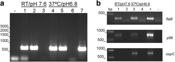
Results: Our results show that BB0173, in contrast to BB0172, is an inner membrane protein, in which the VWFA domain is exposed to the periplasmic space. Further, BB0173 was predicted to have an aerotolerance regulator domain, and expression of BB0173 and the surrounding genes was evaluated under aerobic and microaerophilic conditions, revealing that these genes are downregulated under aerobic conditions. Since the VWFA domain containing proteins of B. burgdorferi are highly conserved, they are likely required for survival of the pathogen through sensing diverse environmental oxygen conditions.

Conclusions: Presently, the complex mechanisms that B. burgdorferi uses to detect and respond to environmental changes are not completely understood. However, studying the mechanisms that allow B. burgdorferi to survive in the highly disparate environments of the tick vector and mammalian host could allow for the development of novel methods of preventing acquisition, survival, or transmission of the spirochete. In this regard, a putative membrane protein, BB0173, was characterized. BB0173 was found to be highly conserved across pathogenic Borrelia, and additionally contains several truly transmembrane domains, and a Bacteroides aerotolerance-like domain. The presence of these functional domains and the highly conserved nature of this protein, strongly suggests a required function of BB0173 in the survival of B. burgdorferi.

Keywords: Aerotolerance; Borrelia burgdorferi; MIDAS motif; Transmembrane; vonWillebrand factor a.


Conflict of interest statement

Ethics approval and consent to participate

Not applicable.

Consent for publication

Not applicable.

Competing interests

The authors declare that they have no competing interests.

Publisher’s Note

Springer Nature remains neutral with regard to jurisdictional claims in published maps and institutional affiliations.

Figures

Fig. 1
Fig. 1
Organization and conservation of bb0173. a Schematic demonstrating the similarity between the Bat region of Borrelia burgdorferi (BB), Borrelia hermsii (BH), Leptospira interrogans (LB), Leptospira biflexa (LBF), and Bacteroides fragilis (BF). Note the similarities across BB0172 through BB0176. b Map demonstrating the pertinent domains of BB0173. The map demonstrates the three transmembrane domains (amino acids: 7–25, 57–77, 310–328), VWFA domain (amino acids: 87–328), MIDAS motif (amino acids: 99–103), BatA domain (amino acids: 9–86), and N-glycosylation sites (amino acids 170–172, 265–267). Additionally, a 30-mer peptide is denoted, BB0173pep, which was used to generate chicken anti-BB0173 antibodies. BB0173T, the truncated BB0173 protein, was also used to generate chicken anti-BB0173 antibodies. c Clustal W (v1.83) alignment of B. burgdorferi B31 BB0173 (bold) against homologues in B. burgdorferi ZS7 (BB0173 BbZS7) Borrelia garinii (BG0172), Borrelia afzelii (BAPKO_0175), and the relapsing fever species Borrelia hermsii (BH0173) and Borrelia turicatae (BT0173). Alignments are also made to B. burgdorferi B31 BB0172 (BB0172 B31), which was found to be very similar in sequence and topology. There is also homology seen to Plasmodium falciparum membrane protein TRAP (Pf TRAP) as well as to the human adhesins LFA-1 (hLFA-1) and CD11b (hCD11b). Conserved residues corresponding to the MIDAS motif are highlighted, including the DXSXS as well as the threonine (T) required for MIDAS function
Fig. 2
Fig. 2
Expression of bb0173 cDNA upon temperature shift. B. burgdorferi B31A3 strain was grown under unfed tick conditions (RT/pH 7.6) to late log phase then shifted to fed tick conditions (37 °C/pH 6.8) before collection of mRNA. The purified mRNA was reverse transcribed to cDNA, and PCR was performed to detect bb0173, flaB, p66, and ospC. Water was used as a negative control (−). a RNA samples were tested for DNA contamination in lanes 3 and 6. Genomic DNA was run in lanes 2 and 5 and served as the positive control. In lanes 1 and 4, cDNA samples were loaded. To confirm functionality of primers, a second genomic DNA sample was applied in lane 7. b The same shifting conditions were used to generate DNA samples as previously. cDNA samples are in lanes 1 and 3, RNA in lanes 2 and 4, and genomic DNA is labeled as (+). Negative control is water, as above. On the left of the figure, the DNA ladder is shown and sizes are denoted in basepairs
Fig. 3
Fig. 3
Expression of bb0170 to bb0176 under decreased-oxygen conditions. Gene expression of the Bat-like genes is quantified under atmospheric oxygen and decreased oxygen conditions. The expression of each gene is determined by comparing the expression under atmospheric oxygen to the expression under low-oxygen growth conditions after normalization to the endogenous control gene, flaB (ΔCT = Ct (goi) – Ct (ref); where “goi” refers to the gene of interest, and “ref” to the reference gene used in the study). All genes tested were found to increase expression under low-oxygen conditions except for bb0174 and rpoN, which both became undetectable in the low-oxygen condition, as denoted by the diamond (♦). For this experiment, rpoN, rrp1, hpk1, and rrp2 have been included as control genes. Statistical analysis was performed using the Holm-Sidak multiple comparisons test with a 95% confidence interval. Each gene was evaluated in triplicate. *Denotes statistical differences: *P value <0.05, **P value <0.01, ***P value <0.001 and ****P value <0.0001
Fig. 4
Fig. 4
Insertion of hydrophobic regions of BB0173 into membranes using Lep as model protein. a The HR sequence in each construct is shown together with the predicted G apparent value, which was estimated using the ∆G prediction algorithm available on the Internet (http://dgpred.cbr.su.se/). Glycosylation acceptor site is shown in bold. b Schematic representation of the Lep construct used to report insertion of hydrophobic regions of BB0173 into endoplasmic reticulum membranes. The TM segment under investigation (HR-tested) was introduced into the P2 domain of Lep, flanked by two artificial glycosylation acceptor sites (G1 and G2). Recognition of the tested sequence as a TM domain by the translocon machinery results in the location of only G1 in the luminal side of the ER membrane, preventing G2 glycosylation (left). The Lep chimera will be doubly glycosylated when the sequence being tested is translocated into the lumen of the microsomes (right). c In vitro translation in the presence of membranes of the different Lep constructs. Constructs containing HR1 (residues 7 to 25; lanes 1–3), HR2 (residues 55 to 77; lanes 4–6), HR3 (residues 163 to 185; lanes 7–9) and HR4 (residues 310 to 328; lines 10–12) were translated in the presence (+) and absence (−) of rough microsomes (RM) and proteinase K (PK). Bands of non- glycosylated proteins are indicated by a white dot; singly and doubly glycosylated proteins are indicated by one and two black dots, respectively. In the case of Lep-HR3 construct a triply glycosylated band was observed (lane 8) due to the presence of an acceptor NGS site (residues 169–171) within the (translocated) hydrophobic region. The protected doubly-glycosylated H2/HR3/P2 fragment is indicated by an arrowhead. Control HRs were used to verify sequence translocation (translocated; lanes 13–15)
Fig. 5
Fig. 5
In vitro analysis of truncated BB0173 constructs. To monitor the membrane orientation of truncated BB0173 molecules a glycosylatable (NSTMSM) tag (white rectangle) was added at position 56 (56mer), 162 (162mer), 278 (278 mer) and 341 (341mer). a Schematic representation of the constructs used in the assay. The position of the glycosylation sites is marked with a Y symbol. The presence of a TM segment identified by the ΔG prediction server (http://dgpred.cbr.su.se/) in each construct and the required linker sequence preceding the glycosylatable tag to allow glycosylation is also included for 341mer truncates. b In vitro translation of the 56mer, 162mer, 278mer and 341mer truncates in the presence (+) or absence (−) of rough microsomes (RM). A white dot marks the non-glycosylated form of the protein while a black dot indicates glycosylation of the C-terminal tag. c In vitro translations in the presence or absence of RM of 162mer truncated constructs were performed bearing an acceptor (NST) or non-acceptor (QST) at N-terminal and/or C-terminal glycosylation tag. White and black dots indicate non-glycosylated and glycosylated molecules respectively, as in panel b. d Schematic representation of the membrane topology of 341mer truncates. A hydrophobic region is noted as a blue box when inserted in the membrane, or as a red box if it is not recognized by the translocon as a TM domain. The position of the glycosylatable tag (white rectangle) and its glycosylation status (white and black dots, represents non-glycosylated and glycosylated respectively) is also shown
Fig. 6
Fig. 6
Localization of BB0173 to the aqueous and inner membrane fractions after treatment with detergent. B. burgdorferi cells disrupted using the detergent Triton X-114 separated into three distinct fractions, the aqueous (AQ), protoplasmic cylinders (PC), and detergent (DT) phases. The phases were separated using SDS-12% PAGE and either stained using Silver Stain Plus (Biorad, Hercules, CA) (a) or were transferred to a PVDF membrane and probed using anti-BB0173T and a secondary anti-chicken HRP-conjugated antibody Lane 1 is B. burgdorferi whole cell lysate. Lane 2 is AQ, Lane 3 is PC, and Lane 4 is DT (b). Controls for outer membrane and inner membrane proteins were OspC and FlaB
Fig. 7
Fig. 7
Protection of BB0173 from protease degradation. Surface proteins of B. burgdorferi are degraded by serine protease Proteinase K (PK). Whole cell lysates were treated with doses ranging from 0 to 200 μg/mL PK prior to separation using SDS-12% PAGE. Gels were either visualized using Coomassie blue staining (a) or transferred to a PVDF membrane and probed with antibodies (b). BB0173 was detected using anti-BB0173pep and anti-chicken HRP-conjugated antibody. Controls for PK mediated degradation and cell integrity during treatment included intercellular protein BosR and periplasmic protein FlaB, as well as outer membrane proteins OspC, VlsE, and P66
Fig. 8
Fig. 8
Localization of the tertiary structures of BB0172 and BB0173 within B. burgdorferi. Models of the tertiary structures of BB0172 and BB0173 were generated and superimposed onto either the inner or outer membrane as predicted from localization studies

References

    1. Centers for Disease C Final 2014 reports of nationally notifiable infectious diseases. MMWR Morb Mortal Wkly Rep. 2015;64(36):1019–1033. doi: 10.15585/mmwr.mm6436a8. - DOI
    1. Kuehn BM. CDC estimates 300,000 US cases of Lyme disease annually. JAMA. 2013;310(11):1110. doi: 10.1001/jama.2013.278331. - DOI - PubMed
    1. Burgdorfer W, Barbour AG, Hayes SF, Benach JL, Grunwaldt E, Davis JP. Lyme disease-a tick-borne spirochetosis? Science. 1982;216(4552):1317–1319. doi: 10.1126/science.7043737. - DOI - PubMed
    1. Stanek G, Wormser GP, Gray J, Strle F. Lyme borreliosis. Lancet. 2012;379(9814):461–473. doi: 10.1016/S0140-6736(11)60103-7. - DOI - PubMed
    1. Coburn J, Magoun L, Bodary SC, Leong JM. Integrins alpha(v)beta3 and alpha5beta1 mediate attachment of lyme disease spirochetes to human cells. Infect Immun. 1998;66(5):1946–1952. - PMC - PubMed

MeSH terms

LinkOut - more resources