. 2017 Nov 22;15(1):86.
doi: 10.1186/s12951-017-0313-2.

Identification of epidermal growth factor receptor-positive glioblastoma using lipid-encapsulated targeted superparamagnetic iron oxide nanoparticles in vitro

Affiliations

Identification of epidermal growth factor receptor-positive glioblastoma using lipid-encapsulated targeted superparamagnetic iron oxide nanoparticles in vitro

Huai-Lu Chen et al. J Nanobiotechnology. .

Abstract

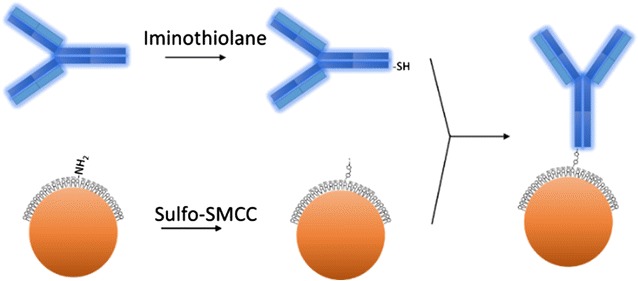
Background: Targeted superparamagnetic iron oxide (SPIO) nanoparticles have emerged as a promising biomarker detection tool for molecular magnetic resonance (MR) image diagnosis. To identify patients who could benefit from Epidermal growth factor receptor (EGFR)-targeted therapies, we introduce lipid-encapsulated SPIO nanoparticles and hypothesized that anti-EGFR antibody cetuximab conjugated of such nanoparticles can be used to identify EGFR-positive glioblastomas in non-invasive T2 MR image assays. The newly introduced lipid-coated SPIOs, which imitate biological cell surface and thus inherited innate nonfouling property, were utilized to reduce nonspecific binding to off-targeted cells and prevent agglomeration that commonly occurs in nanoparticles.

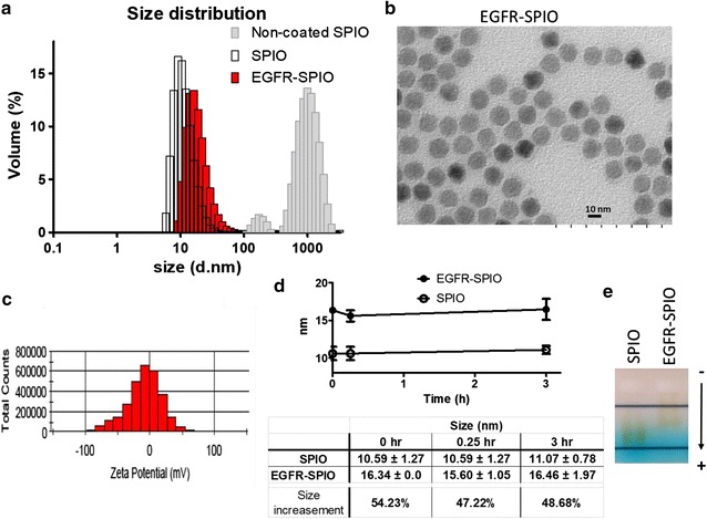
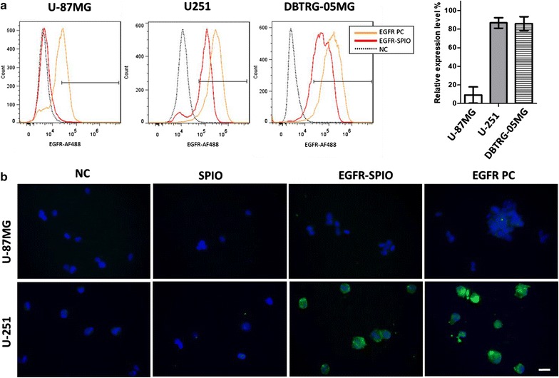
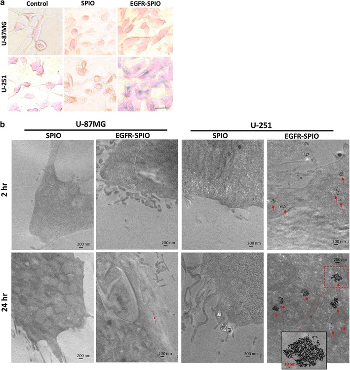
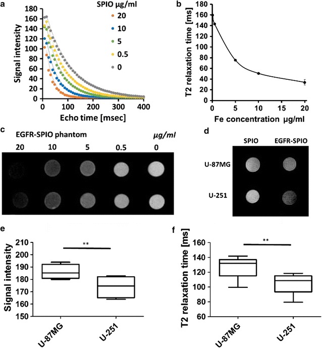
Results: The synthesized targeted EGFR-antibody-conjugated SPIO (EGFR-SPIO) nanoparticles were characterized using dynamic light scattering, zeta potential assays, gel electrophoresis mobility shift assays, transmission electron microscopy (TEM) images, and cell line affinity assays, and the results showed that the conjugation was successful. The targeting efficiency of the synthesized EGFR-SPIO nanoparticles was confirmed through Prussian blue staining and TEM images by using glioblastoma cell lines with high or low EGFR expression levels. The EGFR-SPIO nanoparticles preferentially targeted U-251 cells, which have high EGFR expression, and were internalized by cells in a prolonged incubation condition. Moreover, the T2 MR relaxation time of EGFR-SPIO nanoparticles could be used for successfully identifying glioblastoma cells with elevated EGFR expression in vitro and distinguishing U-251 cells from U-87MG cells, which have low EFGR expression.

Conclusion: These findings reveal that the lipid-encapsulated EGFR-SPIO nanoparticles can specifically target cells with elevated EGFR expression in the three tested human glioblastoma cell lines. The results of this study can be used for noninvasive molecular MR image diagnosis in the future.

Keywords: Epidermal growth factor receptor (EGFR); Glioblastoma; Lipid-encapsulated nanoparticle; Magnetic resonance imaging (MRI); Targeted superparamagnetic iron oxide (SPIO) nanoparticle.


Figures

Fig. 1
Fig. 1
Expression of EGFR in GBM cell lines. a Western blot analysis of EGFR level in GBM cell lines. Cell lysates obtained from the U-87MG, U-251, and DBTRG-05MG GBM cell lines were subjected to Western blot analysis with the anti-EGFR antibody. The β-actin level served as a loading control. b Flow cytometry analysis of EGFR expression profile in untreated GBM cells. The indicated cells were collected and labeled the with anti-EGFR antibody and Alexa Fluore-488 secondary antibody to determine the EGFR level. Cells with EGFR signals that were more than 0.1% stronger than those of the antibody control were categorized as EGFR-positive cells, and the percentage of positive cells is expressed as the relative EGFR expression level. b’ EGFR-positive cells quantified using flow cytometry (n = 3). c IF staining of EGFR-expressing cells. The indicated GBM cells were stained with anti-EGFR (green) and DAPI (blue). Native EGFR was detected in U-251 and DBTRG-05MG cells. Green, anti-EGFR; blue, DAPI. Scale bar, 20 µm
Fig. 2
Fig. 2
Lipid-coated SPIO nanoparticles exhibit superior reduced nonspecific uptake rate compared with uncoated SPIO nanoparticles. TEM images of a non-coated SPIO nanoparticles and b lipid-encapsulated SPIO nanoparticles acquired from a different manufacturer. Scale bar, 10 nm. c Quantification of adsorption rate for non-coated SPIO and lipid-encapsulated SPIO nanoparticles in the U-87MG cell line. Indicated nanoparticles were added to the U-87MG culture medium at final concentrations of 1, 0.5, 0.25, 0.1, 0.05, 0.025, 0.01, and 0 mg/ml before incubating the cells at 37 °C for 1 or 24 h. The cells were then subjected to a colorimetric ferrozine assay to measure the Fe concentration. d The toxicity of lipid-encapsulated SPIO nanoparticles to U-87MG and U-251 cells was evaluated using an MTT viability assay. Lipid-encapsulated SPIO nanoparticles with final Fe concentrations of 0.2, 0.1, 0.05, 0.01, and 0 μg/ml in culture media were incubated with the indicated cells for 24 h before subjecting them to the MTT assay. Error bars, SEM
Fig. 3
Fig. 3
Schematic of anti-EGFR antibody cetuximab conjugated to lipid-encapsulated nonfouling SPIO nanoparticles. Maghemite nanoparticles were modified with maleimide through reaction with sulfo-SMCC; a thiolated antibody was prepared by treating cetuximab with iminothiolane. The maleimide-functionalized SPIO nanoparticles were mixed with the thiolated antibody solution to form antibody-conjugated SPIO nanoparticles. The lipid-coated surface of SPIO nanoparticles provides a nonfouling property
Fig. 4
Fig. 4
Characterization of physical properties of EGFR-SPIO nanoparticles and validation of antibody-SPIO conjugation. a Size distribution of EGFR-SPIO, unconjugated lipid SPIO, and non-coated SPIO nanoparticles were determined through DLS measurement. The size of EGFR-SPIO peaked at 16.34 ± 0.0 nm on volume distribution graph and that of unconjugated lipid SPIO nanoparticles peaked at 10.59 ± 1.27 nm. The non-coated SPIO aggregated and measurement peaked above 1000 nm. b TEM image of EGFR-SPIO nanoparticles. Scale bar, 10 nm. c Zeta potential statistics graph of EGFR-SPIO nanoparticles. The zeta potential of EGFR-SPIO nanoparticles peaked at − 9.24 ± 0.43 mV. d The stability of EGFR-SPIO conjugates was evaluated by measuring the variation of size through DLS. The indicated nanoparticles diluted in PBS (1:12) were incubated for 0, 0.25, and 3 h at room temperature before subjecting to DLS size measurement. The peak of volume distribution from each time point was measured and averaged, and the % of volume incensement after the cetuximab conjugation on lipid SPIO was calculated. The results indicate that EGFR-SPIO nanoparticles were stable over the observation period at room temperature. e Band shift assay of EGFR-SPIO conjugates. Unconjugated lipid SPIO and EGFR-SPIO nanoparticles were subjected to electrophoresis on 1% agarose gel in TAE buffer. Arrowhead, migration direction; −, cathode; +, anode
Fig. 5
Fig. 5
Evaluation of EGFR-SPIO nanoparticle targeting efficiency in GBM cell lines. a The targeting efficiency of EGFR-SPIO nanoparticles was quantified using flow cytometry. Resuspended U-87MG, U-251, and DBTRG-05MG GBM cells were subjected to flow cytometry analysis with EGFR-SPIO nanoparticles or the anti-EGFR antibody and goat anti-human AF-488 antibody. b Evaluate targeting specificity of EGFR-SPIO nanoparticles by IF staining. U-87MG and U-251 cells were fixed and incubated with empty media (negative control, NC), lipid SPIO nanoparticles, EGFR-SPIO nanoparticles, or the cetuximab for positive control (EGFR PC). After PBS wash, the specimens were incubated with goat anti-human AF-488 antibody to detect the presence of cetuximab. Green, cetuximab; blue, DAPI. Scale bar, 20 µm
Fig. 6
Fig. 6
Evaluation of the presence of SPIO nanoparticles in GBM cell lines. a Prussian blue staining for SPIO nanoparticles in GBM cell lines. U-87MG and U-251 cells were incubated with control media, media containing unconjugated lipid SPIO nanoparticles or EGFR-SPIO nanoparticles (100 ng/ml) for 24 h before Prussian blue and nuclear fast red staining. Scale bar, 20 µm. b Lipid SPIO or EGFR-SPIO nanoparticle sorption was detected in U-87MG or U-251 MG cells through TEM. The indicated GBM cells were exposed to EGFR-SPIO or unconjugated lipid SPIO nanoparticle at 0.1 mg/ml for 2 or 24 h before subjecting them to TEM. 20,000× magnification; scale bar, 200 nm; arrowhead, SPIO cluster; red dashed-line box, area with 50,000× magnification; scale bar, 20 nm
Fig. 7
Fig. 7
Detection of EGFR-SPIO nanoparticle binding in U-87MG and U-251 cells in vitro. a Echo time curve fitting of EGFR-SPIO nanoparticle phantoms in various known concentrations was measured using 7T MRI. b T2 relaxation time of EGFR-SPIO nanoparticles in known Fe concentrations (n = 3 for each concentration). c Corresponding signal intensity images of EGFR-SPIO standards measured using T2-weighted MR. Darkest to brightest, 20, 10, 5, 0.5, and 0 μg/ml. d T2-weighted in vitro images of EGFR-SPIO nanoparticle-treated U-87MG and U-251 cells. Cells were incubated with EGFR-SPIO nanoparticles at 0.1 mg/ml at 37 °C for 2 h. A reduction in T2 signal intensity was observed in EGFRhigh U-251 cells compared with EGFRlow U-87MG cells. e Signal intensity of EGFR-SPIO nanoparticle-treated U-87MG and U-251 cells in box plot. f T2 relaxation time of EGFR-SPIO nanoparticle-treated U-87MG and U-251 cells in box plot

References

    1. WHO, GLOBOCAN 2012: estimated cancer incidence, Mortality and Prevalence Worldwide in 2012. http://globocan.iarc.fr/. http://globocan.iarc.fr/Default.aspx. Accessed 20 Feb 2017.
    1. Schwartzbaum JA, Fisher JL, Aldape KD, Wrensch M. Epidemiology and molecular pathology of glioma. Nat Clin Pract Neurol. 2006;2(9):494–503. doi: 10.1038/ncpneuro0289. - DOI - PubMed
    1. Furnari FB, Fenton T, Bachoo RM, et al. Malignant astrocytic glioma: genetics, biology, and paths to treatment. Genes Dev. 2007;21(21):2683–2710. doi: 10.1101/gad.1596707. - DOI - PubMed
    1. Stupp R, Mason WP, van den Bent MJ, et al. Radiotherapy plus concomitant and adjuvant temozolomide for glioblastoma. N Engl J Med. 2005;352(10):987–996. doi: 10.1056/NEJMoa043330. - DOI - PubMed
    1. Brennan CW, Verhaak RGW, McKenna A, et al. The somatic genomic landscape of glioblastoma. Cell. 2013;155(2):462–477. doi: 10.1016/j.cell.2013.09.034. - DOI - PMC - PubMed

MeSH terms