Review
. 2018 Jan 1;98(1):3-58.
doi: 10.1152/physrev.00001.2017.

Endothelial Cell Metabolism

Affiliations
Review

Endothelial Cell Metabolism

Guy Eelen et al. Physiol Rev. .

Abstract

Endothelial cells (ECs) are more than inert blood vessel lining material. Instead, they are active players in the formation of new blood vessels (angiogenesis) both in health and (life-threatening) diseases. Recently, a new concept arose by which EC metabolism drives angiogenesis in parallel to well-established angiogenic growth factors (e.g., vascular endothelial growth factor). 6-Phosphofructo-2-kinase/fructose-2,6-bisphosphatase-3-driven glycolysis generates energy to sustain competitive behavior of the ECs at the tip of a growing vessel sprout, whereas carnitine palmitoyltransferase 1a-controlled fatty acid oxidation regulates nucleotide synthesis and proliferation of ECs in the stalk of the sprout. To maintain vascular homeostasis, ECs rely on an intricate metabolic wiring characterized by intracellular compartmentalization, use metabolites for epigenetic regulation of EC subtype differentiation, crosstalk through metabolite release with other cell types, and exhibit EC subtype-specific metabolic traits. Importantly, maladaptation of EC metabolism contributes to vascular disorders, through EC dysfunction or excess angiogenesis, and presents new opportunities for anti-angiogenic strategies. Here we provide a comprehensive overview of established as well as newly uncovered aspects of EC metabolism.


Figures

FIGURE 1.
FIGURE 1.
General concepts in angiogenesis: formation of a vascular plexus. During vasculogenesis, mesodermal EC progenitors (angioblasts) cluster to form vessel-like endothelial tubes (1) which eventually form a primitive vascular plexus (2). This primitive plexus subsequently undergoes substantial remodeling by vessel intussusception whereby a preexisting capillary splits in two adjacent vessels or by sprouting angiogenesis whereby a new capillary sprouts of a preexisting vessel.
FIGURE 2.
FIGURE 2.
General concepts in angiogenesis: tip versus stalk specification and anastomosis. A: in sprouting angiogenesis, a pro-angiogenic growth factor gradient induces tip and stalk cell formation in a preexisting vessel to form a new sprout. In the tip cell, VEGF binds and activates its receptor VEGFR2, which induces Dll4 expression. In the neighboring ECs, Dll4 binds Notch receptors, which drive expression of the “decoy” VEGF receptor 1 (VEGFR1) while reducing VEGFR2 expression. Ultimately, this increases the VEGFR1/VEGFR2 ratio and lowers EC responsiveness to VEGF, causing the EC to adopt a stalk cell phenotype. Other genetic signals are involved in tip/stalk specification but are not included in the figure for reasons of clarity. B: newly formed sprouts from neighboring vessels meet and fuse through a process termed anastomosis. To allow blood flow, a fully functional, interconnected lumen needs to form in the new sprout; the hemodynamic force of the blood flow itself can cause lumen expansion by causing inverse blebbing of the ECs’ apical membrane. VEGF, vascular endothelial growth factor; VEGFR, vascular endothelial growth factor receptor; Dll4, Delta like 4.
FIGURE 3.
FIGURE 3.
General overview of metabolic pathways in healthy ECs. Schematic representation of metabolic pathways in normal ECs (A, glycolysis; B, TCA cycle; C, pentose phosphate pathway; D, hexosamine biosynthesis pathway; E, polyol pathway; F, uronic cycle; G, glutamine and glutamate metabolism; H, arginine metabolism; I, fatty acid metabolism); to prevent overcrowding of the figure, not all individual steps in each pathway are shown. For the same reason, one-carbon metabolism, the mevalonate pathway, and cysteine-to-H2S metabolism are omitted from this figure and described in detail in FIGURES 10 and 11. Pathways with minimal activity in healthy ECs have lower opacity in the figure. 3DG, 3-deoxyglucosone; 3PG, 3-phosphogylcerate; α-KG, α-ketoglutarate; ALR2, aldose reductase 2; ARG, arginase; ASL, argininosuccinate lyase; ASS, argininosuccinate synthase; BH4, tetrahydrobiopterin; CoA, coenzyme A; CPT1a, carnitine palmitoyltransferase 1a; DHAP, dihydroxyacetone phosphate; eNOS, endothelial nitric oxide synthase; ETC, electron transport chain; F1,6P2, fructose-1,6-bisphosphate; F2,6BP, fructose-2,6-bisphosphate; F6P, fructose-6-phosphate; FA, fatty acid; FAD, flavin adenine dinucleotide; FAO, fatty acid oxidation; FASN, fatty acid synthase; FMN, flavin mononucleotide; G3P, glyceraldehyde-3-phosphate; G6P, glucose-6-phosphate; glucosamine-6-P, glucosamine-6-phosphate; G6PD, glucose-6-phosphate dehydrogenase; GABA, γ-aminobutyric acid; GAPDH, glyceraldehyde-3-phosphate dehydrogenase; GAD, glutamic acid decarboxylase; GDH, glutamate dehydrogenase; GFAT1, glutamine fructose-6-phosphate amidotransferase; GLS, glutaminase; GPAT, glutamine phosphoribosylpyrophosphate amidotransferase; GR, glutathione reductase; GlucN6P, glucosamine-6-phosphate; GLUT1, glucose transporter 1; GS, glutamine synthetase; GSH, reduced glutathione; GSSG, oxidized glutathione; HK, hexokinase; IDH2, isocitrate dehydrogenase 2; LDH, lactate dehydrogenase; NADPH, nicotinamide adenine dinucleotide phosphate; NO, nitric oxide; OAA, oxaloacetate; PGK, phosphoglycerate kinase; PFK1, phosphofructokinase-1; PFKFB3, 6-phosphofructo-2-kinase/fructose-2,6-bisphosphatase 3; PRA, 5-phosphoribosyl-1-amine; PRPP, 5-phosphoribosyl-1-pyrophosphate; ribose-5-P, ribose-5-phosphate; ribulose-5-P, ribulose-5-phosphate; ROS, reactive oxygen species; SORD, sorbitol dehydrogenase; TCA, tricarboxylic acid; THF, tetrahydrofolate; TK, transketolase; UDP-GlcNAc, uridine diphosphate N-acetylglucosamine; UDP-GlcUA, UDP-glucuronic acid; xylulose-5-P, xylulose-5-phosphate.
FIGURE 4.
FIGURE 4.
Glycolytic compartmentalization in ECs. Glycolytic enzymes and glycolytic activity in quiescent ECs are primarily located in perinuclear regions (not shown in figure) but additionally compartmentalize with highly specific cellular structures or components to locally generate ATP or to perform other functions. A: motile ECs colocalize glycolytic enzymes such as PFKFB3 with F-actin fibers in migratory structures like filopodia and lamellipodia. This allows rapid and on-the-spot generation of the energy required by these structures to drive cellular movement and bypasses the need for mitochondria that are too large to fit inside these specialized structures. B: membrane-located transporters, pumps, and channels require ATP to function. This ATP can be delivered in situ by colocalizing glycolytic enzymes. C: in other cell types, PFKFB3 is also present in the nucleus to locally produce F2,6BP which enhances phosphorylation and subsequent proteasomal degradation of the Cdk inhibitor p27Cip/Kip; as a result, cellular proliferation is increased. Whether this also occurs in ECs remains unknown. PFKFB3, 6-phosphofructo-2-kinase/fructose-2,6-bisphosphatase-3; F2,6BP, fructose-2,6-bisphosphate; p27, cyclin-dependent kinase inhibitor 1B.
FIGURE 5.
FIGURE 5.
Compartmentalization of NOX activity in ECs. Schematic and simplified overview of the role and subcellular localization of different NADPH oxidases (NOX) in ECs under normal conditions (left panel) and pathological conditions (right panel). Note that, in contrast to other NOXs, NOX4 activity can have vasculoprotective actions. More details on the role of NOXs in ECs are discussed in the main text. NADPH, nicotinamide adenine dinucleotide phosphate; NOX, NADPH oxidase; ROS, reactive oxygen species, represented in the figure by skull and crossbones.
FIGURE 6.
FIGURE 6.
Metabolic heterogeneity in tip versus stalk cells. A: endothelial tip and stalk cells display differences in metabolic wiring. PFKFB3-driven glycolysis sustains both tip cell competitiveness and stalk cell proliferation, whereas CPT1a-mediated FAO is crucial for stalk cell proliferation but not for tip cell behavior. If and how other metabolic substrates like glutamine (or AAs by extension) drive either tip or stalk cell behavior, or both, requires further study. B: EC metabolism can override genetic tip versus stalk cues. ECs that have been genetically instructed to adopt tip cell behavior by silencing of Notch revert to a stalk cell phenotype upon simultaneous silencing of PFKFB3. C: conversely, overexpression of PFKFB3 provokes tip cell behavior, even in ECs that receive a strong genetic pro-stalk cue from NICD overexpression. PFKFB3, 6-phosphofructo-2-kinase/fructose-2,6-bisphosphatase-3; AA, amino acids; FAO, fatty acid oxidation; CPT1a, carnitine palmitoyltransferase 1a; KD, knock-down; OE, overexpression; NICD, Notch intracellular domain.
FIGURE 7.
FIGURE 7.
Novel role for fatty acid oxidation in differentiation of lymphatic ECs. The dogmatic view on lymphangiogenesis states that at the onset of VEC to LEC differentiation, the transcription factor Prox1 induces the expression of typical lymphangiogenic genes such as VEGFR3. In light of recent findings, this traditional model has been adapted and refined. The binding of Prox1 in the promoter region of lymphangiogenic genes involves an interaction between Prox1 and the histone acetyltransferase p300 to acetylate histones and remodel chromatin. The required acetyl groups are supplied by acetyl-CoA which derives from CPT1a-mediated fatty acid oxidation. Prox1 directly induces the expression of CPT1a to enhance fatty acid oxidation flux. As such, the transcription factor Prox1 teams up with central metabolism to drive lymphatic differentiation. VEC, venous endothelial cell; LEC, lymphatic endothelial cell; Prox1, prospero homeobox protein 1; VEGFR3, vascular endothelial growth factor receptor 3; FAs, fatty acids; CPT1a, carnitine palmitoyltransferase 1a; Ac, acetylated; Ac-CoA, acetyl coenzyme A.
FIGURE 8.
FIGURE 8.
Pathological glycolytic side branches in hyperglycemic dysfunctional ECs. In diabetic ECs, hyperglycemia-induced ROS accumulation (for example caused by impairment of G6PD-mediated PPP flux) causes glycolysis to stall at GAPDH. Upstream glycolytic intermediates pile up and are diverted into pathological glycolytic side branches giving rise to further increases in ROS levels and the formation of noxious AGEs all contributing to the different vascular disease manifestations. 1,3BPG, 1,3-bisphosphoglycerate; 3-DG, 3-deoxyglucosone; ALR2, aldose reductase 2; AGES, advanced glycation end products; DAG, diacylglycerol; DHAP, dihydroxyacetone phosphate; F1,6P2, fructose-1,6-bisphosphate; F6P, fructose-6-phosphate; G3P, glyceraldehyde-3-phosphate; G6P, glucose-6-phosphate; G6PD, glucose-6-phosphate dehydrogenase; GAPDH, glyceraldehyde-3-phosphate dehydrogenase; GFAT1, glutamine fructose-6-phosphate amidotransferase; glucosamine-6-P, glucosamine-6-phosphate; NADPH, nicotinamide adenine dinucleotide phosphate; PARP1, polyADP-ribose polymerase 1; PKC, protein kinase C; UDP-GlcUA, UDP-glucuronic acid; UDP-GlcNAc, uridine diphosphate N-acetylglucosamine.
FIGURE 9.
FIGURE 9.
Different roles of FoxO transcription factors in ECs. Schematic representation of the dual role FoxO transcription factors can play in ECs, either as metabolic gatekeepers to sustain EC quiescence or, conversely, contributing to EC dysfunction. ADMA, asymmetrical dimethyl arginine; BIM, BCL2 like 11; DMA, dimethylamine; FoxO, forkhead box O; NO, nitric oxide.
FIGURE 10.
FIGURE 10.
Metabolic aberrations in ECs in atherosclerosis. In atherosclerosis, ECs display prominent eNOS uncoupling leading to elevated ROS levels and reduced NO-mediated vasodilation. Underlying eNOS uncoupling are decreases in the levels of necessary cofactors (NADPH, CoQ10, BH4) resulting from deregulated metabolic pathways or enzyme activities, and methylation of the eNOS substrate arginine (ADMA). Acetyl-CoA, acetyl-coenzyme A; ADMA, asymmetrical dimethyl arginine; BH2, 7,8-dihydrobiopterin; BH4, tetrahydrobiopterin; CoQ10, coenzyme Q10; DHF, dihydrofolate; DHFR, dihydrofolate reductase; eNOS, endothelial nitric oxide synthase; FPP, farnesylpyrophosphate; GGPP, geranylgeranyl pyrophosphate; GTPCH, GTP cyclohydrolase; hCYS, homocysteine; HMG-CoA, hydroxymethylglutaryl coenzyme A; HMGR, 3-hydroxy-3-methylglutaryl-CoA reductase; HMGS, 3-hydroxy-3-methylglutaryl-CoA synthase; MET, methionine; meTHF, 5,10-methylene-methyltetrahydrofolate; mTHF, 5-methyltetrahydrofolate; MTHFR, methylenetetrahydrofolate reductase; NADPH, nicotinamide adenine dinucleotide phosphate; NO, nitric oxide; NOX, NADPH oxidase; PPP, pentose phosphate pathway; ROS, reactive oxygen species; SAM, S-adenosylmethionine; THF, tetrahydrofolate.
FIGURE 11.
FIGURE 11.
Endothelial cysteine to H2S metabolism in atherosclerosis. The gasotransmitter H2S is an important regulator of cardiovascular homeostasis and has anti-atherogenic effects. In ECs, H2S can be produced from cysteine (via CSE or CAT and 3-MST activity) or in the condensation of cysteine and homocysteine to cystathionine (alternate action of CBS). The anti-atherogenic effects of H2S are listed at the bottom of the image and are discussed in more detail in the main text. 3-MST, 3-mercaptopyruvate sulfurtransferase; CSE, cystathionine-γ-lyase; CBS, cystathionine-β-synthase; CAT, cysteine aminotransferase; eNOS, endothelial NO synthase; LDL, low-density lipoprotein; NH3, ammonia; NO, nitric oxide; vSMC, vascular smooth muscle cell.
FIGURE 12.
FIGURE 12.
Impact of atheroprone versus atheroprotective flow on EC metabolism. Blood flow dynamics critically influence EC metabolism and behavior. Normal atheroprotective blood flow increases the activity of the flow-responsive transcription factor KLF2 which, in addition to increasing VE-cadherin expression and barrier function, transcriptionally represses PFKFB3 expression and lowers glycolytic rates to sustain EC quiescence. Atheroprone turbulent flow lowers KLF2 activity and VE-cadherin levels as well as releases the brake on PFKFB3 expression causing increased glycolysis and EC activation. Additionally, in atheroprone regions, low shear stress and oxLDL induce miR-92a expression to lower KLF2 levels. Finally, normal blood flow sustains a functional EC glycocalyx with protective function. However, atheroprone insults reduce thickness of this glycocalyx through mechanisms discussed in more detail in the main text. PFKFB3, 6-phosphofructo-2-kinase/fructose-2,6-bisphosphatase-3; KLF2, Krüppel-like factor 2; oxLDL, oxidized low-density lipoprotein; VE-cadherin, vascular endothelial cadherin; miR-92a, microRNA-92a.
FIGURE 13.
FIGURE 13.
Reduction of hyperglycolysis in tumor ECs normalizes tumor vessels. Normal, mature blood vessels (top left) have a highly organized endothelial lining to ensure optimal blood flow and barrier function and are stabilized through pericyte coverage. In contrast, tumor vessels (top middle) are highly abnormal and tortuous and have a severely disorganized endothelium and reduced pericyte coverage. As a result, blood flow through these vessels is reduced, leading to a hypoperfused and consequently more aggressive, metastasis-prone tumor. In addition, tumor vessels are leaky which grants cancer cells easy access to the systemic circulation to establish distant metastases. Transcriptomic analysis of ECs isolated from normal vessels (normal ECs or NECs) versus ECs from tumor vessels (tumor ECs or TECs) and subsequent correlation heatmap analysis and hierarchical clustering of 1,255 metabolic genes (bottom left, color coding: high degree of correlation in red; lower degree of correlation in blue; numbers at the bottom and on the right indicate individual samples) shows differential metabolic wiring between NECs and TECs. Further analysis revealed that specifically glycolysis is induced in TECs and has the highest percentage of upregulated enzymes of all central metabolism pathways analyzed (bottom right, green coding indicates increased expression; gray coding indicates unaltered expression levels). Reverting hyperglycolysis in TECs in endothelial PFKFB3 haplodeficient mice leads to tumor vessel normalization featuring restored endothelial barrier function, increased pericyte coverage, and normalized blood flow which sustains tumor perfusion and renders the tumor more benign and less metastasis-prone (top right). [Correlation heatmap (bottom left) and pathway analysis (bottom right) as well as scanning electron microscopy images in the top panels are adapted from Cantelmo et al. (66), with permission from Elsevier.]
FIGURE 14.
FIGURE 14.
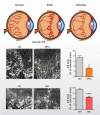
Blocking glycolysis or FAO mitigates pathological ocular neovascularization. In a mouse model for retinopathy of prematurity (ROP), treatment of the mice with 70 mg/kg of the PFKFB3 blocker 3PO significantly reduces the formation of pathological vascular tufts (yellow arrowheads). In the same model, blocking FAO with the CPT1a blocker etomoxir also attenuates tuft formation. The retinal microvasculature is stained with isolectin B4. Pictures and corresponding quantifications of vascular tuft area are from Schoors et al. (481), with permission from Macmillan Publishers Ltd., and Schoors et al. (483), with permission from Elsevier. The drawings at the top of this figure provide schematic overviews to recapitulate these findings. 3PO, 3-(3-pyridinyl)-1-(4-pyridinyl)-2-propen-1-one; eto, etomoxir.
FIGURE 15.
FIGURE 15.
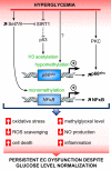
Molecular mechanisms underlying the hyperglycemic memory in ECs. Schematic and simplified scheme showing different (interconnected) molecular mechanisms and their contribution to a hyperglycemic memory in ECs. Hyperglycemic stimuli, even when followed by normal glucose restoration, act on different key mediators such as the methyltransferase Set7/9, the deacetylase SIRT1 and PKC which (epigenetically) change the expression levels/activity of NFκB and the mitochondrial adaptor p66Shc to cause a persistent pro-inflammatory and increased oxidative stress state. More details on the intricate signaling leading to this hyperglycemic memory are provided in the main text. SIRT1, sirtuin 1; H3, histone 3; PKC, protein kinase C; NFκB, nuclear factor-kappa B; NO, nitric oxide; ROS, reactive oxygen species.
FIGURE 16.
FIGURE 16.
Two-way metabolic crosstalk between ECs and other cell types. Schematic and simplified view on possible metabolic crosstalk between ECs and other cell types. ECs use lactate, the end product of glycolysis, to signal to other cell types and thereby influence the immune compartment, cancer cell growth but also vasoconstriction/vasodilation (for details see main text). Conversely, lactate derived from other cell types, for example, cancer cells, can be taken up by ECs through the MCT1 transporter and drive (tumor-)angiogenesis by inducing VEGF/VEGFR2 or NFκB/IL8 signaling. Another way of crosstalk is through the exchange of EVs (containing metabolites, nucleic acids, and proteins) as exemplified here by the bidirectional exchange of EVs between ECs and cardiomyocytes (for more details, see main text). Gln, glutamine; EV, extracellular vesicles; IL8, interleukin 8; IL17A, interleukin 17A; MCT1, monocarboxylate transporter 1; NFκB, nuclear factor-kappa B; Th17, T helper 17 cell; VEGFR2, VEGF receptor 2.

References

    1. Action to Control Cardiovascular Risk in Diabetes Study Group; Gerstein HC, Miller ME, Byington RP, Goff DC Jr, Bigger JT, Buse JB, Cushman WC, Genuth S, Ismail-Beigi F, Grimm RH Jr, Probstfield JL, Simons-Morton DG, Friedewald WT. Effects of intensive glucose lowering in type 2 diabetes. N Engl J Med 358: 2545–2559, 2008. doi:10.1056/NEJMoa0802743. - DOI - PMC - PubMed
    1. Adams RH, Alitalo K. Molecular regulation of angiogenesis and lymphangiogenesis. Nat Rev Mol Cell Biol 8: 464–478, 2007. doi:10.1038/nrm2183. - DOI - PubMed
    1. Adeva-Andany MM, Pérez-Felpete N, Fernández-Fernández C, Donapetry-García C, Pazos-García C. Liver glucose metabolism in humans. Biosci Rep 36: e00416, 2016. doi:10.1042/BSR20160385. - DOI - PMC - PubMed
    1. ADVANCE Collaborative Group; Patel A, MacMahon S, Chalmers J, Neal B, Billot L, Woodward M, Marre M, Cooper M, Glasziou P, Grobbee D, Hamet P, Harrap S, Heller S, Liu L, Mancia G, Mogensen CE, Pan C, Poulter N, Rodgers A, Williams B, Bompoint S, de Galan BE, Joshi R, Travert F. Intensive blood glucose control and vascular outcomes in patients with type 2 diabetes. N Engl J Med 358: 2560–2572, 2008. doi:10.1056/NEJMoa0802987. - DOI - PubMed
    1. Agathocleous M, Harris WA. Metabolism in physiological cell proliferation and differentiation. Trends Cell Biol 23: 484–492, 2013. doi:10.1016/j.tcb.2013.05.004. - DOI - PubMed

Publication types

MeSH terms

LinkOut - more resources