. 2018 Jan 17;92(3):e01965-17.
doi: 10.1128/JVI.01965-17. Print 2018 Feb 1.

β-d- N4-Hydroxycytidine Is a Potent Anti-alphavirus Compound That Induces a High Level of Mutations in the Viral Genome

Affiliations

β-d- N4-Hydroxycytidine Is a Potent Anti-alphavirus Compound That Induces a High Level of Mutations in the Viral Genome

Nadya Urakova et al. J Virol. .

Abstract

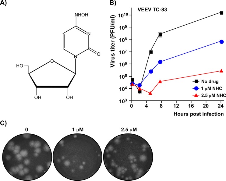
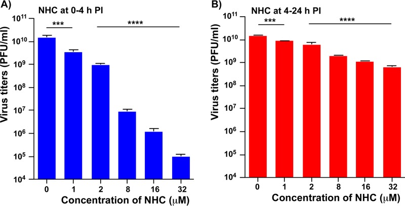
Venezuelan equine encephalitis virus (VEEV) is a representative member of the New World alphaviruses. It is transmitted by mosquito vectors and causes highly debilitating disease in humans, equids, and other vertebrate hosts. Despite a continuous public health threat, very few compounds with anti-VEEV activity in cell culture and in mouse models have been identified to date, and rapid development of virus resistance to some of them has been recorded. In this study, we investigated the possibility of using a modified nucleoside analog, β-d-N4-hydroxycytidine (NHC), as an anti-VEEV agent and defined the mechanism of its anti-VEEV activity. The results demonstrate that NHC is a very potent antiviral agent. It affects both the release of genome RNA-containing VEE virions and their infectivity. Both of these antiviral activities are determined by the NHC-induced accumulation of mutations in virus-specific RNAs. The antiviral effect is most prominent when NHC is applied early in the infectious process, during the amplification of negative- and positive-strand RNAs in infected cells. Most importantly, only a low-level resistance of VEEV to NHC can be developed, and it requires acquisition and cooperative function of more than one mutation in nsP4. These adaptive mutations are closely located in the same segment of nsP4. Our data suggest that NHC is more potent than ribavirin as an anti-VEEV agent and likely can be used to treat other alphavirus infections.IMPORTANCE Venezuelan equine encephalitis virus (VEEV) can cause widespread epidemics among humans and domestic animals. VEEV infections result in severe meningoencephalitis and long-term sequelae. No approved therapeutics exist for treatment of VEEV infections. Our study demonstrates that β-d-N4-hydroxycytidine (NHC) is a very potent anti-VEEV compound, with the 50% effective concentration being below 1 μM. The mechanism of NHC antiviral activity is based on induction of high mutation rates in the viral genome. Accordingly, NHC treatment affects both the rates of particle release and the particle infectivity. Most importantly, in contrast to most of the anti-alphavirus drugs that are under development, resistance of VEEV to NHC develops very inefficiently. Even low levels of resistance require acquisition of multiple mutations in the gene of the VEEV-specific RNA-dependent RNA polymerase nsP4.

Keywords: N-hydroxycytidine; RNA-dependent RNA polymerase; Venezuelan equine encephalitis virus; alphaviruses; antivirals; drug-resistant mutant; lethal mutagenesis.


Figures

FIG 1
FIG 1
NHC has a strong negative effect on VEEV TC-83 replication. (A) Chemical structure of NHC. (B) Vero cells were infected with VEEV TC-83 at an MOI of 0.5 PFU/cell and were either treated with NHC at the indicated concentrations starting from 0 h postinfection (p.i.) throughout the 24 h of incubation period or they remained mock treated. At the indicated times p.i., media were harvested, and viral titers were determined by a plaque assay on Vero cells. Starting from 5 h p.i., virus titers in the samples harvested from the drug-treated cells were significantly different from those collected from the mock-treated cells (P < 0.0001). (C) Morphology of the plaques formed on Vero cells by VEEV TC-83 variants from the samples harvested at 24 h p.i. from mock- and NHC-treated cells. Due to profound differences in infectious titers, different dilutions are presented.
FIG 2
FIG 2
The antiviral effect of NHC depends on its application time. (A) Vero cells were infected with VEEV TC-83 at an MOI of 0.5 PFU/cell. At the time points indicated, cells were treated with 2 μM NHC and then harvested at 24 h p.i. Viral titers were determined using a plaque assay on Vero cells. The data are presented as mean values and standard deviations. In the same samples, the number of genome copies of VEEV TC-83 (GE/ml) was measured by qPCR. (B) GE/PFU ratio in the samples presented in panel A. This experiment was repeated twice with consistent results.
FIG 3
FIG 3
The first 4 h p.i. are a critical time for the antiviral effect of NHC. Vero cells in six-well Costar plates were infected at room temperature with VEEV TC-83 at an MOI of 20 PFU/cell. (A) Cells were incubated with NHC at the concentrations indicated from 0 to 4 h p.i. At 4 h p.i., the cells were washed, and media were replaced with NHC-free medium. (B) Infected cells were incubated in NHC-free medium until 4 h p.i., and then media were replaced by those supplemented with NHC at indicated concentrations. All of the samples indicated in panels A and B were harvested at 24 h p.i., and viral titers were determined by a plaque assay on Vero cells. The experiments were performed in triplicates. The reductions in viral titers in all NHC treated samples were statistically significant, as assessed by the one-way ANOVA.
FIG 4
FIG 4
NHC is a potent anti-VEEV compound with low cytotoxicity. (A) Vero cells were infected with VEEV TC-83 at an MOI of 20 PFU/cell. The indicated concentrations of NHC were applied at 0 h p.i., and media were harvested at 24 h p.i. Infectious titers were determined by a plaque assay on Vero cells and normalized to those in the mock-treated samples. The experiment was performed three times. The standard deviation are too low to be visible on the plot. (B) Vero cells were incubated for 24 h with indicated concentrations of NHC in the media. Cell viability was assessed using the Cell Titer-GLO 2.0 assay. Data were normalized to those generated on the mock-treated cells. RLU, relative light units.
FIG 5
FIG 5
VEEV TC-83 accumulates a large number of mutations when exposed to NHC. (A) VEEV TC-83 was serially passaged in the presence of NHC (see Materials and Methods for details), and the nsP-coding fragments in the genomes of two randomly selected plaque isolates (PP1 and PP2) were sequenced. PP2 isolate was then passaged 20 times in NHC-free media, and the same nsP-coding fragment in the genome of plaque-purified variant PREV1 was sequenced. Only the nonsynonymous mutations are presented. The mutations identified in both PP1 and PP2 are highlighted in gray. Additional mutations identified in PREV1 are highlighted in red. (B) Matrixes of nucleotide substitutions identified in plaque purified variants PP1, PP2, PREV1, and plaque-purified VEEV TC-83 after 20 passages in the absence of NHC.
FIG 6
FIG 6
Drug-resistant VEEV isolate PP2 replicates more efficiently than parental VEEV TC-83 and pseudorevertant PREV1 in the presence, but not in the absence of NHC. Subconfluent Vero cells were infected with parental VEEV TC-83, selected PP2 mutant or PREV1 viruses at an MOI of 1 PFU/cell. Starting from 0 h p.i., cells were incubated either in the absence (A) or in the presence (B) of 2 μM NHC. At the indicated time points, media were harvested, and viral titers were determined using a plaque assay on Vero cells. The experiment was repeated three times. Most of the standard deviations are too small to be visible on the plots. On panel B, starting from 5 h p.i., titers of PP2 and PREV1 are significantly different from those of VEEV TC-83 and from each other (P < 0.0001). (C) In a standard plaque assay, cell monolayers infected with the same dilution samples were overlaid with 0.5% agarose-containing media (see Materials and Methods for details) without or with the indicated concentrations of NHC. Plaques were visualized by crystal violet staining at 2 days p.i. (D) Vero cells were infected with VEEV TC-83 and PP2 mutant at an MOI of 20 PFU/cell. The indicated concentrations of NHC were applied at 0 h p.i., and media were harvested at 24 h p.i. Infectious titers were determined by a plaque assay on Vero cells and normalized to those in the mock-treated samples. The standard deviations are too low to be visible on the plot.
FIG 7
FIG 7
NHC has stronger negative effects on the release and infectivity of VEEV TC-83 and PREV1 particles than those of the PP2 mutant. A total of 5 × 10⁵ Vero cells in six-well plates were infected with VEEV TC-83, PP2, or PREV1 at an MOI of 1 PFU/cell and were incubated with or without 2 μM NHC. At the indicated time points, media were collected, and the infectious titers (PFU/ml) and concentrations of genome-containing viral particles (GE/ml) were determined by a plaque assay on Vero cells and RT-qPCR, respectively. Experiments were performed in triplicates. The standard deviations are too small to be visible on the plots. (B) GE/PFU ratios in virus samples harvested at 24 h p.i. of mock-treated and NHC-treated cells in the experiment presented in panel A. (C) Levels of viral G RNAs in the mock-treated cells at 24 h p.i. The cells were harvested after collecting the medium samples described in panel A at 24 h p.i. Data were normalized to the G RNA levels in VEEV TC-83-infected cells. (D) Ratio of G RNAs in mock-treated versus NHC-treated cells at 24 p.i. with the indicated viruses. Cells were collected in the experiment presented in panel A.
FIG 8
FIG 8
VEEV TC-83 passaging in the presence of increasing concentrations of NHC leads to rapid accumulation of mutations in viral pool. (A) Frequencies of mutations in viral pools at passage 5, 10, 15, and 20 in the absence or presence of NHC, determined by NGS (see Materials and Methods for details). (B) Dynamics of the increase in the frequencies of mutations at the indicated positions of VEEV nsP4.
FIG 9
FIG 9
Accumulation of mutations in the genome of plaque-purified, NHC-resistant VEEV isolate PP2 (see Fig. 5) during its serial passaging in NHC-free media. (A) Frequencies of mutations in viral populations at passage 5, 10, 15, and 20, determined by NGS. (B) Dynamics of increase in mutation frequencies at the indicated positions of nsP4.
FIG 10
FIG 10
The mutations that lead to NHC-resistant and NHC-sensitive phenotypes of VEEV TC-83 are closely located in the 3D structure of the catalytic domain of VEEV nsP4. (A) Structural alignment of the predicted 3D structure of VEEV nsP4 RdRp domain with FMDV 3D polymerase (2E9T). The VEEV RdRp structure is presented as a solid, light red ribbon. The structure of FMDV 3D protein is presented as a solid, light blue ribbon. The amino acids involved in coordination of NTP phosphates are presented as red sticks (VEEV) or blue sticks (FMDV). The amino acids that bind RNA template are presented as turquoise sticks for both polymerases. (B) Positions of mutations identified in nsP4 of NHC-resistant and NHC-sensitive mutants of VEEV TC-83 on the predicted structure of the catalytic domain. The amino acids that were substituted in the NHC-resistant viruses are shown in medium green (I190) and light green (A189 and P187). The amino acids that were substituted in the pool of PP2 virus passaged 20 times in NHC-free media (A201V and V308I, see Fig. 9 for details) are shown in yellow.
FIG 11
FIG 11
nsP4-specific mutations determine the resistance of VEEV replication to NHC. (A) Schematic presentations of VEEV genomes containing the indicated nsP4-specific mutations. (B) In a standard plaque assay performed on the indicated viruses, Vero cells were overlaid with 0.5% agarose-containing medium either with or without 1.5 μM NHC. After 2 days, plaques were visualized by staining with crystal violet. (C) A total of 4 × 10⁵ Vero cells were infected with the indicated viruses at an MOI of 2 PFU/cell. At the indicated times p.i., media were harvested, and viral titers were determined by a plaque assay on Vero cells. (D) A total of 5 × 10⁵ Vero cells were infected with the indicated viruses at an MOI of 2 PFU/cell. Cells were incubated with NHC starting from 0 h p.i. At 8 h p.i., media were harvested, and viral titers were determined by a plaque assay on Vero cells. The titers were normalized to those in the samples harvested from mock-treated cells. This experiment was repeated three times using different concentrations of drug with consistent results. The data from one of the representative experiments are presented in this figure. (E) A total of 5 × 10⁵ Vero cells were infected with the indicated viruses at an MOI of 2 PFU/cell. Cells were incubated with NHC starting from 0 h p.i. At 20 h p.i., media were harvested, and viral titers were determined by a plaque assay on Vero cells. Titers were normalized to those in the samples harvested from mock-treated cells.
FIG 12
FIG 12
A201V mutation in VEEV nsP4 reverts the NHC-resistant phenotype of the VEEV/3x mutant to drug sensitive. (A) Schematic presentation of VEEV genomes containing the indicated nsP4 mutations. (B) Vero cells were infected with indicated mutant viruses at an MOI of 1 PFU/cell. Cells were incubated in the presence of 2 μM NHC starting from 0 h p.i. At 8 h p.i., media were harvested, and viral titers were determined by a plaque assay on Vero cells. (C) Titers in samples from NHC-treated cells were normalized to those in samples from mock-treated cells. This experiment was repeated three times using different concentrations of drug with consistent results. The data from one of the representative experiments are presented in this figure.

References

    1. Strauss JH, Strauss EG. 1994. The alphaviruses: gene expression, replication, evolution. Microbiol Rev 58:491–562. - PMC - PubMed
    1. Zacks MA, Paessler S. 2010. Encephalitic alphaviruses. Vet Microbiol 140:281–286. doi:10.1016/j.vetmic.2009.08.023. - DOI - PMC - PubMed
    1. Weaver SC, Ferro C, Barrera R, Boshell J, Navarro JC. 2004. Venezuelan equine encephalitis. Annu Rev Entomol 49:141–174. doi:10.1146/annurev.ento.49.061802.123422. - DOI - PubMed
    1. Gibbs EP. 1976. Equine viral encephalitis. Equine Vet J 8:66–71. doi:10.1111/j.2042-3306.1976.tb03293.x. - DOI - PubMed
    1. Gibney KB, Robinson S, Mutebi JP, Hoenig DE, Bernier BJ, Webber L, Lubelczyk C, Nett RJ, Fischer M. 2011. Eastern equine encephalitis: an emerging arboviral disease threat, Maine, 2009. Vector Borne Zoonotic Dis 11:637–639. doi:10.1089/vbz.2010.0189. - DOI - PubMed

Publication types