. 2018 Jan 30;92(4):e01890-17.
doi: 10.1128/JVI.01890-17. Print 2018 Feb 15.

Glycine Zipper Motifs in Hepatitis C Virus Nonstructural Protein 4B Are Required for the Establishment of Viral Replication Organelles

Affiliations

Glycine Zipper Motifs in Hepatitis C Virus Nonstructural Protein 4B Are Required for the Establishment of Viral Replication Organelles

David Paul et al. J Virol. .

Abstract

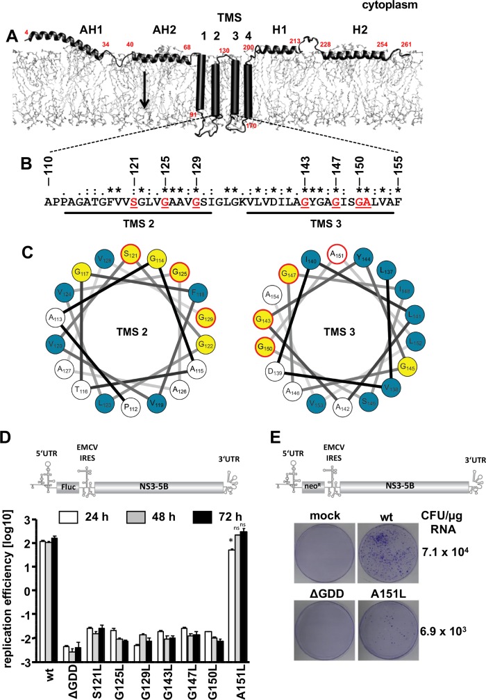
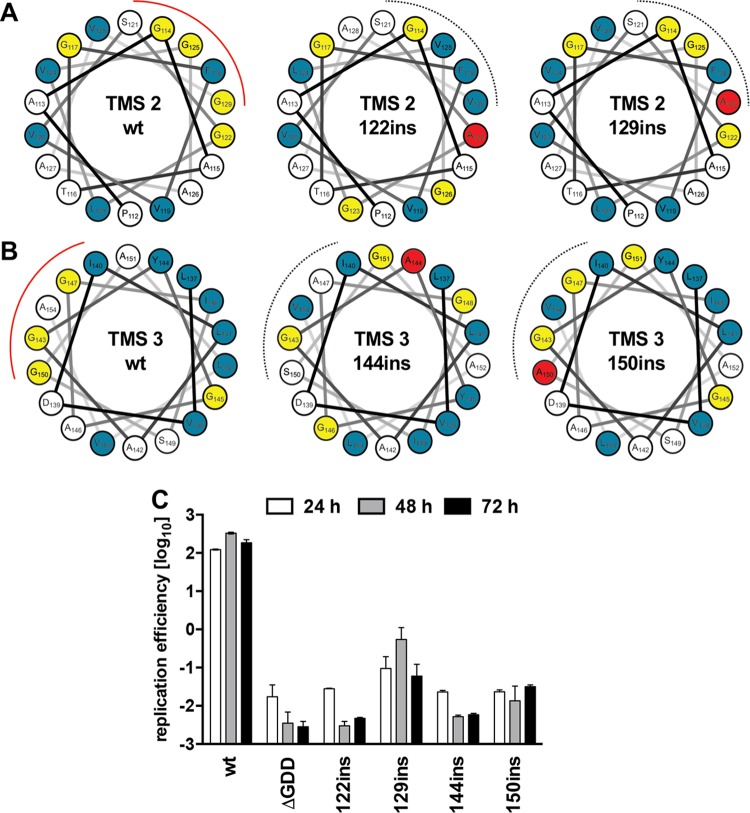
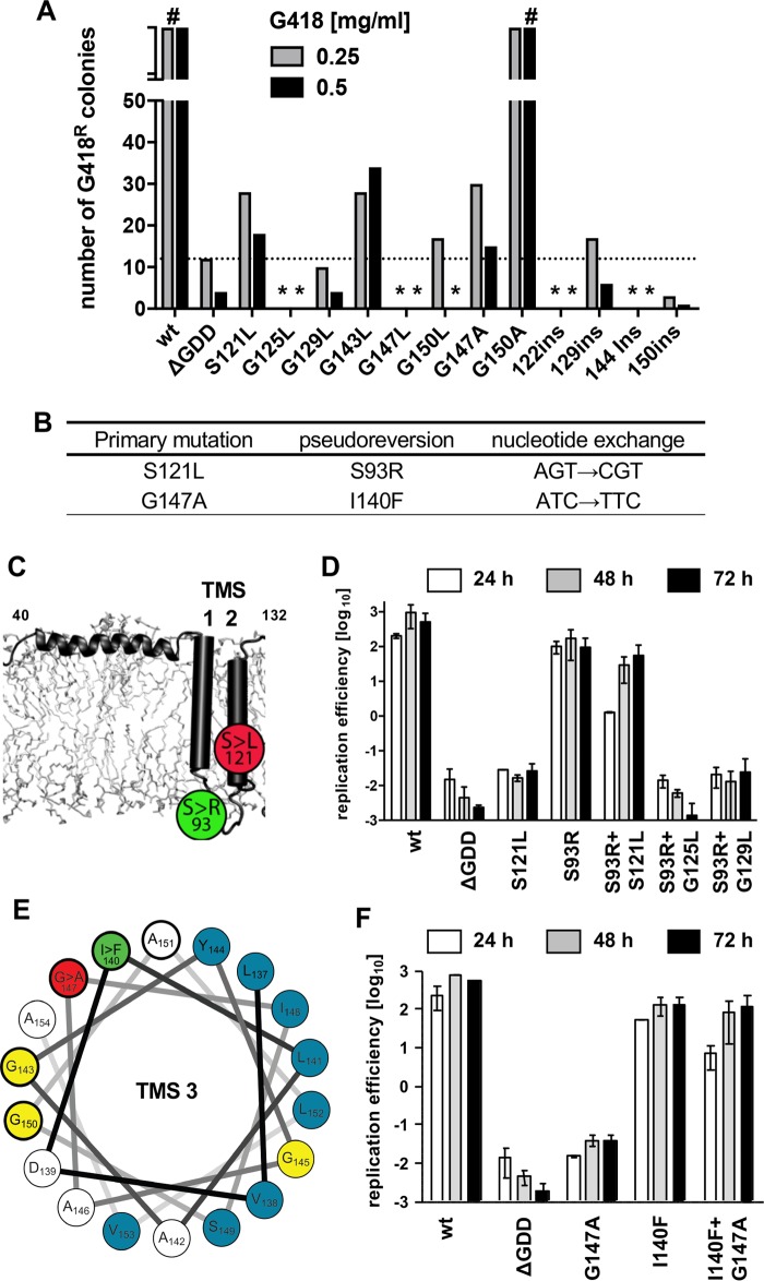
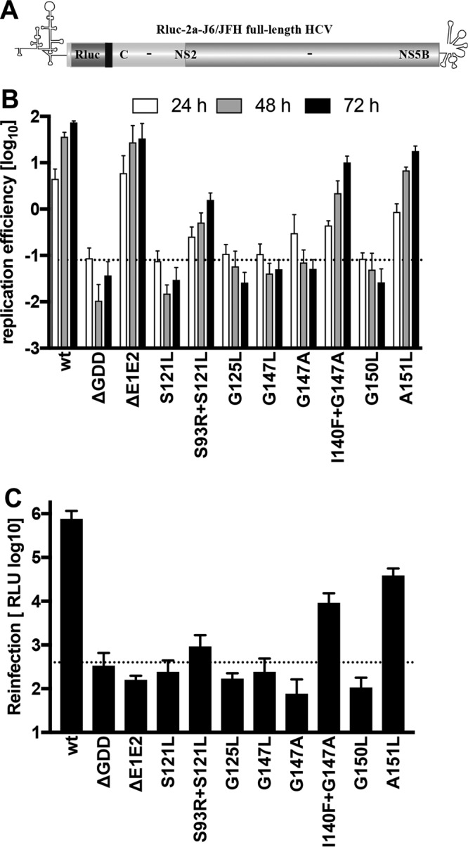
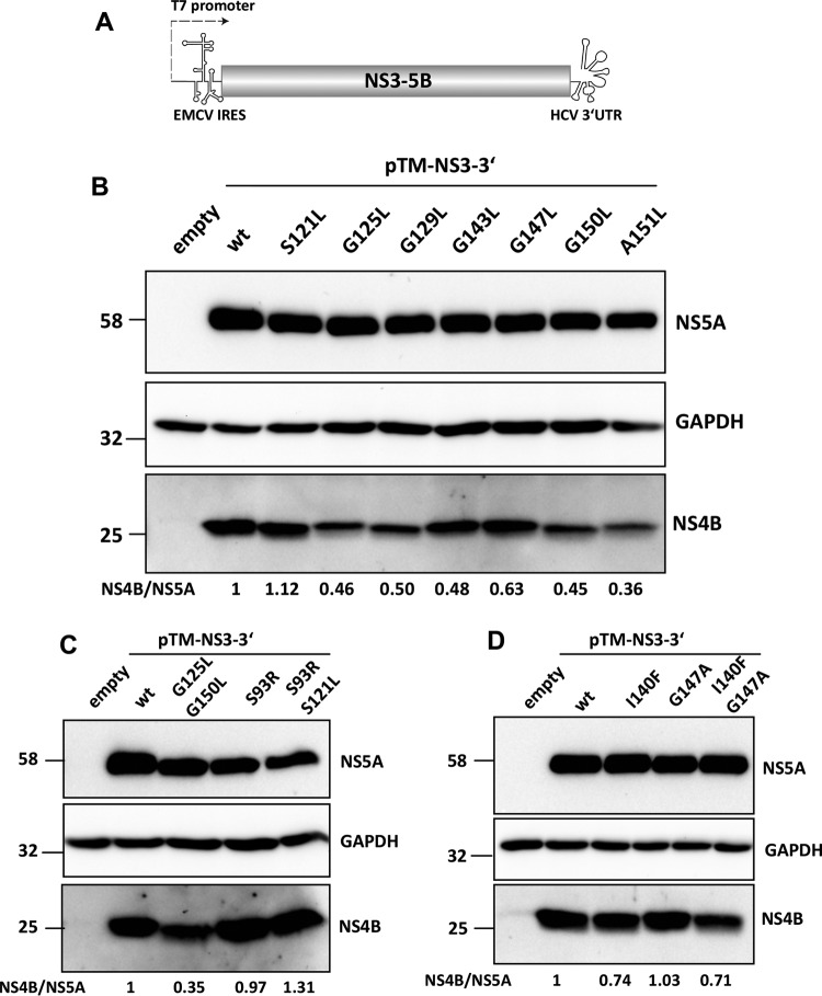
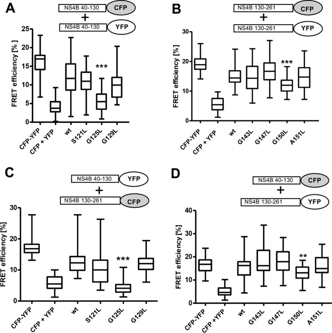
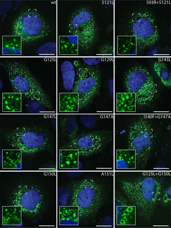
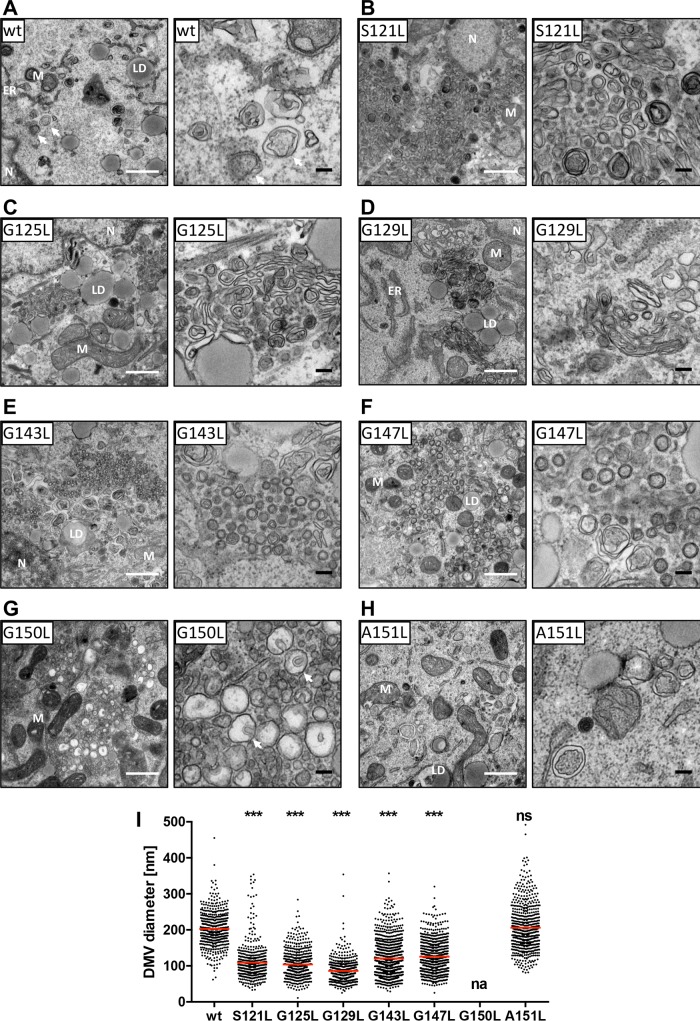
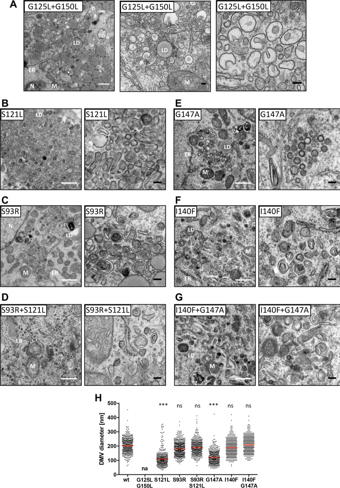
Hepatitis C virus (HCV) RNA replication occurs in tight association with remodeled host cell membranes, presenting as cytoplasmic accumulations of single-, double-, and multimembrane vesicles in infected cells. Formation of these so-called replication organelles is mediated by a complex interplay of host cell factors and viral replicase proteins. Of these, nonstructural protein 4B (NS4B), an integral transmembrane protein, appears to play a key role, but little is known about the molecular mechanisms of how this protein contributes to organelle biogenesis. Using forward and reverse genetics, we identified glycine zipper motifs within transmembrane helices 2 and 3 of NS4B that are critically involved in viral RNA replication. Foerster resonance energy transfer analysis revealed the importance of the glycine zippers in NS4B homo- and heterotypic self-interactions. Additionally, ultrastructural analysis using electron microscopy unraveled a prominent role of glycine zipper residues for the subcellular distribution and the morphology of HCV-induced double-membrane vesicles. Notably, loss-of-function NS4B glycine zipper mutants prominently induced single-membrane vesicles with secondary invaginations that might represent an arrested intermediate state in double-membrane vesicle formation. These findings highlight a so-far-unknown role of glycine residues within the membrane integral core domain for NS4B self-interaction and functional as well as structural integrity of HCV replication organelles.IMPORTANCE Remodeling of the cellular endomembrane system leading to the establishment of replication organelles is a hallmark of positive-strand RNA viruses. In the case of HCV, expression of the nonstructural proteins induces the accumulation of double-membrane vesicles that likely arise from a concerted action of viral and coopted cellular factors. However, the underlying molecular mechanisms are incompletely understood. Here, we identify glycine zipper motifs within HCV NS4B transmembrane segments 2 and 3 that are crucial for the protein's self-interaction. Moreover, glycine residues within NS4B transmembrane helices critically contribute to the biogenesis of functional replication organelles and, thus, efficient viral RNA replication. These results reveal how glycine zipper motifs in NS4B contribute to structural and functional integrity of the HCV replication organelles and, thus, viral RNA replication.

Keywords: NS4B; glycine zipper; hepatitis C virus; membrane proteins; membranous web; positive-strand RNA virus; replication organelle.


Figures

FIG 1
FIG 1
Glycine zipper constituting residues in NS4B transmembrane segments 2 and 3 are required for HCV RNA replication. (A) NS4B membrane topology. The N-terminal amphipathic α-helices AH1 and AH2 and the C-terminal α-helices H1 and H2 are depicted, while the four transmembrane segments (TMS1-4) are shown as cylinders. Red numbers give NS4B amino acid residues. The arrow indicates the reported dual topology of the N-terminal domain, resulting from the posttranslational flip of the N terminus into the ER lumen. (B) Amino acid sequence of NS4B TMS2 and TMS3 of the HCV isolate JFH-1. Fully conserved amino acid residues are marked with asterisks. Residues targeted in this study are underlined and highlighted in red. (C) Helical wheel representations of NS4B TMS2 (left) and TMS3 (right) are depicted. Bulky hydrophobic amino acids are shown in blue, glycine residues are depicted in yellow, and red circles highlight positions targeted in this study. (D) The design of the subgenomic luciferase reporter replicon used for transient replication assays is shown in the top panel. The firefly luciferase (Fluc) coding region is translated under the control of the HCV internal ribosome entry site (IRES) contained in the 5′ untranslated region (UTR). The second cistron (NS3 to NS5B) is translated via the IRES of the encephalomyocarditis virus (EMCV-IRES). Huh7-Lunet cells were transfected with in vitro-transcribed replicon RNAs specified on the bottom. Cells were lysed 4, 24, 48, and 72 h after transfection, and luciferase activity in cell lysates was determined. Data were normalized for transfection efficiency using the 4-h values. The active-site NS5B polymerase mutant (ΔGDD) indicates the assay background. Mean values of two independent experiments, each measured in duplicate are shown. Error bars indicate standard deviations. The significance of differences between wild-type (wt) and mutant A151L for each time point was calculated by using the unpaired, two-tailed Student's t test (*, P = 0.0056; ns, not significant). (E) The design of the subgenomic replicon used for selection assays is shown in the top panel. It is analogous to the one shown in panel D but encodes the neomycin phosphotransferase (neoR) instead of luciferase. Huh7-Lunet cells were transfected with replicon RNAs specified above each image and subjected to selection with G418. After 3 to 4 weeks, single-cell clones were stained and counted. CFU per μg RNA are given.
FIG 2
FIG 2
Single-amino-acid residue insertions into NS4B TMS2 and TMS3 abolish HCV RNA replication. Helical wheel representations of NS4B TMS2 (A) and TMS3 (B) are depicted. Bulky hydrophobic amino acids are indicated in blue, glycine residues are depicted in yellow, and red lines highlight the glycine zipper interface. Alanine insertions are highlighted in red and disrupt the glycine zipper interfaces, as indicated by the dashed black lines. (C) Huh7-Lunet cells were transfected with subgenomic luciferase reporter replicon RNAs specified on the bottom. Cells were lysed 4, 24, 48, and 72 h after transfection, and luciferase activity in cell lysates was determined. Data were normalized for transfection efficiency by using the 4-h values. The active-site NS5B polymerase mutant (ΔGDD) was used to determine the assay background. Mean values of two independent experiments are shown. Error bars indicate standard deviations.
FIG 3
FIG 3
Pseudoreversions located in close proximity of the primary mutation in NS4B TMS2 and TMS3 rescue replication of glycine zipper mutants. (A) Huh7-Lunet cells were transfected with G418-selectable subgenomic replicon RNAs encoding either the wild-type (wt) replicase (NS3-5B) or a replicase containing a mutation in NS4B as specified at the bottom. Transfected cells were cultured in medium containing either 0.25 or 0.5 mg/ml G418, as specified at the top, and after 3 to 4 weeks single-cell clones were counted. #, confluence of the cell layer; *, no G418-resistant cell clones obtained. (B) Single-cell clones were expanded, and compensatory mutations contained in replicon RNAs in these cells were identified by nucleotide sequence analysis of replicon RNAs after amplification by reverse transcription-PCR. (C) Membrane topology of the NS4B N-terminal fragment (amino acids 40 to 132) indicating the positions of the primary mutation (S121L) in TMS2 and the second-site mutation (S93R) in the ER-luminal loop between TMS1 and TMS2. (D) Huh7-Lunet cells were transfected with subgenomic luciferase reporter replicon RNAs specified at the bottom. Cells were lysed 4, 24, 48, and 72 h after transfection, and luciferase activity in cell lysates was determined. Data were normalized for transfection efficiency using the 4-h values. The active-site NS5B polymerase mutant (ΔGDD) was used to determine the assay background. Mean values from two independent experiments are shown. Error bars indicate standard deviations. (E) The helical wheel representation of TMS3 highlights bulky hydrophobic amino acid residues in blue, glycine residues in yellow, the primary mutation G147A in TMS2 in red, and the corresponding pseudoreversion I140F, localizing two turns up in the same helical TMS, in green. (F) HCV RNA replication kinetics of NS4B mutants as determined by transient assays specified for panel D. Note the replication rescue of the G147A mutant by the I140F pseudoreversion.
FIG 4
FIG 4
Impact of mutations and pseudoreversions in TMS2 and TMS3 on full-length HCV RNA replication and particle production. (A) Huh7-Lunet cells were transfected with in vitro-transcribed HCV full-length RNAs derived from the reporter virus genome specified at the bottom (JcR2a) (28). (B) Cells were lysed 4, 24, 48, and 72 h after transfection, and luciferase activity in cell lysates was determined. Data were normalized for transfection efficiency using the 4-h values. The active-site NS5B polymerase mutant (ΔGDD) indicates the assay background. Mean values from two independent experiments, each measured in duplicate, are shown. Error bars indicate standard deviations. (C) Culture supernatants harvested 72 h after transfection of cells described for panel B were used to infect naive Huh7.5 cells. After 72 h cells were lysed and luciferase activity in cell lysates was determined. The envelope-deficient mutant (ΔE1E2) indicates the assay background (dotted line). Mean values from two independent experiments, each measured in duplicate, are shown. Error bars indicate standard deviations.
FIG 5
FIG 5
Impact of mutations in TMS2 and TMS3 on NS4B protein stability. (A to C) Huh7-Lunet/T7 cells were transfected with the pTM expression vector (empty) or the same vector encoding the NS3-5B polyprotein fragment, schematically depicted in panel A, corresponding to wild-type (wt) NS4B or an NS4B mutant specified on the top. Cells were harvested 24 h posttransfection, and proteins were detected by Western blotting using antibodies monospecific for NS4B, NS5A, or GAPDH. Numbers below each lane refer to the relative abundance of NS4B as determined by densitometry. After subtraction of the background and normalization for equal loading by using GAPDH, NS4B-specific signals were normalized to those obtained for NS5A. Representative results of two independent experiments are shown in each panel.
FIG 6
FIG 6
Impact of mutations in glycine zipper motifs on NS4B homo- and heterotypic self-interactions. U2OS cells were transfected with plasmids encoding NS4B N- and C-terminal fragments, each C-terminally fused to cyan fluorescent protein (CFP) or yellow fluorescent protein (YFP), as indicated at the top of each panel. The NS4B coding sequence was either wild type (wt) or contained the mutation specified at the bottom of the diagrams. Coexpression of CFP and YFP only (CFP+YFP) and expression of a CFP-YFP fusion protein (CFP-YFP) served as negative and positive controls, respectively. Twenty-four hours after transfection, the interaction of protein fragments was assessed by FRET acceptor photobleaching. Box-and-whisker diagrams show the median FRET efficiency (middle line) from at least 30 measurements from 2 independent experiments. Bottom and top lines of the boxes represent the 25th and 75th percentile, respectively. Vertical lines indicate minimum and maximum values. (A) Impact of mutations in TMS2 on homotypic interaction of the NS4B N-terminal fragment. (B) Influence of mutations in TMS3 on homotypic interaction of the NS4B C-terminal half. The heterotypic interaction of the NS4B N-terminal with the C-terminal fragment, containing mutations in TMS2 and TMS3, are shown in panels C and D, respectively. The significance of differences compared to the wild type was calculated by using the unpaired, two-tailed Student's t test (***, P < 0.0001; **, P < 0.001).
FIG 7
FIG 7
Influence of mutations in NS4B TMS2 and TMS3 on subcellular localization of NS4B. Huh7-Lunet/T7 cells were transfected with the pTM expression vector encoding NS3-5B of either wild-type (wt) sequence or containing mutations in NS4B specified at the top right of each panel. After 24 h, cells were fixed and NS4B was detected by immunofluorescence. Representative images for each condition are displayed. Magnified views of dashed-line boxed areas are shown in the lower left of each panel. The scale bars represent 25 μm.
FIG 8
FIG 8
Effects of mutations in NS4B TMS2 and TMS3 on subcellular distribution and morphology of HCV-induced replication organelles. Huh7-Lunet/T7 cells were transfected with the pTM expression vector encoding NS3-5B of either the wild-type (wt) sequence or containing NS4B mutations specified at the top left of each panel. After 24 h cells were fixed, processed, and analyzed by TEM as described in Materials and Methods. (A to H) Subcellular distribution and morphology of DMVs induced by expression of wild-type NS3-5B or NS3-5B containing a mutation in NS4B as specified at the top left corner. White and black scale bars in each panel represent 500 nm and 100 nm, respectively. Localization of cellular organelles is indicated. ER, endoplasmic reticulum; LD, lipid droplet; M, mitochondrion; N, nucleus. White arrows in panels A and G refer to regular DMVs and SMVs, respectively, with stalled invaginations. (I) Quantitation of DMV diameter. The scatter plot depicts the size (diameter) of at least 500 individual DMVs from at least 10 different transfected cells per condition. Horizontal lines indicate mean values; na, not applicable. Each data set was compared to that of the wild type, and significance of differences was calculated by using the unpaired, two-tailed Student's t test (***, P < 0.0001; ns, nonsignificant).
FIG 9
FIG 9
Pseudoreversions restore regular subcellular distribution and morphology of HCV replication factories of glycine zipper mutants. Huh7-Lunet/T7 cells were transfected with the NS3-5B expression vector containing either the wild type (wt) or given mutation(s) in NS4B. After 24 h cells were fixed, processed, and analyzed by TEM as described in Materials and Methods. (A) Cells transfected with the NS3-5B expression vector containing mutations G125L and G150L in NS4B exhibited prominent accumulations of small single-membrane vesicles around lipid droplets (middle), in addition to bigger single-membrane vesicles with secondary invaginations. Subcellular distribution and DMV morphology of primary mutant S121L, the psdeudorevertant S93R, and the double mutant is shown in panels B to D and for primary mutant G147A, the psdeudorevertant I140F, and the double mutant is shown in panels E to G. White and black scale bars in each panel represent 500 nm and 100 nm, respectively. (H) Quantitation of DMV diameters. The scatter plot depicts the size (diameter) of at least 500 individual DMVs from at least 10 different transfected cells per condition. Data sets for the wild-type and mutant S121L are the same as those described for Fig. 8I and are shown for comparison. Horizontal lines indicate mean values; na, not applicable. Each data set was compared to the wild type, and significance of differences was calculated by using the unpaired, two-tailed Student's t test (***, P < 0.0001; ns, nonsignificant).

Similar articles

Cited by

References

    1. Yamane D, McGivern DR, Masaki T, Lemon SM. 2013. Liver injury and disease pathogenesis in chronic hepatitis C. Curr Top Microbiol Immunol 369:263–288. - PubMed
    1. International Committee on Taxonomy of Viruses (ICTV). 2016. The online (10th) report of the International Committee on Taxonomy of Viruses. https://talk.ictvonline.org/ictv-reports/ictv_online_report/positive-sen....
    1. Bartenschlager R, Penin F, Lohmann V, Andre P. 2011. Assembly of infectious hepatitis C virus particles. Trends Microbiol 19:95–103. doi:10.1016/j.tim.2010.11.005. - DOI - PubMed
    1. Lindenbach BD, Rice CM. 2013. The ins and outs of hepatitis C virus entry and assembly. Nat Rev Microbiol 11:688–700. doi:10.1038/nrmicro3098. - DOI - PMC - PubMed
    1. Ding Q, von Schaewen M, Ploss A. 2014. The impact of hepatitis C virus entry on viral tropism. Cell Host Microbe 16:562–568. doi:10.1016/j.chom.2014.10.009. - DOI - PMC - PubMed

Publication types

LinkOut - more resources