. 2018 Jan 15;29(2):137-153.
doi: 10.1091/mbc.E17-08-0510. Epub 2017 Nov 22.

DRC2/CCDC65 is a central hub for assembly of the nexin-dynein regulatory complex and other regulators of ciliary and flagellar motility

Affiliations

DRC2/CCDC65 is a central hub for assembly of the nexin-dynein regulatory complex and other regulators of ciliary and flagellar motility

Raqual Bower et al. Mol Biol Cell. .

Abstract

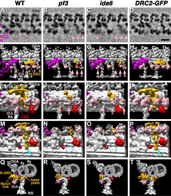
The nexin-dynein regulatory complex (N-DRC) plays a central role in the regulation of ciliary and flagellar motility. In most species, the N-DRC contains at least 11 subunits, but the specific function of each subunit is unknown. Mutations in three subunits (DRC1, DRC2/CCDC65, DRC4/GAS8) have been linked to defects in ciliary motility in humans and lead to a ciliopathy known as primary ciliary dyskinesia (PCD). Here we characterize the biochemical, structural, and motility phenotypes of two mutations in the DRC2 gene of Chlamydomonas Using high-resolution proteomic and structural approaches, we find that the C-terminal region of DRC2 is critical for the coassembly of DRC2 and DRC1 to form the base plate of N-DRC and its attachment to the outer doublet microtubule. Loss of DRC2 in drc2 mutants disrupts the assembly of several other N-DRC subunits and also destabilizes the assembly of several closely associated structures such as the inner dynein arms, the radial spokes, and the calmodulin- and spoke-associated complex. Our study provides new insights into the range of ciliary defects that can lead to PCD.


Figures

FIGURE 1:
FIGURE 1:
Both ida6 and sup-pf5 are drc2 mutations. (A) Diagram of genes contained within BAC clone 31N21. This clone was digested with DraI and SpeI to release a genomic fragment encoding DRC2 (FAP250). (B) Diagram of the subclone containing DRC2 showing the location of the sup-pf5 and ida6 mutations and the site of the HA or GFP epitope tags. (C) Diagram of the predicted secondary structure of the DRC2 polypeptide showing the four coiled-coil domains in the wild-type (WT) sequence, the addition of 101 amino acids to the C-terminus of the ida6 sequence, and the truncation of the third coiled-coil domain in the sup-pf5 sequence. (D) The aligned amino acid sequences of the third coiled-coil domains in the Chlamydomonas (Cr) and Homo sapiens (Hs) DRC2 polypeptides. The asterisks indicate the site of the Chlamydomonas sup-pf5 mutation (top) and the H. sapiens mutation (bottom) linked to PCD by Austin-Tse et al. (2013). (E) Western blot of WT and sup-pf5 axonemes probed with antibodies to different polypeptides reveals that truncation of DRC2 (indicated by an asterisk in the DRC2 panel) reduces the assembly of multiple N-DRC subunits, the CSC (FAP61), and tektin, but does not alter assembly of ODA subunits (IC2/IC69), RS subunits (RSP16), or the molecular ruler CCDC39 (FAP59).
FIGURE 2:
FIGURE 2:
Transformation with HA- or GFP-tagged DRC2 rescues ida6 and sup-pf5. (A) Live cell images of an ida6 strain rescued with DRC2-GFP transgene as viewed by DIC (left) or confocal (right) microscopy show the distribution of DRC2 in the basal body region and along the length of both flagella. (B) Images of a fixed sup-pf5 cell (bottom) or DRC2-HA rescued cell (top) stained with an antibody to the HA epitope tag as viewed by DIC (left) or fluorescence (right) microscopy demonstrate the presence of DRC2-HA in the flagella of the rescued strain. The basal body region is out of the plane of focus in this image. (C, D) Measurements of motility as viewed by phase contrast microscopy indicate that transformation of either ida6 (C) or sup-pf5 (D) with WT or epitope-tagged DRC2 transgenes increased forward swimming velocities to near WT levels. All strains indicated by asterisks were slower than WT, but the DRC2 rescued strains were significantly faster than ida6 or sup-pf5 (p < 0.05). (E, F) Western blots of axonemes from WT, mutant, and rescued strains were probed with antibodies against DRC subunits and tektin. Loss or truncation of DRC2 was correlated with major decreases in DRC1 and tektin (see also Figure 1E), but changes in DRC4 were less obvious. DRC1 and tektin were increased to WT levels in DRC2-rescued strains. (A portion of the blot shown in E was previously shown in Austin-Tse et al., 2013.) (G) A WT cell was mated to an ida6 strain that had been rescued with DRC2-GFP, and the resulting quadriflagellate dikaryon was fixed 60 min after mating and viewed by DIC (left) and fluorescence (right) microscopy. GFP signal was visible in the two DRC2-GFP flagella but not in the two flagella from the WT parent. (H) An ida6 cell was mated to an ida6 strain that had been rescued with DRC2-GFP, and the resulting quadriflagellate dikaryon was fixed 30 min after mating and viewed by DIC (left) and fluorescence (right) microscopy. GFP signal was visible in all four flagella. All scale bars are 5 μm.
FIGURE 3:
FIGURE 3:
The distribution of N-DRC subunits in different strains identifies ida6(drc2) and pf3(drc1) as members of a distinct subclass of motility mutants. (A) Western blot of axonemes isolated from WT, ida6, DRC2-HA, and pf3 strains was probed with multiple antibodies against DRC subunits and other axonemal proteins. DRC1, DRC2, DRC5, and DRC11 were clearly reduced or missing in ida6 and pf3. DRC3, DRC4, DRC7, DRC8, and CaM-IP3 were slightly reduced. All were restored to WT levels in the DRC2-HA rescued strain. Subunits of the ODA (IC2/IC69), the I1 dynein (IC140), the RS (RSP16), and other doublet microtubule-associated proteins (FAP59) served as loading controls and were not noticeably altered in any strain. (B) Western blot of whole cell lysates obtained from WT and ida6 cells was probed with antibodies against several DRC subunits. Both DRC1 and DRC2 were reduced in ida6 cell lysates, but DRC5, DRC7, and DRC11 were present at WT levels in ida6. (C) Western blot of WT and motility mutants that disrupt other axoneme substructures (outer arms, inner arms, B-tubule beaks, and radial spokes) was probed with multiple antibodies against DRC subunits. FAP59/CCDC39 and IC69/IC2 served as loading controls.
FIGURE 4:
FIGURE 4:
The assembly of a subset of IDA and RS subunits is altered in ida6. (A) The inner arm DHCs present in axonemes isolated from WT, ida6, and DRC2 rescued strains (HA- and GFP-tagged) were fractionated by SDS–PAGE, digested with trypsin, and analyzed by mass spectrometry. The total number of spectra for each DHC was compared with the total number of spectra for the 1-alpha and 1-beta DHCs of the I1 dynein in each sample. Significant decreases were observed in ida6 for DHC2, DHC6, DHC7, DHC8, and DHC9. DHC11 was increased in ida6. All DHCs were restored to WT levels in the DRC2 rescued strains. For each DHC, the alternate name (dynein a–g) and the proposed inner arm position (IA2-6/X or proximal minor dynein, see also Figure 6A) are listed. (B) Western blot of isolated axonemes from WT, ida6, and a DRC2-HA rescued strain was probed with antibodies to different dynein subunits. (C) Western blot of isolated axonemes from WT, ida6, and a DRC2-HA rescued strain was probed with antibodies to different RS subunits.
FIGURE 5:
FIGURE 5:
DRC1 and DRC2 form a distinct subcomplex whose loss alters the extraction or sedimentation behavior of other subunits of the N-DRC. (A) Whole axonemes (WA) were isolated from WT, three drc mutants (ida6, sup-pf5, and pf2), and three rescued strains (ida6 rescued with DRC2-HA or DRC2-GFP and pf2 rescued with DRC4-GFP) and then subjected to sequential extraction with 0.6 M NaCl (HS), followed by 0.2, 0.4, and 0.6 M NaI. The resulting extracts and final pellets of outer doublets (OD) were analyzed on Western blots probed with antibodies to DRC subunits. Note that DRC4 is more readily extracted from isolated axonemes in the absence of DRC1 and DRC2 and vice versa. (B–F) Nal extracts (0.5 M) of axonemes from WT (B), ida6 (C), sup-pf5 (D), pf14 (E), and an ida6 strain rescued with DRC2-HA (F) were fractionated by sucrose density gradient centrifugation and analyzed on Western blots probed with antibodies to different DRC subunits. Note the changes in sedimentation behavior of other DRC subunits, especially DRC4, in the absence of DRC1 and DRC2 in ida6 and sup-pf5.
FIGURE 6:
FIGURE 6:
Cryo-ET reveals defects in the assembly of multiple structures in ida6 and pf3 axonemes. (A–T) Tomographic slices (A–D) and isosurface renderings (E–T) show the 3D structures of averaged 96-nm repeats of axonemes isolated from WT, pf3 (drc1), ida6 (drc2), and the DRC2-GFP rescued strain. Longitudinal views (A–P) show an overview of the entire repeat (A–H) and close-ups of the N-DRC (I–P) viewed from the front (E–L) and the bottom (looking from the CP toward the microtubule doublet, M–P). Cross-sectional views (Q–T) show the axonemal repeat viewed from the distal end of the N-DRC. Most structures, such as the ODAs or the I1 dynein, including α- and β-motor domains as well as the ICLC complex, appear very similar in all strains. However, some IDAs are reduced in both drc mutants: IA4 is missing, and IA5 and IA6 are reduced in both pf3 and ida6 (see rose arrowheads in B, C, F, G, J, K). Additionally, ida6 shows a reduction in IA2 (C, G). Note that IAX is only present in a subset of doublets and is therefore more variable and weaker than the other IDAs (A–H). The N-DRC is severely reduced in both drc mutants: the linker region and most of the base plate are missing in pf3 and ida6, and only a small portion of the N-DRC remains (J, K, N, O, R, S). In WT, the B-tubule has a small hole (cyan arrow in M) at the inner junction between the A- and B-tubules (At, Bt), which is not present in pf3 and ida6 (dashed cyan circles in N, O). All of the structural defects (IDAs, N-DRC, B-tubule hole) were repaired in the DRC2-GFP rescued strain, which appears indistinguishable from WT. The CSC (red) is located between the base of RS2 and the shorter RS3S and shows no obvious differences in the averages of all repeats. Scale bar (D) is 20 nm. Similar views of the WT and pf3 data were previously published (Heuser et al., 2009).
FIGURE 7:
FIGURE 7:
Class averaging reveals defects in RS assembly in both ida6 and pf3 axonemes. A principal component–based classification of RS2 revealed heterogeneity in the 96-nm repeats of pf3 and ida6 axonemes (A–H). RS2 was present at its regular location in most 96-nm repeats but is missing from this site in ∼25% of the repeats, and so the two classes of repeats were averaged separately. The tomographic slices show the center of the A-tubule (At) in longitudinal view. The proximal (prox) and distal (dist) ends of the repeats and the location of the CSC containing the base of RS2 and RS3S (outlined in red dashes) are shown in A for easier orientation. RS2 and RS3S are present in the Class 1 averages of pf3 (B) and ida6 (C) and look similar to WT (A). The Class 2 averages show that most of RS2 is missing (green arrowheads), except maybe a small remnant at the base, and RS3S is reduced (purple arrowheads) in both pf3 (F) and ida6 (G). In WT and the rescued DRC2-GFP strain, the classification approach yielded two almost identical classes without any RS defects. No defects in the assembly of RS1 were observed in any of the class averages. Scale bar (H) is 20 nm. See Supplemental Figure S1 for raw tomographic slices of intact axonemes showing the individual 96-nm repeats.

Similar articles

Cited by

References

    1. Alford LM, Stoddard D, Li JH, Hunter EL, Tritschler D, Bower R, Nicastro D, Porter ME, Sale WS. The nexin link and B-tubule glutamylation maintain the alignment of outer doublets in the ciliary axoneme. Cytoskeleton (Hoboken) 2016;73:331–340. - PMC - PubMed
    1. Armbrust EV, Berges JA, Bowler C, Green BR, Martinez D, Putnam NH, Zhou S, Allen AE, Apt KE, Bechner M, et al. The genome of the diatom Thalassiosira pseudonana: ecology, evolution, and metabolism. Science. 2004;306:79–86. - PubMed
    1. Austin-Tse C, Halbritter J, Zariwala MA, Gilberti RM, Gee HY, Hellman N, Pathak N, Liu Y, Panizzi JR, Patel-King RS, et al. Zebrafish ciliopathy screen plus human mutational analysis identifies C21orf59 and CCDC65 defects as causing primary ciliary dyskinesia. Am J Hum Genet. 2013;93:672–686. - PMC - PubMed
    1. Awata J, Song K, Lin J, King SM, Sanderson MJ, Nicastro D, Witman GB. DRC3 connects the N-DRC to dynein g to regulate flagellar waveform. Mol Biol Cell. 2015;26:2788–2800. - PMC - PubMed
    1. Blackburn K, Bustamante-Marin X, Yin W, Goshe MB, Ostrowski LE. Quantitative proteomic analysis of human airway cilia identifies previously uncharacterized proteins of high abundance. J Proteome Res. 2017;16:1579–1592. - PMC - PubMed

Publication types

LinkOut - more resources