. 2017 Nov 22;9(417):eaan8462.
doi: 10.1126/scitranslmed.aan8462.

Large-scale proteomics identifies MMP-7 as a sentinel of epithelial injury and of biliary atresia

Affiliations

Large-scale proteomics identifies MMP-7 as a sentinel of epithelial injury and of biliary atresia

Chatmanee Lertudomphonwanit et al. Sci Transl Med. .

Abstract

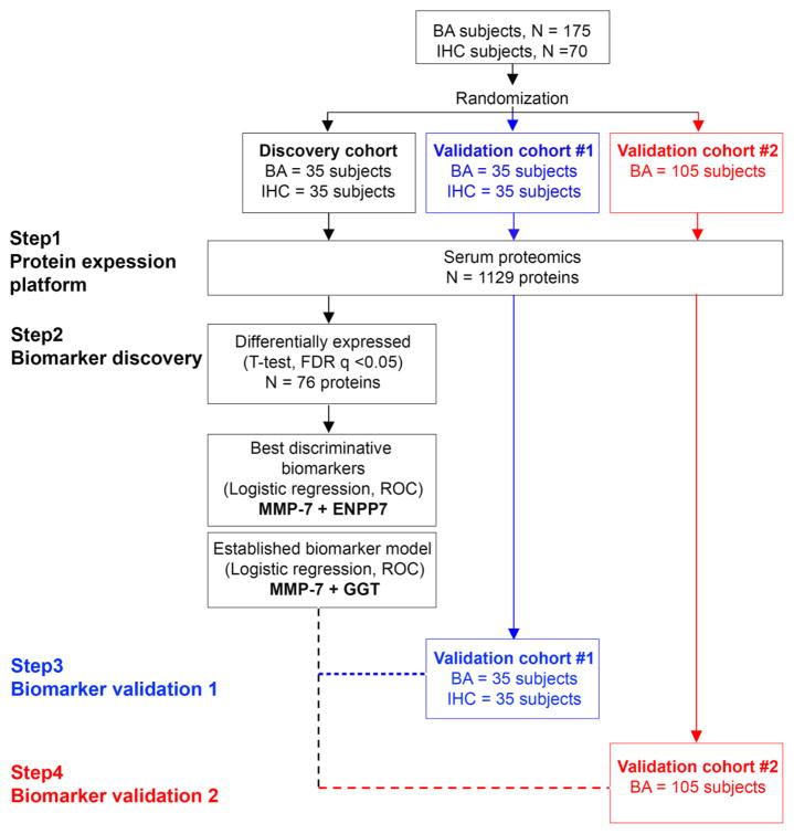
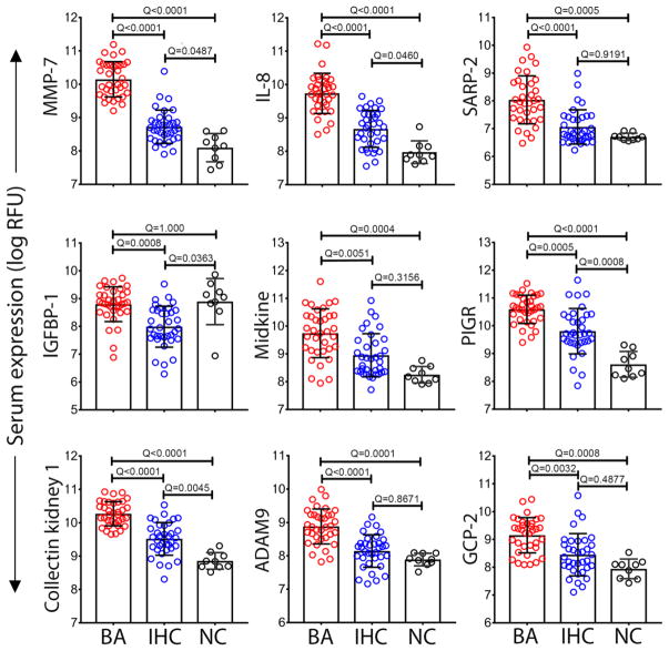
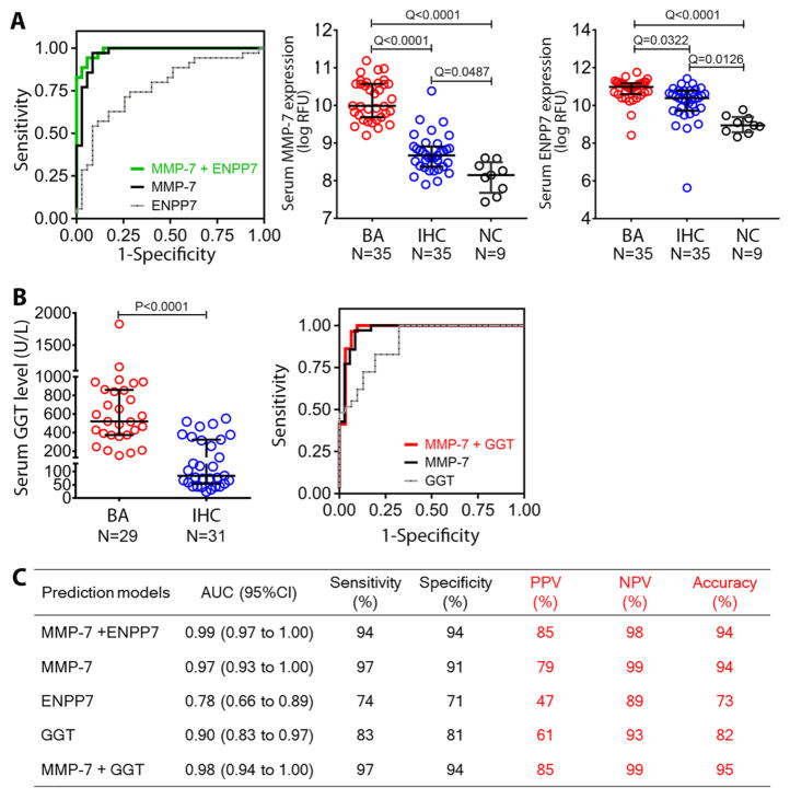
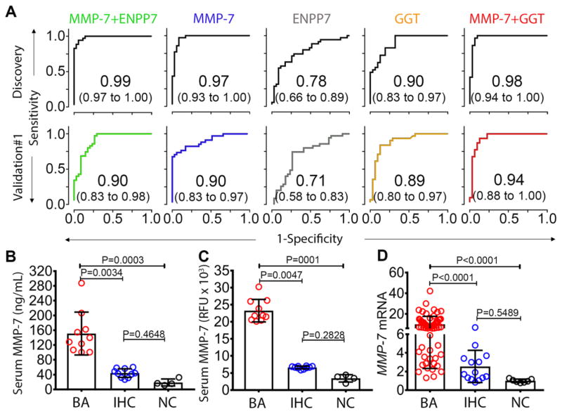
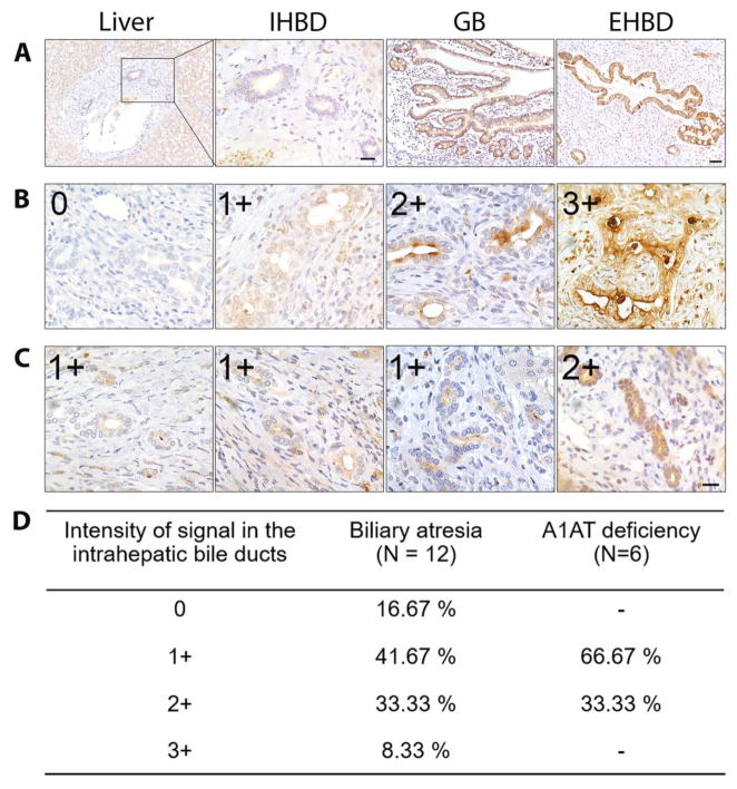
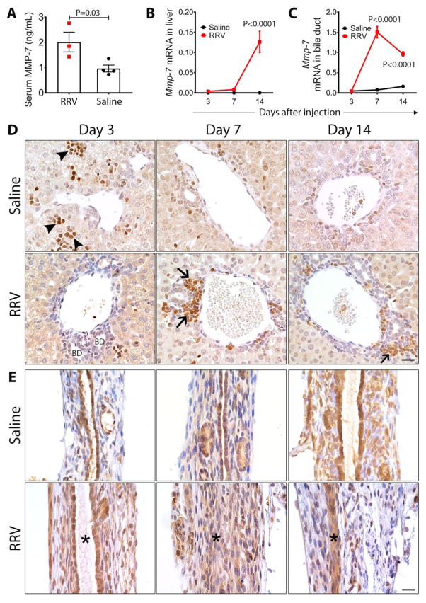
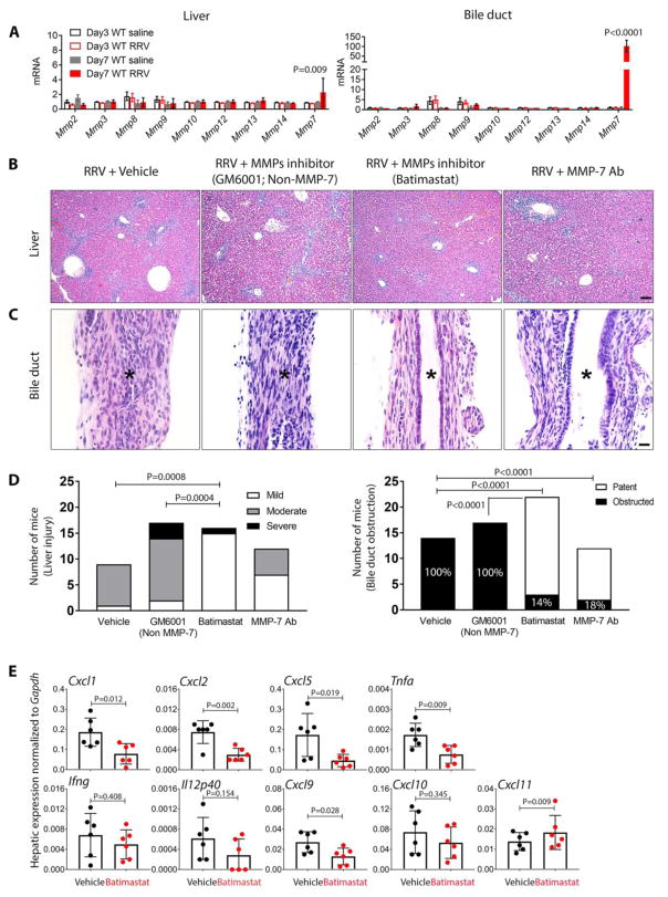
Biliary atresia is a progressive infantile cholangiopathy of complex pathogenesis. Although early diagnosis and surgery are the best predictors of treatment response, current diagnostic approaches are imprecise and time-consuming. We used large-scale, quantitative serum proteomics at the time of diagnosis of biliary atresia and other cholestatic syndromes (serving as disease controls) to identify biomarkers of disease. In a discovery cohort of 70 subjects, the lead biomarker was matrix metalloproteinase-7 (MMP-7), which retained high distinguishing features for biliary atresia in two validation cohorts. Notably, the diagnostic performance reached 95% when MMP-7 was combined with γ-glutamyltranspeptidase (GGT), a marker of cholestasis. Using human tissue and an experimental model of biliary atresia, we found that MMP-7 is primarily expressed by cholangiocytes, released upon epithelial injury, and promotes the experimental disease phenotype. Thus, we propose that serum MMP-7 (alone or in combination with GGT) is a diagnostic biomarker for biliary atresia and may serve as a therapeutic target.


Conflict of interest statement

Competing Interests

The authors declare no competing interests.

Figures

Fig. 1
Fig. 1. Study design
A total of 175 biliary atresia (BA) subjects and 70 subjects with intrahepatic cholestasis (IHC) were randomized into the discovery cohort (BA=35, IHC=35), validation cohort #1 (BA=35, IHC=35) and validation cohort #2 (BA=105). Serum proteins were measured by high-throughput proteomic assay (SOMAscan®) separately for individual cohorts. FDR, false discovery rate; ROC, receiver operating characteristic.
Fig. 2
Fig. 2. Top 9 proteins differentially expressed between BA and IHC
Data from the discovery cohort categorized into biliary atresia (BA, N=35), intrahepatic cholestasis (IHC, N=35), and age-matched normal controls (NC, N=9). Dots represent the log of relative fluorescent unit (RFU) for individual serum proteins. Bars and whiskers represent median and interquartile range. Q-values from ANOVA with multiple hypothesis correction (Benjamini-Hochberg procedure).
Fig. 3
Fig. 3. Predictive features of MMP-7, ENPP7 and GGT for biliary atresia
(A) ROC curves for serum MMP-7 and/or ENPP7 in distinguishing biliary atresia (BA) from intrahepatic cholestasis (IHC) in multivariable logistic regression analyses and dot plots of the proteins in the discovery cohort. Data are shown as log of relative fluorescent units (RFU); mid horizontal lines and whiskers represent median and interquartile range. (B) Serum GGT levels in subjects with BA and IHC; normal range is 5–59 U/L. The right panel shows ROC curves from MMP-7 and/or GGT. (C) Prediction models for MMP-7, ENPP7 and GGT. Sensitivity, specificity, positive and negative predictive values were calculated at selected optimal cut-off. Q values from ANOVA with multiple hypothesis correction (Benjamini-Hochberg procedure) for (A) and P-value from Mann Whitney test for (B). AUC, area under the ROC curve; NPV, negative predictive value; PPV, positive predictive value; ROC, receiver operating characteristic.
Fig. 4
Fig. 4. MMP-7 biomarker validation and hepatic expression
(A) ROC curves in discovery and validation cohort #1 for MMP-7, ENPP7, and GGT. (B) Scatter plot of serum MMP-7 of randomly selected samples from biliary atresia (BA: N=10), intrahepatic cholestasis (IHC; N=10), and normal controls (NC; N=4) measured by antibody-based enzyme-linked immunosorbent assay compared with (C) the values measured by SOMAScan®. Data is presented as median and interquartile range. (D) Hepatic mRNA expression of MMP-7 for BA (N=64), IHC (N=14), and NC (N=7). Expression levels are normalized to normal controls and are presented as fold change. P-values from Kruskal-Wallis test.
Fig. 5
Fig. 5. MMP-7 expression in human liver, extrahepatic bile duct, and gallbladder
(A) Immunohistochemical staining of normal human liver shows minimal or no expression of MMP-7 in hepatocytes and intrahepatic bile ducts (IHBD), but intense expression in the biliary epithelium of gallbladder (GB) and extrahepatic bile duct (EHBD). Scale bars = 50 μm in liver, GB and EHBD and 20 μm in IHBD. (B) Increased expression of MMP-7 in the cytoplasm of intrahepatic biliary epithelium of biliary atresia subjects with variable intensity (0–3+). (C) Livers of patients with alpha-1 antitrypsin deficiency had lower MMP-7 expression in the intrahepatic bile ducts. Scale bar = 20 μm in (B) and (C). (D) Percentage of livers according to the intensity and extension of MMP-7 immunostaining in livers from patients with biliary atresia and alpha-1 antitrypsin deficiency.
Fig. 6
Fig. 6. Mmp-7 expression in experimental biliary atresia
(A) Serum Mmp-7 measured 7 days after rotavirus (RRV) or saline injection (N=3–4 per group). Mean±SEM, P-values from unpaired t-test. (B) Mmp-7 mRNA expression in the livers and extrahepatic bile ducts (C) at 3, 7 and 14 days after RRV or saline injection (N= 3–4 per group and per time point). Values are normalized to Gapdh and are expressed as mean±SEM; P-values were between two groups at each time point (ANOVA). (D) Immunohistochemistry detects Mmp-7 in hematopoietic cells (arrowheads) predominantly at day 3, and inflammatory cells (arrows) infiltrating the portal tracts at day 7 and 14 after RRV injection. There was no staining of intrahepatic bile duct epithelium (BD). (E) Mmp-7 is detected in the duct epithelium in RRV and saline injected groups. At days 7 and 14 after injection, Mmp-7 is also detected in inflammatory cells and injured epithelium. Asterisks depict the duct lumen (or lack thereof); scale bars = 20 μm.
Fig. 7
Fig. 7. Suppression of tissue injury, inflammation, and cytokine expression by Batimastat and MMP-7 antibody in experimental biliary atresia
Neonatal BALB/c mice were injected with RRV in the first day of life, then were injected with Batimastat (RRV + Batimastat; N=22) or GM6001 (Non-MMP-7, RRV + GM6001; N=17), anti-MMP-7 antibody (RRV + MMP-7 Ab; N=12) or vehicle in the control group (RRV + vehicle; N=14). (A) mRNA expression of tissue matrix metalloproteinases (Mmps) at 3 and 7 days after viral injection. (B) Representative liver sections showing variable degrees of portal inflammation and hepatic necrosis in different groups. (C) Representative sections of extrahepatic bile ducts in each group. Asterisks depict the duct lumen (or lack thereof); scale bars = 100 μm for (B) and 20 μm for (C). (D) Graphs depict the frequency of mice with liver injury, according to the degree of inflammation and hepatocellular necrosis, and frequency of mice with obstructed lumen. (E) Hepatic mRNA expression for cytokines/chemokines 12 days after RRV injection and treatment with Batimastat (inhibitor) or vehicle (as controls). P-values from ANOVA for (A), Chi-square for (D) and unpaired T-test for (E).

Comment in

References

    1. Liu C, Aronow BJ, Jegga AG, Wang N, Miethke A, Mourya R, Bezerra JA. Novel resequencing chip customized to diagnose mutations in patients with inherited syndromes of intrahepatic cholestasis. Gastroenterology. 2007;132:119–126. - PMC - PubMed
    1. Yang Y, Muzny DM, Reid JG, Bainbridge MN, Willis A, Ward PA, Braxton A, Beuten J, Xia F, Niu Z, Hardison M, Person R, Bekheirnia MR, Leduc MS, Kirby A, Pham P, Scull J, Wang M, Ding Y, Plon SE, Lupski JR, Beaudet AL, Gibbs RA, Eng CM. Clinical whole-exome sequencing for the diagnosis of mendelian disorders. N Engl J Med. 2013;369:1502–1511. - PMC - PubMed
    1. Boyapati RK, Kalla R, Satsangi J, Ho GT. Biomarkers in Search of Precision Medicine in IBD. Am J Gastroenterol. 2016;111:1682–1690. - PubMed
    1. Hyman DM, Taylor BS, Baselga J. Implementing Genome-Driven Oncology. Cell. 2017;168:584–599. - PMC - PubMed
    1. Bessho K, Bezerra JA. Biliary atresia: will blocking inflammation tame the disease? Annu Rev Med. 2011;62:171–185. - PMC - PubMed