. 2018 Jan 10;38(2):379-397.
doi: 10.1523/JNEUROSCI.2110-17.2017. Epub 2017 Nov 22.

A Critical Role for Dopamine D5 Receptors in Pain Chronicity in Male Mice

Affiliations

A Critical Role for Dopamine D5 Receptors in Pain Chronicity in Male Mice

Salim Megat et al. J Neurosci. .

Abstract

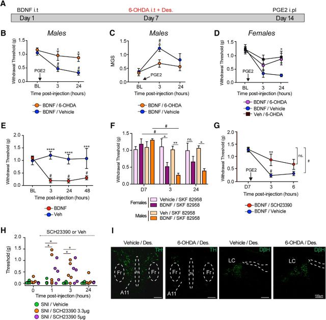
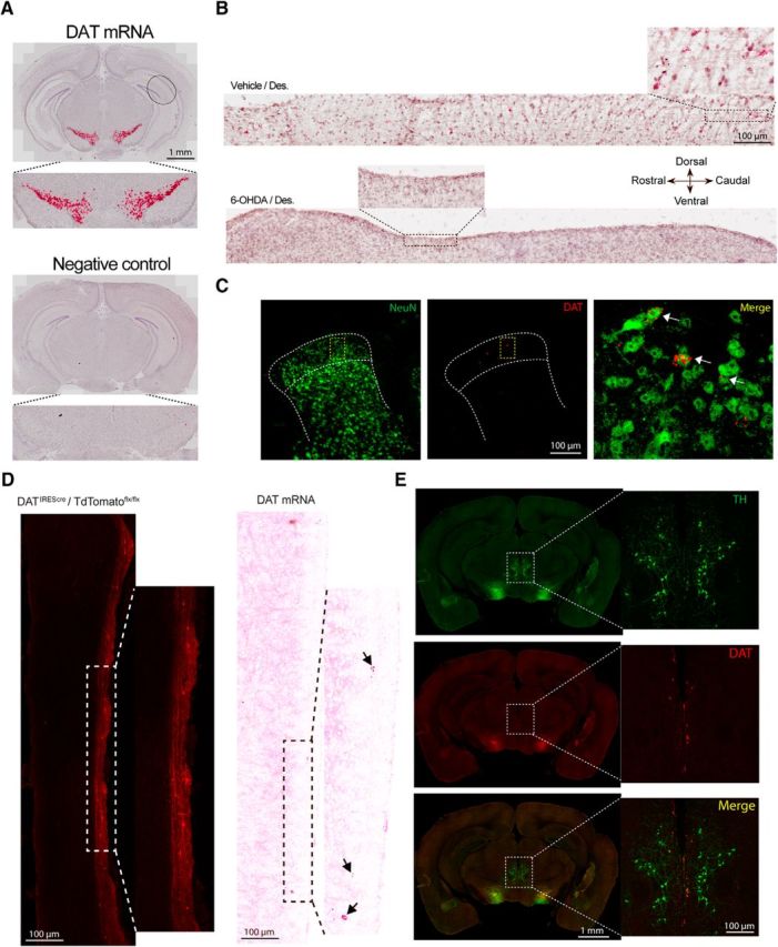
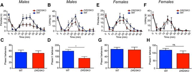
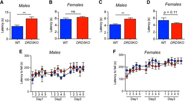
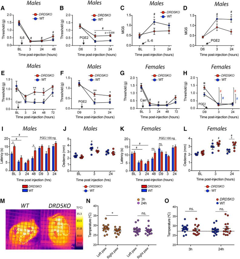
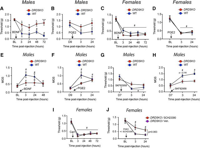
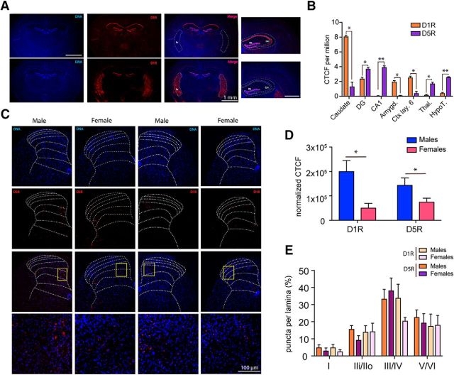
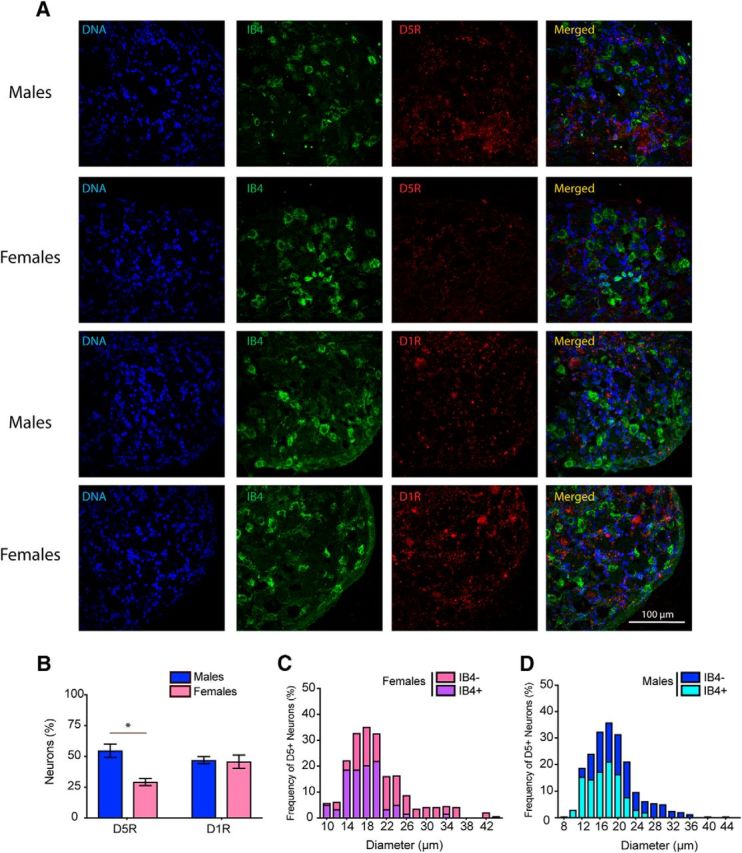
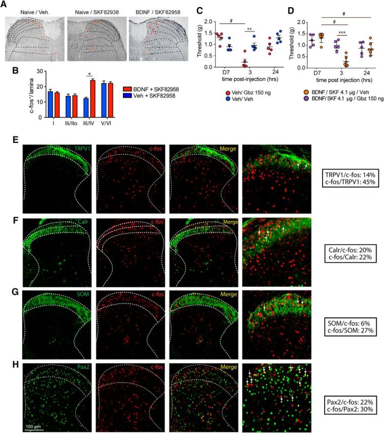
Dopaminergic modulation of spinal cord plasticity has long been recognized, but circuits affected by this system and the precise receptor subtypes involved in this modulation have not been defined. Dopaminergic modulation from the A11 nucleus of the hypothalamus contributes to plasticity in a model of chronic pain called hyperalgesic priming. Here we tested the hypothesis that the key receptor subtype mediating this effect is the D5 receptor (D5R). We find that a spinally directed lesion of dopaminergic neurons reverses hyperalgesic priming in both sexes and that a D1/D5 antagonist transiently inhibits neuropathic pain. We used mice lacking D5Rs (DRD5KO mice) to show that carrageenan, interleukin 6, as well as BDNF-induced hyperalgesia and priming are reduced specifically in male mice. These male DRD5KO mice also show reduced formalin pain responses and decreased heat pain. To characterize the subtypes of dorsal horn neurons engaged by dopamine signaling in the hyperalgesic priming model, we used c-fos labeling. We find that a mixed D1/D5 agonist given spinally to primed mice activates a subset of neurons in lamina III and IV of the dorsal horn that coexpress PAX2, a transcription factor for GABAergic interneurons. In line with this, we show that gabazine, a GABA-A receptor antagonist, is antihyperalgesic in primed mice exposed to spinal administration of a D1/D5 agonist. Therefore, the D5R, in males, and the D1R, in females, exert a powerful influence over spinal cord circuitry in pathological pain likely via modulation of deep dorsal horn GABAergic neurons.SIGNIFICANCE STATEMENT Pain is the most prominent reason why people seek medical attention, and chronic pain incidence worldwide has been estimated to be as high as 33%. This study provides new insight into how descending dopamine controls pathological pain states. Our work demonstrates that dopaminergic spinal projections are necessary for the maintenance of a chronic pain state in both sexes; however, D5 receptors seem to play a critical role in males whereas females rely more heavily on D1 receptors, an effect that could be explained by sexual dimorphisms in receptor expression levels. Collectively, our work provides new insights into how the dopaminergic system interacts with spinal circuits to promote pain plasticity.

Keywords: A11 nucleus; D5 receptor; descending modulation; dopamine; hyperalgesic priming; sex differences.


Figures

Figure 1.
Figure 1.
Spinal DA projections control BDNF-induced hyperalgesic priming and contribute to neuropathic pain. A, Timeline diagram showing 6-OHDA injection in primed mice. B, Subsequent intraplantar injection of PGE2 in mice that were previously treated with BDNF caused mechanical hypersensitivity that lasted for 24 h, whereas DA lesion 7 d after BDNF injection attenuated hyperalgesic priming. N = 6–8 mice per group. C, DA lesion also attenuated PGE2-induced increase in MGS scores in BDNF-primed mice. N = 6–8 mice per group. D, DA lesion also attenuated PGE2-induced allodynia in females BDNF-primed mice. N = 6 or 7 mice per group. E, Intrathecal injection of BDNF produces mechanical hypersensitivity that lasted for at least 48 h. N = 5 or 6 mice per group. F, At day 7, after complete recovery, spinal injection of the D1/D5 agonist in males and females, SKF82958 only reveals priming in animals previously treated with BDNF. N = 5–7 mice per group. G, Intrathecal injection of the D1/D5 antagonist SCH23390 at the time of PGE2 blocks mechanical hypersensitivity in BDNF-primed mice. N = 5 or 6 mice per group. H, Intrathecal injection of the D1/D5 antagonist transiently attenuates neuropathic mechanical allodynia at 1 and 3 h after intrathecal injection. N = 5 or 6 mice per group. I, Intrathecal injection of 6-OHDA reduces A11 hypothalamic DA neurons (6-OHDA + desipramine) without disrupting noradrenergic neurons in the locus ceruleus. Images are representative of N = 3 mice per group. *p < 0.05, comparing WT versus DRD5KO (two-way ANOVA with Bonferroni post hoc test). **p < 0.01, comparing WT versus DRD5KO (two-way ANOVA with Bonferroni post hoc test). ***p < 0.001, comparing WT versus DRD5KO (two-way ANOVA with Bonferroni post hoc test). ****p < 0.0001, comparing WT versus DRD5KO (two-way ANOVA with Bonferroni post hoc test). #p < 0.05, compared with baseline (two-way ANOVA with Bonferroni post hoc test).
Figure 2.
Figure 2.
Spinal DA lesion reduces the number of TH-immunoreactive neurons in A11 and spinal cord DAT expression. A, DAT in situ hybridization probe shows a specific signal in the substantia nigra, whereas the negative control probe shows no signal. B, Parasagittal sections of the spinal cord show DAT mRNA expression in the dorsal horn. Red punctate are visible throughout the rostrocaudal axis, whereas spinal DA lesion drastically decreases the level of DAT mRNA expression. N = 3 per group. C, Coronal section of the spinal cord showing DAT mRNA expression in the dorsal horn mainly localized in superficial laminae and overlapped with a neuronal marker (NeuN). N = 3 per group. D, Illustration of the DAT expression pattern in a DATIREScre/ROSA-LSL-tdTomato animal as well as DAT mRNA expression using RNAscope. E, Representative coronal section of the brain showing a high level of TH immunoreactivity in the A11 nucleus, whereas DAT mRNA signal is not readily detected in this region. Mice in these experiments were female and male.
Figure 3.
Figure 3.
Formalin-induced nocifensive behavior is attenuated in males DRD5KO mice. A, B, Formalin-induced flinching and licking behavior is reduced in DRD5KO male mice. C, D, The second phase of the formalin test is significantly blunted in male DRD5KO compared with their WT littermates. N = 6 mice per group. E, F, DRD5KO does not affect formalin-induced flinching or licking behavior in female mice. G, H, No significant differences were observed in the formalin test phases in female WT and DRD5KO mice. N = 6 mice per group. *p < 0.05 (two-way ANOVA with Bonferroni post hoc test).
Figure 4.
Figure 4.
Male DRD5KO mice exhibit thermal hypoalgesia. A, Thermal sensitivity is significantly impaired in male DRD5KO compared with WT mice in the Hargreaves test. N = 6–8 mice per group. B, Baseline thermal sensitivity is similar between female DRD5KO and WT mice in the Hargreaves test. N = 5–7 mice per group. C, Male DRD5KO show an increased tail-withdrawal latency compared with WT mice in the tail flick test at 49°C. N = 6 mice per group. D, No differences were observed in thermal sensitivity between female DRD5KO and WT. N = 6 mice per group. E, F, Motor skill learning and motor coordination are not impaired in male or female DRD5KO mice. N = 6–8 mice per group. **p < 0.01 (two-way ANOVA with Bonferroni post hoc test).
Figure 5.
Figure 5.
Carrageenan- and IL-6-induced hyperalgesic priming is reduced in male, but not female, DRD5KO mice. A, IL-6 injection induces mechanical hypersensitivity in male WT and DRD5KO mice that lasts for at least 48 h. N = 6 mice per group. B, Subsequent injection of PGE2 reveals hyperalgesic priming in male WT mice less in DRD5KO mice. N = 6 mice per group. C, Similarly, IL-6 failed to induce an increase in the mouse grimace scores in DRD5KO mice at 3 h. N = 6 mice per group. D, Finally, DRD5KO mice exhibit a significant reduction in MGS scores compared with WT mice after injection of PGE2 at 3 and 24 h. N = 6 mice per group. E, Intrathecal injection of carrageenan (1%) produces a strong mechanical hypersensitivity in male WT mice that is significantly reduced in DRD5KO mice. F, Moreover, PGE2-induced mechanical hypersensitivity is observed in primed DRD5KO mice. N = 6 mice per group. G, Intrathecal injection of carrageenan produces a robust mechanical hypersensitivity in female WT and DRD5KO mice that lasted for 72 h. N = 5 or 6 mice per group. H, Injection of PGE2 reveals the presence of priming in both genotypes. N = 6 mice per group. I, Carrageenan-induced thermal hypersensitivity is significantly attenuated in male DRD5KO mice at 48 h. N = 5 or 6 mice per group. J, However, the size of the edema is comparable between WT and DRD5KO mice. N = 6 mice per group. K, Carrageenan-induced thermal hypersensitivity is similar between WT and DRD5KO female mice. N = 6 mice per group. L, Yet, a significant increase in the size of the edema in the female DRD5KO mice was observed 24 h after treatment. N = 6 mice per group. M, Changes in paw temperature as a measure of inflammation observed in the left paw injected with carrageenan compared with the right paw. N, Carrageenan induces a significant increase in temperature of the ipsilateral compared with the contralateral paw at 3 h, but not at 24 h. N = 12–14 per group. O, No differences were observed in the temperature of the injected paw between WT and DRD5KO mice at 3 and 24 h. *p < 0.05, comparing WT versus DRD5KO (two-way ANOVA with Bonferroni post hoc test). **p < 0.01, comparing WT versus DRD5KO (two-way ANOVA with Bonferroni post hoc test). #p < 0.05, compared with baseline (two-way ANOVA with Bonferroni post hoc test).
Figure 6.
Figure 6.
Spinal D5Rs and D1Rs are predominantly involved in the maintenance of hyperalgesic priming in males and females, respectively. A, BDNF fails to produce mechanical hypersensitivity in male DRD5KO mice but produces robust effects in WT mice. N = 6 mice per group. B, Subsequent priming revealed by PGE2 is blocked in male DRD5KO mice at 24 h. N = 6 mice per group. C, In female mice, BDNF produces a robust mechanical hypersensitivity in WT and DRD5KO mice that lasts for at least 72 h. D, Again, injection of PGE2 reveals priming in female DRD5KO and WT mice. N = 5 or 6 mice per group. E, In males, the lack of D5 receptors blocks the development of an affective pain state after an intrathecal injection of BDNF. F, PGE2-induced grimacing in BDNF-primed mice is attenuated in male DRD5KO mice at 3 h after injection. N = 6 mice per group. G, Spinal injection of the D1LR agonist SKF82958 revealed a smaller magnitude of hyperalgesic priming induced by an intraplantar injection of IL-6 in DRD5KO mice, suggesting only a small role for D1Rs in priming in males. N = 6 mice per group. H, Likewise, male DRD5KO mice primed with IL-6 exhibited an attenuation of an affective pain state after spinal injection of the D1RL agonist compared with their WT littermates. N = 5 or 6 mice per group. I, Intraplantar injection of carrageenan 1% produces a strong mechanical hypersensitivity in female DRD5KO and WT mice. J, Intraplantar injection of a D1RL antagonist blocks hyperalgesic priming revealed by PGE2 injection in female DRD5KO mice. N = 6 mice per group. *p < 0.05, WT versus DRD5KO (two-way ANOVA with Bonferroni post hoc test). ***p < 0.001, WT versus DRD5KO (two-way ANOVA with Bonferroni post hoc test). #p < 0.05, compared with baseline (two-way ANOVA with Bonferroni post hoc test).
Figure 7.
Figure 7.
D1R and D5R mRNAs are highly expressed in the lamina III/IV of the dorsal horn of the spinal cord. A, Image showing the pattern of D1R and D5R mRNA expression in the brain. B, D1R mRNA expression is significantly higher in the caudate compared with D5R mRNA, whereas D5R mRNA is predominantly expressed in CA1 where D1R mRNA is almost completely absent. Images are representative of 3 mice. C, Images showing the pattern of expression of the D1R and D5R mRNAs in the spinal cord. D, CTCF analysis demonstrates a significant higher level of D1R and D5R mRNAs in the spinal cord of male compared with female mice. E, Moreover, D1R and D5R are highly enriched in the lamina III/IV of the spinal cord in male and female mice. N = 3 mice per group. *p < 0.05 (t test with correction for multiple comparisons). **p < 0.01 (t test with correction for multiple comparisons).
Figure 8.
Figure 8.
Differential expression of D5R mRNA in lumbar DRGs of male and female mice. A, Images represent the expression of D1R and D5R mRNAs in female and male lumbar DRGs. B, Histogram represents the percentage of DRG neurons that are positive for D1R and D5R mRNA. Images are representative of 4 or 5 mice per group. C, Histogram showing the relative frequency of D5R mRNA-positive neurons as a function of the diameter of female lumbar DRG neurons. D, Histogram showing the relative frequency of D5R mRNA-positive neurons as a function of the diameter of neurons in male lumbar DRGs. N = 4 or 5 mice per group. *p < 0.05 (t test with correction for multiple comparisons).
Figure 9.
Figure 9.
D1LR activation promotes hyperalgesic priming via a GABAergic mechanism in the lamina III of the spinal cord. A, Coronal section of the spinal cord showing an increase of c-fos-positive neurons in lamina III/IV. B, Spinal injection of the D1LR agonist SKF82958 in BDNF-primed mice induces a significant increase of c-fos-positive neurons in the lamina III and IV. N = 3 mice per group. C, Intrathecal injection of the GABA-AR antagonist gabazine produces a strong mechanical hypersensitivity in nonprimed mice that lasts for 24 h, whereas the D1LR agonist has no effect. N = 5 or 6 mice per group. D, Intrathecal injection of gabazine in BDNF-primed mice completely reverses D1LR agonist-induced mechanical hypersensitivity. N = 5 or 6 mice per group. **p < 0.01, WT versus DRD5KO (two-way ANOVA with Bonferroni post hoc test). ***p < 0.001, WT versus DRD5KO (two-way ANOVA with Bonferroni post hoc test). #p < 0.05, compared with baseline (two-way ANOVA with Bonferroni post hoc test). E, In BDNF-primed mice, 45% of the c-fos-immunoreactive neurons colocalize with TRPV1 afferent terminal staining in lamina I/II (N = 3/group). F, A total of 20% of the c-fos-immunoreactive neurons also express calretinin in lamina II (N = 3/group). G, In lamina I-III, 27% of the c-fos-immunoreactive neurons express somatostatin (N = 3/group). H, Finally, in BDNF-primed mice, 30% of the c-fos neurons express PAX2, a marker of GABAergic interneurons. *p < 0.05 (two-way ANOVA with Bonferroni post hoc test). ***p < 0.001 (two-way ANOVA with Bonferroni post hoc test).

Similar articles

Cited by

References

    1. Abdallah K, Artola A, Monconduit L, Dallel R, Luccarini P (2013) Bilateral descending hypothalamic projections to the spinal trigeminal nucleus caudalis in rats. PLoS One 8:e73022. 10.1371/journal.pone.0073022 - DOI - PMC - PubMed
    1. Abdallah K, Monconduit L, Artola A, Luccarini P, Dallel R (2015) GABAAergic inhibition or dopamine denervation of the A11 hypothalamic nucleus induces trigeminal analgesia. Pain 156:644–655. 10.1097/j.pain.0000000000000091 - DOI - PubMed
    1. Aira Z, Barrenetxea T, Buesa I, García Del Caño G, Azkue JJ (2016a) Dopamine D1-like receptors regulate constitutive, -opioid receptor-mediated repression of use-dependent synaptic plasticity in dorsal horn neurons: more harm than good? J Neurosci 36:5661–5673. 10.1523/JNEUROSCI.2469-15.2016 - DOI - PMC - PubMed
    1. Aira Z, Barrenetxea T, Buesa I, Martínez E, Azkue JJ (2016b) Spinal D1-like dopamine receptors modulate NMDA receptor-induced hyperexcitability and NR1 subunit phosphorylation at serine 889. Neurosci Lett 618:152–158. 10.1016/j.neulet.2016.03.006 - DOI - PubMed
    1. Asiedu MN, Tillu DV, Melemedjian OK, Shy A, Sanoja R, Bodell B, Ghosh S, Porreca F, Price TJ (2011) Spinal protein kinase M underlies the maintenance mechanism of persistent nociceptive sensitization. J Neurosci 31:6646–6653. 10.1523/JNEUROSCI.6286-10.2011 - DOI - PMC - PubMed

Publication types

Substances

LinkOut - more resources