Review
. 2018 Aug;596(15):3029-3042.
doi: 10.1113/JP274357. Epub 2017 Dec 27.

Interdependent feedback regulation of breathing by the carotid bodies and the retrotrapezoid nucleus

Affiliations
Review

Interdependent feedback regulation of breathing by the carotid bodies and the retrotrapezoid nucleus

Patrice G Guyenet et al. J Physiol. 2018 Aug.

Abstract

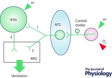
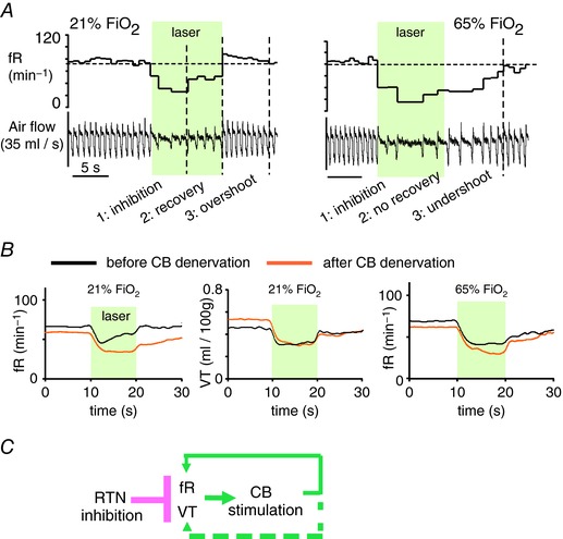
The retrotrapezoid nucleus (RTN) regulates breathing in a CO2 - and state-dependent manner. RTN neurons are glutamatergic and innervate principally the respiratory pattern generator; they regulate multiple aspects of breathing, including active expiration, and maintain breathing automaticity during non-REM sleep. RTN neurons encode arterial PCO2 /pH via cell-autonomous and paracrine mechanisms, and via input from other CO2 -responsive neurons. In short, RTN neurons are a pivotal structure for breathing automaticity and arterial PCO2 homeostasis. The carotid bodies stimulate the respiratory pattern generator directly and indirectly by activating RTN via a neuronal projection originating within the solitary tract nucleus. The indirect pathway operates under normo- or hypercapnic conditions; under respiratory alkalosis (e.g. hypoxia) RTN neurons are silent and the excitatory input from the carotid bodies is suppressed. Also, silencing RTN neurons optogenetically quickly triggers a compensatory increase in carotid body activity. Thus, in conscious mammals, breathing is subject to a dual and interdependent feedback regulation by chemoreceptors. Depending on the circumstance, the activity of the carotid bodies and that of RTN vary in the same or the opposite directions, producing additive or countervailing effects on breathing. These interactions are mediated either via changes in blood gases or by brainstem neuronal connections, but their ultimate effect is invariably to minimize arterial PCO2 fluctuations. We discuss the potential relevance of this dual chemoreceptor feedback to cardiorespiratory abnormalities present in diseases in which the carotid bodies are hyperactive at rest, e.g. essential hypertension, obstructive sleep apnoea and heart failure.

Keywords: carotid body; central respiratory chemoreceptor; optogenetics.


Figures

Figure 1
Figure 1. Anatomy of the retrotrapezoid nucleus
A, cluster of RTN neurons containing mRNA transcripts for VGlut2 and NMB. These neurons are immunoreactive for eGFP, the latter denoting the presence of Phox2b (transverse section, JX‐99 – Phox2b‐eGFP mouse; dashed line identifies the ventral medullary surface (VMS). Scale bar = 25 μm. Unpublished work of R. L. Stornetta. Every neuron is triple‐labelled. B, computer‐assisted mapping of RTN neurons (NMB+) in one mouse. Neurons identified in a one‐in‐three series of 30 μm‐thick transverse sections between 6.65 and 5.63 mm behind bregma. Scale bar = 500 μm (from Shi et al. 2017, with modifications). C, Venn diagram representing the various cell populations located in the parafacial region of the mouse. RTN neurons are defined as positive for Phox2b, VGlut2 and NMB. A least 90% of these neurons contain putative proton sensor TASK‐2 and ∼82% contain GPR4 (Shi et al. 2017). D, location and dendritic structure of two RTN neurons from rat recorded and labelled juxtacellularly in vivo. Note presence of extensive dendrites in the marginal layer (ML) regardless of the position of the cell body (adapted from Mulkey et al. 2004).
Figure 2
Figure 2. Retrotrapezoid nucleus: axonal projections and CO2 sensing mechanisms
A, brain regions targeted by RTN neurons. Schematic summary of results from Bochorishvili et al. (2012). B, CO2 detection by RTN neurons. Four mechanisms are thought to contribute to the exquisite sensitivity of RTN neurons to hypercapnia in vivo: (1) direct effect of proton on RTN neurons, (2) paracrine effect of protons mediated via acid‐sensitive astrocytes, (3) excitatory inputs from other pH‐sensitive neurons, (4) CO2‐induced vasoconstriction. The relative importance of each of the 4 mechanisms is unsettled. C, cell autonomous and paracrine mechanisms underlying the CO2 sensitivity of RTN neurons. The cell‐autonomous neuronal response to protons is mediated by TASK‐2 (Kcnk5) and GPR4. RTN is surrounded by astrocytes that are depolarized by local acidification. The depolarization activates an electrogenic sodium–bicarbonate transporter (NBCe) which alkalizes the glial cytoplasm and simultaneously acidifies the extracellular space. This extracellular acidification likely potentiates the intrinsic response of RTN neurons to a given change in arterial pH/PCO2. Sodium entry via NBCe activation also stimulates sodium–calcium exchange. The rise in intracellular calcium causes exocytosis of gliotransmitters such as ATP. ATP may also be released through connexin26 hemichannels opened by carbamylation. ATP recruits neighbouring astrocytes and may depolarize RTN neurons. Acid‐induced ATP release (from astrocytes or other unidentified cells) causes vasoconstriction, reduced wash‐out of metabolically produced CO2 and further acidification. Key supporting references: Mulkey et al. (2004); Erlichman & Leiter, (2010); Gestreau et al. (2010); Gourine et al. (2010); Huckstepp et al. (2010); Wenker et al. (2010); Wang et al. (2013); Kumar et al. (2015); Turovsky et al. (2016); Hawkins et al. (2017).
Figure 3
Figure 3. RTN‐dependent and ‐independent activation of breathing by the carotid bodies
A, single‐unit recording evidence that carotid body stimulation can activate RTN. This RTN neuron was activated by hypercapnia (eeCO2: end‐expiratory CO2) and by brief hypoxia (magenta). Administration of kynurenate i.c.v. (orange arrow), a blocker of excitatory glutamatergic synapses, eliminated the effect of hypoxia selectively whereas the effect of hypercapnia, which is primarily mediated by cell‐autonomous and paracrine effects of CO2, persisted (redrawn from Mulkey et al. 2004). B, evidence that the carotid bodies activate breathing via a pathway that bypasses RTN. Bilateral opto‐inhibition of RTN neurons (conscious rats) reduces breathing under normoxia (21% FI,O2) but has no effect under hypoxia (12% FI,O2). Breathing rate and amplitude increased under hypoxia despite RTN being silent, demonstrating that the CBs activated breathing via a pathway that bypasses RTN. The addition of a small amount of CO2 restored the breathing reduction caused by RTN inhibition, suggesting that hypoxia‐induced hyperventilation had inhibited RTN via the resultant respiratory alkalosis. Average plasma pH identified in 6 rats is shown below the representative examples (modified from Basting et al. 2015). C, breathing reduction elicited by bilateral optoinhibition of RTN is reduced under hypoxia. Left panel, minute volume before (black bars) and during RTN inhibition (green bars); right panel change in breathing frequency elicited by RTN inhibition. At 12% FI,O2, breathing is activated but RTN no longer contributes to breathing (right panel). At 15% FI,O2 breathing is unchanged from room air but, as indicated in the right panel, a much smaller portion of the respiratory drive originates from RTN (modified from Basting et al. 2015). D, relationship between arterial pH and effect of RTN inhibition on breathing frequency (f R) and plasma pH was manipulated with hypoxia. Respiratory alkalosis was compensated by adding CO2 (blue symbols) or by inducing metabolic acidosis (acetazolamide, yellow symbols). Above pHa 7.5, RTN inhibition has no effect on breathing consistent with single‐unit evidence that the neurons are silent (e.g. panel B). Below pH 7.5, the breathing reduction elicited by RTN inhibition (change in breathing frequency is depicted) is a linear function of arterial pH, consistent with single‐unit evidence (not shown) that RTN neurons are increasingly active (modified from Basting et al. 2015).
Figure 4
Figure 4. RTN activates multiple breathing parameters in a state‐dependent fashion
A, unilateral phasic optogenetic activation of RTN (channelrhodopsin‐2; trains of 3 light pulses; trains delivered at a rate slightly above resting breathing frequency) during quiet waking entrains the respiratory pattern generator, increases inspiratory amplitude, produces active expiration (note the distinctive peak‐expiratory flow, PEF, during late expiratory phase, E2) and activates the expiratory brake (note reduced expiratory flow during early expiration, E1). During non‐REM sleep (same rat), RTN activation still entrained the pattern generator and increased inspiratory amplitude but active expiration and the expiratory brake were no longer elicited (from Burke et al. 2015, with slight modifications). B, bilateral optogenetic inhibition of RTN (archaerhodopsin, 10 s light pulses; green bars) reduces respiratory frequency (f R) and tidal volume (V T) during quiet resting and non‐REM sleep. During REM sleep RTN inhibition has no effect on f R but still reduces V T, albeit to a lesser degree. This evidence indicates that RTN controls breathing frequency only when the pattern generator is auto‐rhythmic (slow wave sleep or quiet awake state) but not when it is externally driven, as in REM sleep. Adapted from Basting et al. 2015). C, schematic representation of the four known effects of RTN on breathing and their differential regulation by the state of vigilance.
Figure 5
Figure 5. RTN‐dependent (1) and RTN‐independent pathways (2) from the carotid bodies to the respiratory pattern generator (RPG)
Figure 6
Figure 6. Carotid body activation rapidly restores breathing during RTN inhibition
A, representative breathing response to instant bilateral optogenetic inhibition of RTN in a conscious rat. Under normoxia, the nadir occurs within 5 s and is followed by a gradual recovery. A brief overshoot occurs when the laser is switched off. Under hyperoxia, to silence the carotid bodies, the recovery phase is not observed and breathing inhibition persists for some time after the light is switched off. Recovery and overshoot of f R are therefore attributable to carotid body stimulation. Adapted from Basting et al. (2016). B, mean breathing response to bilateral opto‐inhibition of RTN in conscious rats (N = 6). Left panel: frequency response before and 7 days after bilateral CB denervation (same 6 rats). Middle panel: tidal volume (V T) response before and after CB denervation. Note that, contrary to f R, V T inhibition is unaffected by CB denervation. Right panel: under hyperoxia, the breathing response to RTN opto‐inhibition is no longer affected by CB denervation. From Basting et al. (2016). Confidence intervals removed for clarity. C, sequence of events (schematic): RTN inhibition reduces both f R and V T causing a decrease in alveolar ventilation and subsequent carotid body activation via the ensuing changes in blood gases. In rats, the carotid bodies stimulate breathing by increasing breathing frequency primarily; this effect quickly mitigates the hypoventilation elicited by RTN inhibition.

Similar articles

Cited by

References

    1. Abbott SB, Stornetta RL, Coates MB & Guyenet PG (2011). Phox2b‐expressing neurons of the parafacial region regulate breathing rate, inspiration, and expiration in conscious rats. J Neurosci 31, 16410–16422. - PMC - PubMed
    1. Abdala AP, Rybak IA, Smith JC & Paton JF (2009). Abdominal expiratory activity in the rat brainstem–spinal cord in situ: patterns, origins, and implications for respiratory rhythm generation. J Physiol 587, 3539–3559. - PMC - PubMed
    1. Barna BF, Takakura AC & Moreira TS (2014). Acute exercise‐induced activation of Phox2b‐expressing neurons of the retrotrapezoid nucleus in rats may involve the hypothalamus. Neuroscience 258, 355–363. - PubMed
    1. Basting TM, Abe C, Viar KE, Stornetta RL & Guyenet PG (2016). Is plasticity within the retrotrapezoid nucleus responsible for the recovery of the PCO2 set‐point after carotid body denervation in rats? J Physiol 594, 3371–3390. - PMC - PubMed
    1. Basting TM, Burke PG, Kanbar R, Viar KE, Stornetta DS, Stornetta RL & Guyenet PG (2015). Hypoxia silences retrotrapezoid nucleus respiratory chemoreceptors via alkalosis. J Neurosci 35, 527–543. - PMC - PubMed

Publication types