. 2017 Dec;9(12):1229-1234.
doi: 10.1038/nchem.2817. Epub 2017 Jul 10.

UV-light-driven prebiotic synthesis of iron-sulfur clusters

Affiliations

UV-light-driven prebiotic synthesis of iron-sulfur clusters

Claudia Bonfio et al. Nat Chem. 2017 Dec.

Abstract

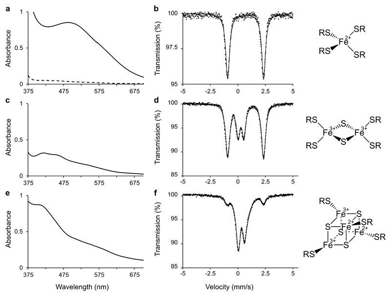
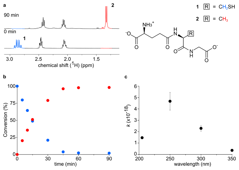
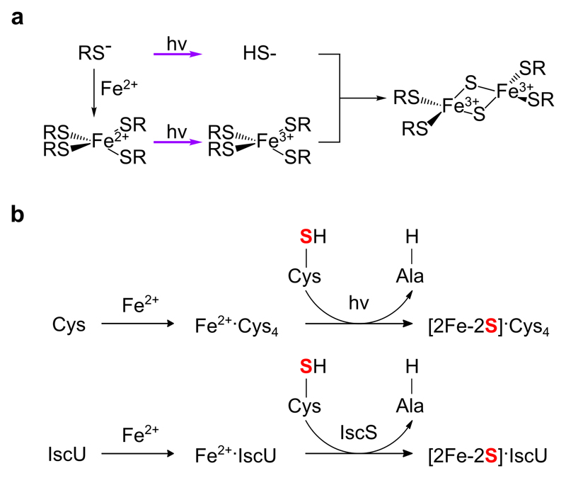
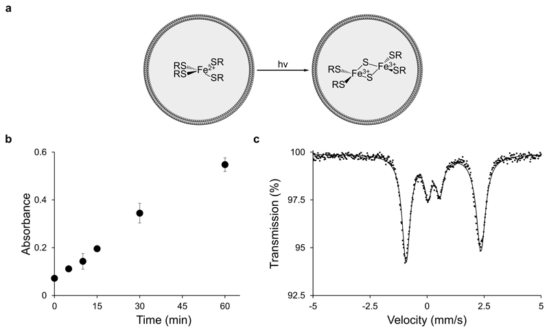
Iron-sulfur clusters are ancient cofactors that play a fundamental role in metabolism and may have impacted the prebiotic chemistry that led to life. However, it is unclear whether iron-sulfur clusters could have been synthesized on prebiotic Earth. Dissolved iron on early Earth was predominantly in the reduced ferrous state, but ferrous ions alone cannot form polynuclear iron-sulfur clusters. Similarly, free sulfide may not have been readily available. Here we show that UV light drives the synthesis of [2Fe-2S] and [4Fe-4S] clusters through the photooxidation of ferrous ions and the photolysis of organic thiols. Iron-sulfur clusters coordinate to and are stabilized by a wide range of cysteine-containing peptides and the assembly of iron-sulfur cluster-peptide complexes can take place within model protocells in a process that parallels extant pathways. Our experiments suggest that iron-sulfur clusters may have formed easily on early Earth, facilitating the emergence of an iron-sulfur-cluster-dependent metabolism.


Figures

Figure 1
Figure 1. Light driven, prebiotic synthesis of iron-sulfur peptides.
The addition of Fe3+ to glutathione results in an initial UV-visible spectrum consistent with a Fe3+-glutathione complex (a, solid line), which is then rapidly (over 180 s) reduced to a Fe2+-glutathione complex (a, dashed line, and b). Irradiation at 254 nm results in the appearance of a [2Fe-2S] cluster (c and d) and then a [4Fe-4S] cluster (e and f). a, c, and e: UV-visible absorption spectra; b, d, and f: Mössbauer spectra. d shows a mixture of mononuclear Fe2+ and [2Fe-2S]2+. f shows a mixture of mononuclear Fe2+, [2Fe-2S]2+, and [4Fe-4S]2+.
Figure 2
Figure 2. UV light desulfurization of the cysteine of glutathione to an alanine.
a, Conversion of the thiolate 1 (blue) to the corresponding desulfurized species 2 (red) shown by 1H NMR, consistent with the release of sulfide ions. b, Time course of conversion as obtained by the integration of NMR peaks for the irradiated solution of glutathione in the presence of hypophosphite. Blue, glutathione; red, alanyl-analogue. c, Dependence of the conversion rate constant on the irradiation wavelength. The data are congruent with the electronic transitions of thiolates. The rate constant (expressed as s-1) was normalized by the photon flux (expressed as s). Data represent n ≥ 3 replicates (mean and SEM).
Figure 3
Figure 3. Prebiotic pathways for the synthesis of iron-sulfur clusters.
a, Proposed mechanism of photooxidation and photolysis for the synthesis of a [2Fe-2S] cluster from a thiolate solution in the presence of ferrous ions. b, A comparison between model prebiotic (top) and extant, biosynthetic (bottom) pathways for the donation of sulfide during the synthesis of an iron-sulfur cluster.
Figure 4
Figure 4. [2Fe-2S] cluster formation in situ within fatty acid vesicles.
a, Schematic representation of light-driven, [2Fe-2S] cluster synthesis within fatty acid vesicles. b, A methylene blue assay detected the release of sulfide within oleate vesicles induced by UV light, demonstrating the compatibility of the photolysis of entrapped thiolates with fatty acid vesicles. Colorimetric data represent mean and SEM, n ≥ 3 replicates. c, Mössbauer spectrum of the [2Fe-2S]2+ cluster, minor component, formed within oleate vesicles by UV light.

Comment in

References

    1. Eck RV, Dayhoff MO. Evolution of the Structure of Ferredoxin Based on Living Relics of Primitive Amino Acid Sequences. Science. 1966;152:363–366. - PubMed
    1. Goldford JE, Hartman H, Smith TF, Segrè D. Remnants of an Ancient Metabolism without Phosphate. Cell. 2017;168:1126–1134. - PubMed
    1. Martin W, Russell MJ. On the origins of cells: a hypothesis for the evolutionary transitions from abiotic geochemistry to chemoautotrophic prokaryotes, and from prokaryotes to nucleated cells. Philos Trans R Soc B Biol Sci. 2003;358:59–85. - PMC - PubMed
    1. Martin WF, Sousa FL, Lane N. Energy at life’s origin. Science. 2014;344:1092–1093. - PubMed
    1. Maurel M, Leclerc F. From Foundation Stones to Life: Concepts and Results. Elements. 2016;12:407–412.

Publication types

MeSH terms

Substances

LinkOut - more resources