. 2017 Dec;9(12):1181-1190.
doi: 10.1038/nchem.2826. Epub 2017 Jul 31.

Global profiling of lysine reactivity and ligandability in the human proteome

Affiliations

Global profiling of lysine reactivity and ligandability in the human proteome

Stephan M Hacker et al. Nat Chem. 2017 Dec.

Abstract

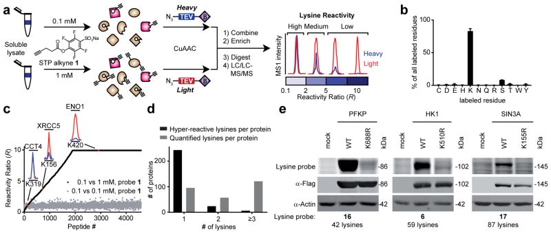
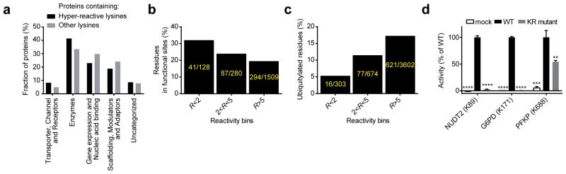
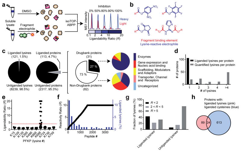
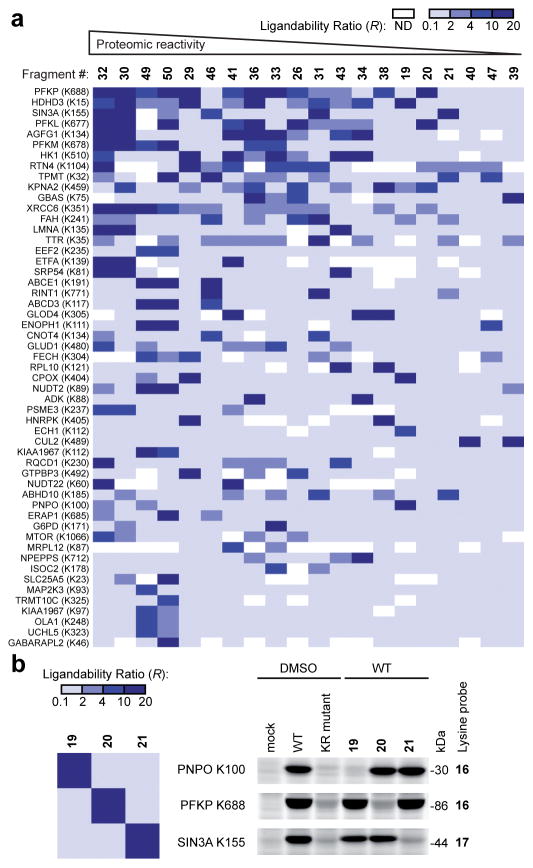
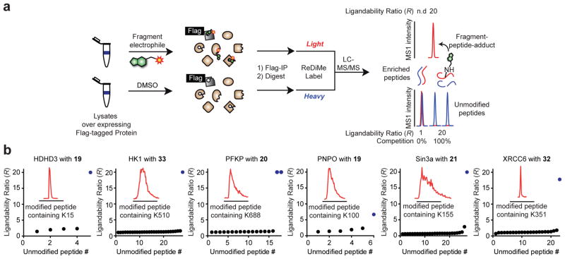
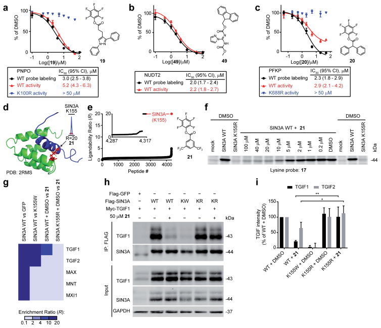
Nucleophilic amino acids make important contributions to protein function, including performing key roles in catalysis and serving as sites for post-translational modification. Electrophilic groups that target amino-acid nucleophiles have been used to create covalent ligands and drugs, but have, so far, been mainly limited to cysteine and serine. Here, we report a chemical proteomic platform for the global and quantitative analysis of lysine residues in native biological systems. We have quantified, in total, more than 9,000 lysines in human cell proteomes and have identified several hundred residues with heightened reactivity that are enriched at protein functional sites and can frequently be targeted by electrophilic small molecules. We have also discovered lysine-reactive fragment electrophiles that inhibit enzymes by active site and allosteric mechanisms, as well as disrupt protein-protein interactions in transcriptional regulatory complexes, emphasizing the broad potential and diverse functional consequences of liganding lysine residues throughout the human proteome.


Conflict of interest statement

Competing financial interests

The authors declare competing financial interests. Dr. Cravatt is a founder and advisor to Vividion Therapeutics, a biotechnology company interested in using chemical proteomic methods to develop small-molecule drugs to treat human disease.

Figures

Figure 1
Figure 1
Proteome-wide quantification of lysine reactivity. a, General protocol for lysine reactivity profiling by isoTOP-ABPP. Cellular lysates are labeled with an amine-reactive STP alkyne probe (1) at different concentrations. Labeled samples are conjugated to isotopically-differentiated TEV protease-cleavable biotin tags [heavy (blue) and light (red) for 0.1 and 1.0 mM probe 1 treatment groups, respectively] by CuAAC) mixed, and 1-labeled proteins enriched by streptavidin-conjugated beads and digested stepwise with trypsin and TEV to yield 1-labeled peptides for LC-MS analysis. Isotopic ratios, or R values reflect the relative MS1 chromatographic peak intensities for 1-labeled peptides in light versus heavy samples. b, Probe 1 preferentially labels lysine residues in human cell proteomes. Residues labeled by 1 were assigned by differential modification analysis of all quantified peptides identified in three replicate experiments comparing 0.1 vs 0.1 mM probe 1 treatments of MDA-MB-231 cell lysates. Peptides were required to feature no missed cleavage sites on unmodified lysine residues. Data represent mean values ± standard deviation for three experiments. c, R values for probe 1-labeled peptides from human cancer cell proteomes (MDA-MB-231, Ramos and Jurkat) treated with 0.1 vs 1.0 mM (black) or 0.1 vs 0.1 mM (gray) of probe 1. Representative MS1 chromatographic peaks for lysines of different reactivity categories are shown as insets (high, or hyper-reactive, R < 2.0 (K319 of CCT4); medium, 2.0 < R < 5.0 (K156 of XRCC5); and low, > 5.0 (K420 of ENO1)). d, Number of hyper-reactive and quantified lysines per protein shown for proteins found to contain at least one hyper-reactive lysine. e, Hyper-reactive lysines are site-selectively labeled by activated ester probes. HEK 293T cells expressing representative proteins with hyper-reactive lysines (or the corresponding lysine-to-arginine mutant) as FLAG epitope-tagged proteins were treated with the indicated lysine-reactive probe and analyzed by gel-based ABPP.
Figure 2
Figure 2
Global and specific assessments of the functionality of lysine reactivity. a, Distribution of functional classes of proteins that contain hyper-reactive lysines compared to other quantified proteins lacking hyper-reactive lysines. b, Hyper-reactive lysines are enriched proximal to (within 10 Å of) annotated functional sites for proteins that have x-ray or NMR structures in the Protein Data Bank (see Supplementary Methods for further details). c, Hyper-reactive lysines are less likely to be ubiquitylated than lysines of lower reactivity (ubiquitylated lysines were defined as those with ≥ 10 reported ubiquitylation events in public databases). d, Mutation of hyper-reactive lysines blocks the catalytic activity of NUDT2 and G6PD and reduces the activity of PFKP. Data represent mean values ± standard deviation for three experiments. Statistical significance was calculated with unpaired students t-tests in comparison to the WT activity; **, p < 0.01, ***, p < 0.001, ****, p < 0.0001.
Figure 3
Figure 3
Proteome-wide screening of lysine-reactive fragment electrophiles. a, General protocol for competitive isoTOP-ABPP. Competition ratios, or R values, are measured by quantifying the relative MS1 chromatographic peak intensities for 1-labeled peptides in DMSO- (heavy, or blue) versus fragment-treated (light, or red) samples. An R value of ≥ 4 was used to define a fragment liganding event for a quantified lysine. b, General structures of a lysine-reactive, electrophilic fragment library. See Supplementary Fig. 4 for chemical structures of library members. c, Fraction of total quantified lysines and proteins that were liganded by fragment electrophiles (left panel); of the liganded proteins, the fraction that is found in Drugbank (middle panel); functional classes of liganded Drugbank and non-Drugbank proteins (right panel). d, Number of liganded and quantified lysines per protein. Analysis was applied to proteins containing at least one liganded lysine. e, R values for ten lysines in PFKP, identifying K688 as the only liganded lysine in this protein. Each point represents a distinct fragment-lysine interaction quantified by isoTOP-ABPP. The red dashed line marks an R value of 4 used to define a fragment liganding event. f, Comparison of the ligandability of lysine residues as a function of reactivity with probe 1 (as measured in Fig. 1). Individual lysines are plotted on the x-axis sorted by reactivity, which is shown on the left y-axis, with lower R values correlating with elevated reactivity. A histogram with a bin-size of 200 is shown in blue for the percentage of liganded lysines within each reactivity bin (percent values shown on the right y-axis). g, Lysine reactivity distribution for both liganded and unliganded lysine residues labeled by probe 1. h, Overlap of proteins harboring liganded lysines and liganded cysteines. Cysteine ligandability was taken from reference .
Figure 4
Figure 4
Analysis of fragment-lysine interactions. a, Heat-map showing R values for representative lysines and fragments organized by relative proteomic reactivity of the fragments (high to low, left to right) and number of fragment hits for individual lysines (high to low, top to bottom). b, Fragment SAR determined by competitive isoTOP-ABPP is recapitulated by gel-based ABPP of recombinant proteins. Left panel, heat-map depicts R values for the indicated fragment-lysine interactions determined by competitive isoTOP-ABPP. Right panel, HEK 293T cells recombinantly expressing representative liganded proteins (or the corresponding lysine-to-arginine (KR) mutants) as FLAG epitope-tagged proteins were treated with fragment electrophiles (50 μM, 1 h) followed by the indicated lysine-reactive probes and analyzed by gel-based ABPP. SIN3A corresponds to a.a. 1-400 of SIN3A.
Figure 5
Figure 5
Confirmation of site-specific fragment-lysine reactions by MS-based proteomics. a, Schematic workflow for direct measurement of lysine-fragment reactions on proteins by quantitative proteomics. Flag epitope-tagged proteins are recombinantly expressed in HEK 293T cells and the cellular lysates treated with DMSO or a fragment ligand and immunoprecipitated with anti-Flag agarose resin. The enriched proteins are eluted from the resin, digested with trypsin, and tryptic peptides from the DMSO and fragment-treated samples isotopically labeled by reductive dimethylation (ReDiMe) with heavy (blue) and light (red) formaldehyde,, respectively. The DMSO and fragment-treated samples are then combined and analyzed by LC-MS. Lysine-fragment reactions were confirmed by both: i) detection of the peptide-fragment adduct exclusively in the fragment-treated sample (top trace); and ii) depletion of the parent unlabeled tryptic peptides containing the indicated lysine or having the lysine at a proteolytic cleavage site (bottom trace). b, R values for all detected, unmodified lysine-containing tryptic peptides for representative liganded proteins after treatment with the indicated compounds at 50 μM for 1 h. Unmodified peptides that contain the liganded lysine or have it at a proteolytic cleavage site are shown as blue dots. MS1 chromatographic peaks for fragment-peptide adducts are shown in the inset traces.
Figure 6
Figure 6
Fragment-lysine reactions inhibit the function of diverse proteins. ac, Fragments targeting active site (PNPO and NUDT2) and allosteric (PFKP) lysines in metabolic enzymes block enzymatic activity in a concentration-dependent manner with apparent IC50 values comparable to those measured by gel-based ABPP with lysine-reactive probes (probe labeling). Data represent mean values ± standard deviation for at least three experiments. CI, confidence interval. d, The liganded lysine K155 in SIN3A (red) is located at the protein-protein interaction site of the PAH1 domain (green) (shown in the NMR structure (PDB ID: 2RMS) to interact with the SID domain of SAP25 (blue)). e, Fragment 21 (50 μM) fully competes probe 1 labeling of K155 of SIN3A as determined by isoTOP-ABPP of human cancer cell proteomes. See inset in part d, for a representative MS1 chromatographic peak of the tryptic peptide containing K155 (R = 20, >95% blockade of probe 1 labeling by 21). f, Gel-based ABPP confirms that 21 blocks probe 17 labeling of SIN3A at K155 in a concentration-dependent manner. g, Heat-map showing the enrichment of SIN3A-interacting proteins in co-immunoprecipitation-MS-based proteomic experiments and blockade of SIN3A-TGIF1 interaction by 21 (50 μM, 1 h). h, i, Western blot analysis of Flag-SIN3A or the indicated Flag-SIN3A mutants, or Flag-GFP, co-expressed in HEK 293T cells with Myc-TGIF1 or Myc-TGIF2. Cellular lysates were treated with DMSO or 21 (50 μM, 1 h), and proteins immunoprecipitated prior to western blot analysis (h). Quantification of western blotting data for four biological replicates (i). Data represent mean values ± standard deviation for four experiments. Statistical significance was calculated with unpaired students t-tests comparing SIN3A WT + 21 to K155R + 21 groups; *, p < 0.05, **, p < 0.01. For panels fi, Flag-SIN3A or the indicated Flag-SIN3A mutants correspond to a.a. 1-400 of SIN3A.

Similar articles

Cited by

References

    1. Weiss WA, Taylor SS, Shokat KM. Recognizing and exploiting differences between RNAi and small-molecule inhibitors. Nat Chem Biol. 2007;3:739–744. - PMC - PubMed
    1. Makley LN, Gestwicki JE. Expanding the Number of “Druggable” Targets: Non-Enzymes and Protein-Protein Interactions. Chem Biol Drug Des. 2013;81:22–32. - PMC - PubMed
    1. Singh J, Petter RC, Baillie TA, Whitty A. The resurgence of covalent drugs. Nat Rev Drug Discov. 2011;10:307–317. - PubMed
    1. Bachovchin DA, Cravatt BF. The Pharmacological Landscape and Therapeutic Potential of Serine Hydrolases. Nat Rev Drug Discov. 2012;11:52–68. - PMC - PubMed
    1. Kato D, et al. Activity-based probes that target diverse cysteine protease families. Nat Chem Biol. 2005;1:33–38. - PubMed

Publication types