. 2017 Dec;9(12):1157-1164.
doi: 10.1038/nchem.2846. Epub 2017 Aug 21.

De novo design of a hyperstable non-natural protein-ligand complex with sub-Å accuracy

Affiliations

De novo design of a hyperstable non-natural protein-ligand complex with sub-Å accuracy

Nicholas F Polizzi et al. Nat Chem. 2017 Dec.

Abstract

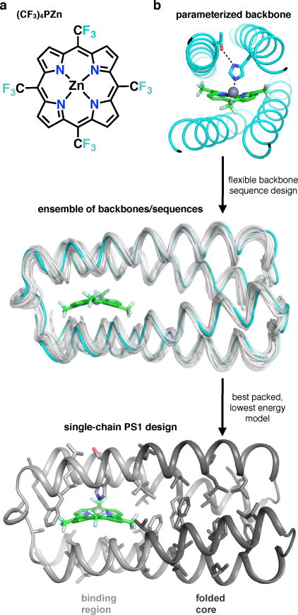
Protein catalysis requires the atomic-level orchestration of side chains, substrates and cofactors, and yet the ability to design a small-molecule-binding protein entirely from first principles with a precisely predetermined structure has not been demonstrated. Here we report the design of a novel protein, PS1, that binds a highly electron-deficient non-natural porphyrin at temperatures up to 100 °C. The high-resolution structure of holo-PS1 is in sub-Å agreement with the design. The structure of apo-PS1 retains the remote core packing of the holoprotein, with a flexible binding region that is predisposed to ligand binding with the desired geometry. Our results illustrate the unification of core packing and binding-site definition as a central principle of ligand-binding protein design.


Conflict of interest statement

The authors declare no competing financial interests.

Figures

Extended Data Figure 1
Extended Data Figure 1. PS1 design metrics
a, PS1 design ensemble resulting from flexible backbone sequence design. b, Residues (Cα atoms shown as blue and green spheres) within the PS1 design that were allowed to vary from the SCRPZ-2 sequence. 40 of the 108 residues were allowed to vary, and, of the 40 residues, 28 were mutated (blue) and 12 were retained (green) from the original SCRPZ-2 sequence as a result of the computational design process.
Extended Data Figure 2
Extended Data Figure 2. Analytical ultracentrifugation and gel filtration analysis show that apo- and holo-PS1 are monomeric in solution
a, Analytical ultracentrifugation. Solutions of apo- and holo-PS1 were centrifuged at speeds ranging from 25,000 r.p.m. to 45,000 r.p.m. and monitored by absorbance at 280 nm. Parameters were globally fit to the data. Single-species fitting agrees well with the data over the entire range and yields the molecular weight of apo-PS1 15.81 ± 0.09 kD and holo-PS1 12.24 ± 0.91 kD, which agrees well with the 12.86 kD weight of PS1. At high concentration, the fit for apo-PS1 is not ideal, suggesting a small degree of aggregation. Partial specific volumes were estimated from SEDNTERP, for amino acid side chains. b, Analytical gel filtration analysis of apo- and holo-PS1. Detection wavelengths are labeled as the same color as their respective curves. Apo shows a small degree (< 5%) of dimerization (1.35 ml elution volume) relative to the monomer peak (1.62 ml elution volume). The small peak near 1.05 ml elution volume in holo-PS1 is unbound (excess), aggregated porphyrin eluting in the void volume of the superdex 75 5/150 column. Samples were run at concentrations of 100 μM and 37 μM for apo and holo, respectively, in 50 mM NaPi, 150 mM NaCl, pH 7.0 buffer.
Extended Data Figure 3
Extended Data Figure 3. Electronic absorption and emission spectra of apo- and holo-PS1
The insets show normalized emission spectra: (left) electronic excitation of Trp68 at 280 nm, monitoring Trp68 emission; (right) electronic excitation of (CF3)4PZn at 405 nm, monitoring porphyrin emission. OD = 0.1 at excitation wavelength, buffer = 100 mM NaCl, 50 mM NaPi, pH 7.5.
Extended Data Figure 4
Extended Data Figure 4. Temperature and GnHCl induced unfolding of apo- and holo-PS1 show the designed protein is extremely thermostable
a, Circular dichroism (CD) spectra of apo-PS1 in 50 mM NaPi, 100 mM NaCl, pH 7.5 (No denaturant), as a function of temperature. b, CD spectra at 222 nm of apo-PS1 as a function of temperature and denaturant (Guanidine HCl, GnHCl) concentration in 50 mM NaPi, 100 mM NaCl, pH 7.5 buffer. c, CD spectra (blue) at 222 nm of holo-PS1 as a function of temperature in 50 mM NaPi, 100 mM NaCl, pH 7.5 (No denaturant), and fraction folded (black), defined as [MRE@222nm (T)- unfolded@222nm (T)]/[folded@222nm (T)- unfolded@222nm (T)], where unfolded(T) and folded(T) are unfolded and folded baselines, respectively, and T is temperature. The transitions appear reversible based on the fact that the spectra are identical after cooling to room temperature. The midpoint for GnHCl-induced unfolding at 95 °C was approximately 4.5 M.
Extended Data Figure 5
Extended Data Figure 5. 2D 1H-15N HSQC spectra acquired for apo- and holo-PS1
Experimental conditions: 0.78 mM at 298K, 50 mM NaPi, 100 mM NaCl, pH 7.5, in 5% D2O. Resonance assignments are indicated using the one-letter amino acid code. Signals arising from side chains (Asn HD2/ND2, Gln HE2/NE2, Arg HE/NE and Trp HE1/NE1) are also labeled. The residues belonging to the binding region and folded core are color-coded as in Fig. 4a. Non-helical residues are labeled in cyan font face. The inset in the HSQC spectrum of apo-PS1 shows the chemical shift of the indole proton of Trp68 near 10.2 ppm. A dashed box surrounds 90% of the backbone resonances of apo-PS1 and is also placed at the same position in the holo-PS1 spectrum. Arrows point to resonances of residues within the binding region that change dramatically upon binding of the cofactor.
Extended Data Figure 6
Extended Data Figure 6. Ab initio folding predictions of PS1 sequence
The Rosetta folding algorithm predicts a shallow folding funnel for the binding region (light gray) and a deep folding funnel shifted toward lower RMSD for the folded core (dark gray) of apo-PS1. The RMSD (root mean squared deviation) in Å is against the helical residues within these regions in the designed model. Energy is in Rosetta energy units (r.e.u.).
Extended Data Figure 7
Extended Data Figure 7. The NMR structural ensemble of apo-PS1 contains two clusters of conformations, closed and open
Above, color mapping of the pairwise backbone RMSD matrix of each NMR ensemble member of apo-PS1. Apo models with high structural similarity in the region of residues 61-67 and 99-105 (labeled in the open structure shown below) are blue in the plot. Models that are structurally dissimilar (large RMSD) are red in the plot. Below, the model centroids representing the closed and open structures (models 1 and 18, respectively, in the deposited NMR structure). The porphyrin (CF3)4PZn is shown in green, and the holo centroid (orange) is also drawn for comparison.
Extended Data Figure 8
Extended Data Figure 8. HDX protection factors for apo- and holo-PS1, as described in Table S5
Note that “68 indole” denotes the indole N of Trp68 side chain.
Extended Data Figure 9
Extended Data Figure 9. Molecular dynamics simulations show the binding region of apo-PS1 is more accessible to solvent
Histogram of number of waters within 3.5 Å of any heavy atom of each buried amino acid side chain (an A or D position of the heptad repeat), from 1000 snapshots of a 1 μs trajectory of apo-PS1. All histograms are drawn to the same scale and show number of solvating waters normalized by side chain surface area. Binding region shown in light gray, and folded core in dark gray.
Figure 1
Figure 1. The design strategy
a, Structures of natural cofactor-binding proteins show a folded core supporting a cofactor-binding region. b, Examples of previously designed tetra-helical porphyrin-binding proteins; all but PS1 (this work) lack a folded core (dark red). α2 protein is from ref ; the remainder are described in the text. c, The design process starts with a parameterized backbone, which undergoes simultaneous optimization of packing of core residues (shown as spheres) in the binding region (light color) and folded core (dark color), with flexible backbone. The resultant holo-protein (red) is tightly packed both in the binding region and in the folded core, whereas the apo-protein (purple) is tightly packed only in the folded core, which anchors the under-packed binding region to bind the cofactor. cyt b562, cytochrome b562 (pdb 256b), DHFR, dihydrofolate reductase (pdb 8dfr), flavodoxin (pdb 1czu).
Figure 2
Figure 2. The computational design workflow for optimized core packing
a, The abiological porphyrin cofactor, (CF3)4PZn. b, The constrained, parameterized backbone of SCRPZ-2 (cyan) feeds into a flexible backbone design protocol that allows the interior side chains and backbone to simultaneously conform to the porphyrin (CF3)4PZn (green).
Figure 3
Figure 3. The structure of holo-PS1 agrees closely with the design
a, The structure of holo-PS1 (orange) superimposed on the design (gray), with mean helical backbone RMSD of 0.8 ± 0.1 Å. The holo-PS1 model shown is the centroid of the NMR structural ensemble. b,c, shows ~10 Å slices of the holo-PS1 NMR centroid and design in the binding region and folded core, respectively. d, compares observed (green) vs. designed (gray) orientations. All hydrophobic and helical backbone heavy atoms within 4 Å of porphyrin heavy atoms in the design were used for alignment (0.9 ± 0.1 Å all-atom RMSD). e, Pump-probe transient absorption spectra of (CF3)4PZn bound in the interior of holo-PS1 at 21 °C and 100 °C. The black spectrum shows characteristic S1→SN absorptions of (CF3)4PZn, which smoothly transitions into the gold spectrum showing characteristic T1→TN absorptions of (CF3)4PZn. Inset exemplifies identical transient dynamics (primarily intersystem crossing from S1 to T1) at ΔAbs. = 482 nm (scaled). Experimental conditions: solvent = 50 mM NaPi, 100 mM NaCl, pH 7.5; excitation wavelength = 600 ± 5 nm; magic-angle polarization between pump and probe pulses; pump-probe cross-correlation of ~250 fs.
Figure 4
Figure 4. Apo- and holo-PS1 share similar folded cores and differ in the binding region
a, Solution NMR structures of apo-PS1 (purple) and holo-PS1 (orange). The structures were aligned to the backbone of the helical folded core of the lowest energy holo-PS1 model. Terminal residues 1, 108, and 109 are not shown for clarity. b, Hydrogen-deuterium exchange protection factors (PF) measured for apo- and holo-PS1, mapped onto the centroid structure of holo-PS1. Backbone amide nitrogens of residues with determined PFs are shown as spheres. Not shown: N of Trp68 indole sidechain is protected in holo, but not apo. c–e, Backbone alignment of the holo- and apo-centroids at the folded core shows, e, agreement of side chain rotamer states far from the binding site and, d, differences in first-shell rotamers (e.g., Trp68, Leu98) accompanied by changes in backbone of the binding region. Centroids are from NMR structural ensembles clustered via RMSD of core side chain heavy atoms.

References

    1. Roy S, et al. A Protein Designed by Binary Patterning of Polar and Nonpolar Amino Acids Displays Native-like Properties. J Am Chem Soc. 1997;119:5302–5306.
    1. Kuhlman B, et al. Design of a novel globular protein fold with atomic-level accuracy. Science. 2003;302:1364–1368. - PubMed
    1. Nanda V, Koder RL. Designing artificial enzymes by intuition and computation. Nat Chem. 2010;2:15–24. - PMC - PubMed
    1. Peacock AFA. Incorporating metals into de novo proteins. Curr Opin Chem Biol. 2013;17:934–939. - PubMed
    1. Huang PS, et al. High thermodynamic stability of parametrically designed helical bundles. Science. 2014;346:481. - PMC - PubMed

Publication types