

Dental enamel defect diagnosis through different technology-based devices
- PMID: 29168574
- PMCID: PMC9378886
- DOI: 10.1111/idj.12350
Dental enamel defect diagnosis through different technology-based devices
Abstract
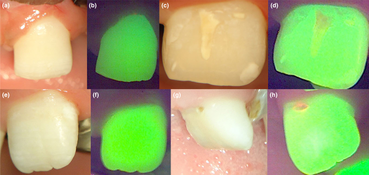
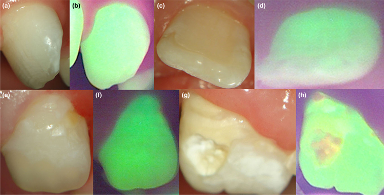
Introduction: Dental enamel defects (DEDs) are faulty or deficient enamel formations of primary and permanent teeth. Changes during tooth development result in hypoplasia (a quantitative defect) and/or hypomineralisation (a qualitative defect).
Objective: To compare technology-based diagnostic methods for detecting DEDs.
Material and methods: Two-hundred and nine dental surfaces of anterior permanent teeth were selected in patients, 6-11 years of age, with cleft lip with/without cleft palate. First, a conventional clinical examination was conducted according to the modified Developmental Defects of Enamel Index (DDE Index). Dental surfaces were evaluated using an operating microscope and a fluorescence-based device. Interexaminer reproducibility was determined using the kappa test. To compare groups, McNemar's test was used. Cramer's V test was used for comparing the distribution of index codes obtained after classification of all dental surfaces.
Results: Cramer's V test revealed statistically significant differences (P < .0001) in the distribution of index codes obtained using the different methods; the coefficients were 0.365 for conventional clinical examination versus fluorescence, 0.961 for conventional clinical examination versus operating microscope and 0.358 for operating microscope versus fluorescence. The sensitivity of the operating microscope and fluorescence method was statistically significant (P = .008 and P < .0001, respectively). Otherwise, the results did not show statistically significant differences in accuracy and specificity for either the operating microscope or the fluorescence methods.
Conclusion: This study suggests that the operating microscope performed better than the fluorescence-based device and could be an auxiliary method for the detection of DEDs.
Keywords: Tooth enamel; dental operating microscope; diagnosis; fluorescence.
© 2017 FDI World Dental Federation.
Figures
Similar articles
-
Structural color changes in permanent enamel of patients with cleft lip and palate: a case-control study.J Orofac Orthop. 2016 Jan;77(1):45-51. doi: 10.1007/s00056-015-0007-z. J Orofac Orthop. 2016. PMID: 26744208
-
A comparison of information recorded using the Thylstrup Fejerskov index, Tooth Surface Index of Fluorosis and Developmental Defects of Enamel index.Int Dent J. 1994 Dec;44(6):628-36. Int Dent J. 1994. PMID: 7851996
-
Prevalence of enamel defects in permanent teeth of patients with complete cleft lip and palate.Cleft Palate Craniofac J. 2013 Jul;50(4):394-9. doi: 10.1597/11-200. Epub 2012 Jan 31. Cleft Palate Craniofac J. 2013. PMID: 22292803
-
Clinical diagnosis of enamel defects: pitfalls and practical guidelines.Int Dent J. 1997 Jun;47(3):173-82. doi: 10.1002/j.1875-595x.1997.tb00783.x. Int Dent J. 1997. PMID: 9448804 Review.
-
Hypomineralisation or hypoplasia?Br Dent J. 2019 Oct;227(8):683-686. doi: 10.1038/s41415-019-0782-9. Br Dent J. 2019. PMID: 31654000 Review.
Cited by
-
Er:YAG laser for the aesthetic treatment of developmental enamel defects in frontal teeth: a case report.Laser Ther. 2019 Sep 30;28(3):199-202. doi: 10.5978/islsm.28_19-CR-02. Laser Ther. 2019. PMID: 32009734 Free PMC article.
-
Characterization of hypomineralization second primary molar and molar incisor hypomineralization in patients with orofacial clefts.Clin Oral Investig. 2025 Apr 25;29(5):269. doi: 10.1007/s00784-025-06311-7. Clin Oral Investig. 2025. PMID: 40278923
-
Microinvasive esthetic approach for deep enamel white spot lesion.Dent Res J (Isfahan). 2022 Mar 21;19:29. eCollection 2022. Dent Res J (Isfahan). 2022. PMID: 35432791 Free PMC article.
-
Etiology Study of Acquired Developmental Defects of Enamel and Their Association with Dental Caries in Children between 3 and 19 Years Old from Dolj County, Romania.Children (Basel). 2022 Sep 14;9(9):1386. doi: 10.3390/children9091386. Children (Basel). 2022. PMID: 36138695 Free PMC article.
-
Maxillary incisor enamel defects in individuals born with cleft lip/palate.PLoS One. 2020 Dec 28;15(12):e0244506. doi: 10.1371/journal.pone.0244506. eCollection 2020. PLoS One. 2020. PMID: 33370403 Free PMC article.
References
-
- FDI A review of the developmental defects of enamel index (DDE Index). commission on oral health, research & epidemiology. report of an FDI working group. Int Dent J. 1992;42:411–426. - PubMed
-
- Jalevik B, Noren JG. Enamel hypomineralization of permanent first molars: a morphological study and survey of possible aetiological factors. Int J Paediatr Dent. 2000;10:278–289. - PubMed
-
- Seow WK, Ford D, Kazoullis S, et al. Comparison of enamel defects in the primary and permanent dentitions of children from a low-fluoride District in Australia. Pediatr Dent. 2011;33:207–212. - PubMed
-
- Vitor LLR, Kobayashi TY, Carrara CFC, et al. Do technology-based devices improve carious lesion detection in children with oral cleft? Braz Dent Sci. 2016;19:24–31.
Publication types
MeSH terms
LinkOut - more resources
Full Text Sources
Other Literature Sources
