Review
. 2017 Nov 23;22(12):2045.
doi: 10.3390/molecules22122045.

Regulation of G2/M Transition by Inhibition of WEE1 and PKMYT1 Kinases

Affiliations
Review

Regulation of G2/M Transition by Inhibition of WEE1 and PKMYT1 Kinases

Matthias Schmidt et al. Molecules. .

Abstract

In the cell cycle, there are two checkpoint arrests that allow cells to repair damaged DNA in order to maintain genomic integrity. Many cancer cells have defective G1 checkpoint mechanisms, thus depending on the G2 checkpoint far more than normal cells. G2 checkpoint abrogation is therefore a promising concept to preferably damage cancerous cells over normal cells. The main factor influencing the decision to enter mitosis is a complex composed of Cdk1 and cyclin B. Cdk1/CycB is regulated by various feedback mechanisms, in particular inhibitory phosphorylations at Thr14 and Tyr15 of Cdk1. In fact, Cdk1/CycB activity is restricted by the balance between WEE family kinases and Cdc25 phosphatases. The WEE kinase family consists of three proteins: WEE1, PKMYT1, and the less important WEE1B. WEE1 exclusively mediates phosphorylation at Tyr15, whereas PKMYT1 is dual-specific for Tyr15 as well as Thr14. Inhibition by a small molecule inhibitor is therefore proposed to be a promising option since WEE kinases bind Cdk1, altering equilibria and thus affecting G2/M transition.

Keywords: G2/M transition; PKMYT1; WEE1.


Conflict of interest statement

The authors declare no conflict of interest.

Figures

Figure 1
Figure 1
Cell cycle control: DNA damage checkpoint (modified from Aarts et al. [55]). Reproduced with permission from Nick Turner, Current Opinion in Pharmacology; published by Elsevier, 2013.
Figure 2
Figure 2
Superposition of crystal structures from the Protein Data Bank (PDB) of WEE1 (PDB ID: 1X8B) and PKMYT1 (PDB ID: 3P1A). The protein ribbon is colored as following: N-Terminus: gold; C-Terminus: magenta; hinge region: dark green; activation loop: cyan; P-loop: yellow; PKMYT1 residues: white; WEE1 residues: green.
Figure 3
Figure 3
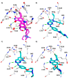
Interaction of WEE1 and PKMYT1 inhibitors observed in reported crystal structures. Hydrogen bonds with hinge region residues are displayed as dashed green lines. (A) Superposition of the X-ray structures of WEE1 in complex with pyrrolocarbazoles and pyrroloindoles (colored magenta); (B) WEE1 in complex with the diaminopyrimidine derivative MK1775 (colored cyan, PDB ID: 5V5Y); (C) WEE1 in complex with the pyridopyrimidine derivative PD-0166285 (colored cyan, PDB ID: 5VC5); (D) WEE1 in complex with Bosutinib (colored cyan, PDB ID: 5VC3); (E) PKMYT1 in complex with Dasatinib (colored yellow, PDB ID: 5VCV); (F) PKMYT1 in complex with Saracatinib (colored yellow, PDB ID: 5VCX); (G) PKMYT1 in complex with the diaminopyrimidine derivative MK1775 (colored yellow, PDB ID: 5VD0); (H) PKYMT1 in complex with the 4-aminquinoline derivative Bosutinib Isomer (colored yellow, PDB ID: 5VCZ).
Figure 3
Figure 3
Interaction of WEE1 and PKMYT1 inhibitors observed in reported crystal structures. Hydrogen bonds with hinge region residues are displayed as dashed green lines. (A) Superposition of the X-ray structures of WEE1 in complex with pyrrolocarbazoles and pyrroloindoles (colored magenta); (B) WEE1 in complex with the diaminopyrimidine derivative MK1775 (colored cyan, PDB ID: 5V5Y); (C) WEE1 in complex with the pyridopyrimidine derivative PD-0166285 (colored cyan, PDB ID: 5VC5); (D) WEE1 in complex with Bosutinib (colored cyan, PDB ID: 5VC3); (E) PKMYT1 in complex with Dasatinib (colored yellow, PDB ID: 5VCV); (F) PKMYT1 in complex with Saracatinib (colored yellow, PDB ID: 5VCX); (G) PKMYT1 in complex with the diaminopyrimidine derivative MK1775 (colored yellow, PDB ID: 5VD0); (H) PKYMT1 in complex with the 4-aminquinoline derivative Bosutinib Isomer (colored yellow, PDB ID: 5VCZ).
Figure 4
Figure 4
Superposition of the ATP-bindings pocket of WEE1 (PDB ID: 1X8B, green) and PKMYT1 (PDB ID: 3P1A, white residues). Ribbons have been colored as mentioned in Figure 2. The co-crystallized inhibitor PD0407824 of WEE1 is shown in cyan. Hydrogen bonds are displayed as dashed lines.
Figure 5
Figure 5
Chemical structure of WEE1 inhibitors.
Figure 6
Figure 6
Chemical structures of potent PKMYT1 inhibitors.

Similar articles

Cited by

References

    1. Manning G., Whyte D.B., Martinez R., Hunter T., Sudarsanam S. The Protein Kinase Complement of the Human Genome. Science. 2002;298:1912–1934. doi: 10.1126/science.1075762. - DOI - PubMed
    1. Brognard J., Hunter T. Protein kinase signaling networks in cancer. Curr. Opin. Genet. Dev. 2011;21:4–11. doi: 10.1016/j.gde.2010.10.012. - DOI - PMC - PubMed
    1. Domingo-Sananes M.R., Kapuy O., Hunt T., Novak B. Switches and latches: A biochemical tug-of-war between the kinases and phosphatases that control mitosis. Philos. Trans. R. Soc. Lond. B Biol. Sci. 2011;366:3584–3594. doi: 10.1098/rstb.2011.0087. - DOI - PMC - PubMed
    1. Lindqvist A., Rodriguez-Bravo V., Medema R.H. The decision to enter mitosis: Feedback and redundancy in the mitotic entry network. J. Cell Biol. 2009;185:193–202. doi: 10.1083/jcb.200812045. - DOI - PMC - PubMed
    1. Pomerening J.R. Positive-feedback loops in cell cycle progression. FEBS Lett. 2009;583:3388–3396. doi: 10.1016/j.febslet.2009.10.001. - DOI - PMC - PubMed

MeSH terms