

Evaluating the Benefit of Elevated Acoustic Output in Harmonic Motion Estimation in Ultrasonic Shear Wave Elasticity Imaging
- PMID: 29169880
- PMCID: PMC5743577
- DOI: 10.1016/j.ultrasmedbio.2017.10.003
Evaluating the Benefit of Elevated Acoustic Output in Harmonic Motion Estimation in Ultrasonic Shear Wave Elasticity Imaging
Abstract
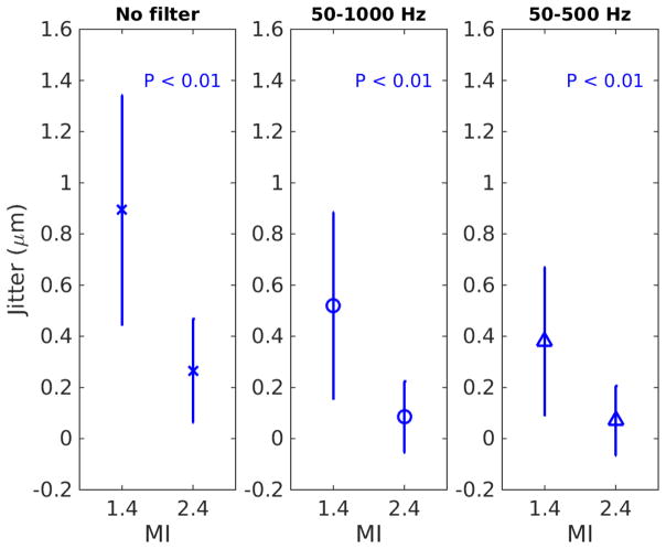
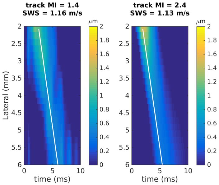
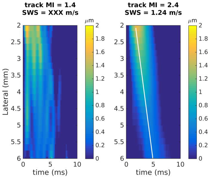
Harmonic imaging techniques have been applied in ultrasonic elasticity imaging to obtain higher-quality tissue motion tracking data. However, harmonic tracking can be signal-to-noise ratio and penetration depth limited during clinical imaging, resulting in decreased yield of successful shear wave speed measurements. A logical approach is to increase the source pressure, but the in situ pressures used in diagnostic ultrasound have been subject to a de facto upper limit based on the Food and Drug Administration guideline for the mechanical index (MI <1.9). A recent American Institute of Ultrasound in Medicine report concluded that an in situ MI up to 4.0 could be warranted without concern for increased risk of cavitation in non-fetal tissues without gas bodies if there were a concurrent clinical benefit. This work evaluates the impact of using an elevated MI in harmonic motion tracking for hepatic shear wave elasticity imaging. The studies indicate that high-MI harmonic tracking increased shear wave speed estimation yield by 27% at a focal depth of 5 cm, with larger yield increase in more difficult-to-image patients. High-MI tracking improved harmonic tracking data quality by increasing the signal-to-noise ratio and decreasing jitter in the tissue motion data. We conclude that there is clinical benefit to use of elevated acoustic output in shear wave tracking, particularly in difficult-to-image patients.
Keywords: Elevated acoustic output; Harmonic imaging; Liver; Mechanical index; Shear wave elasticity imaging.
Copyright © 2018 World Federation for Ultrasound in Medicine and Biology. Published by Elsevier Inc. All rights reserved.
Figures
References
-
- American Institute of Ultrasound in Medicine/National Electrical Manufacturers Association (AIUM/NEMA) Acoustic output measurement standard for diagnostic ultrasound equipment. 1998.
-
- Bercoff J, Tanter M, Fink M. Supersonic shear imaging: A new technique for soft tissue elasticity mapping. IEEE Transactions on Ultrasonics, Ferroelectrics, and Frequency Control. 2004;51:396–409. - PubMed
-
- Bradley C. Mechanisms of image quality improvement in tissue harmonic imaging. AIP Conference Proceedings; 2006. pp. 247–254.
-
- Center for Devices and Radiological Health (CDRH) 510(k) Guide for Measuring and Reporting Acoustic Output of Diagnostic Ultrasound Medical Devices. US Dept of Health and Human Services; 1985. Rev. 1993, 1994.
Publication types
MeSH terms
Grants and funding
LinkOut - more resources
Full Text Sources
Other Literature Sources
