. 2017 Nov 23;8(1):1720.
doi: 10.1038/s41467-017-01865-x.

R-Ras-Akt axis induces endothelial lumenogenesis and regulates the patency of regenerating vasculature

Affiliations

R-Ras-Akt axis induces endothelial lumenogenesis and regulates the patency of regenerating vasculature

Fangfei Li et al. Nat Commun. .

Abstract

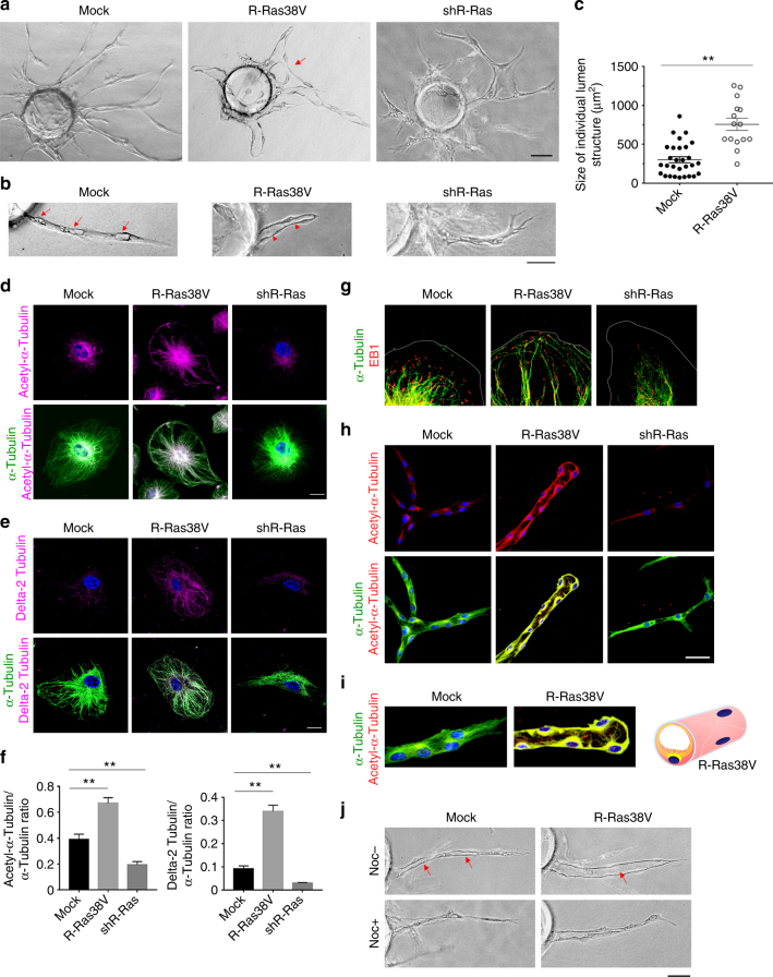
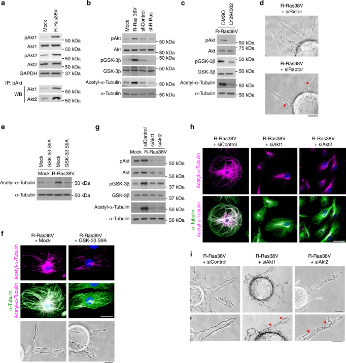
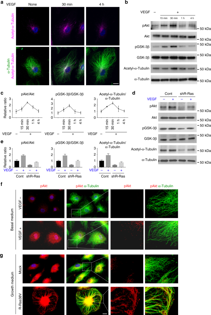
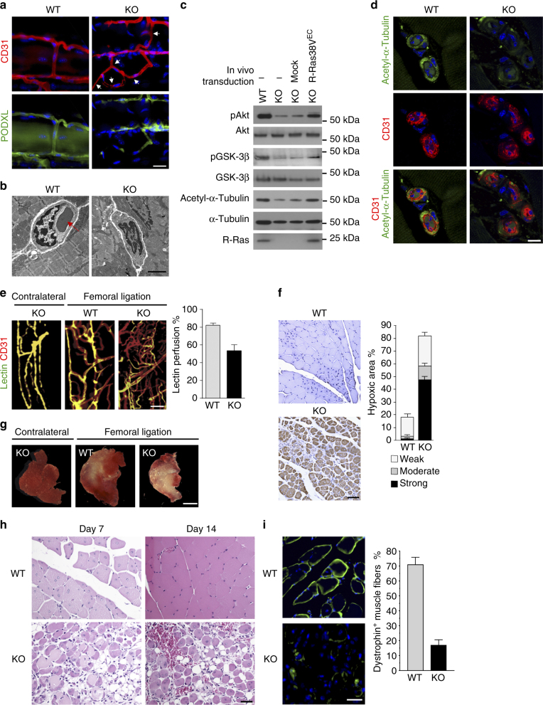
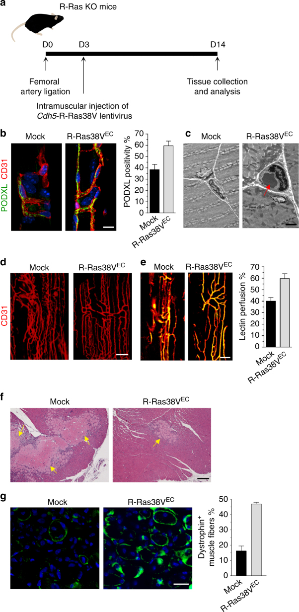
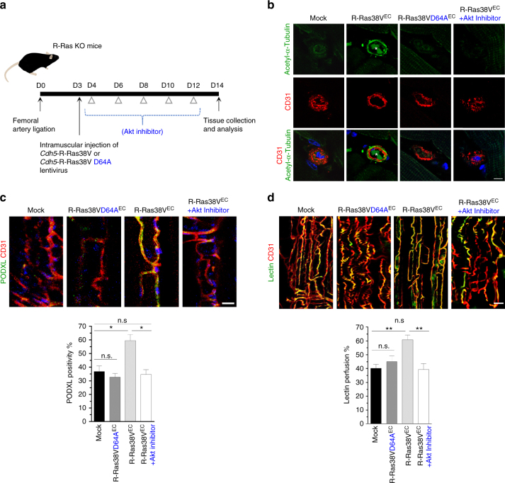
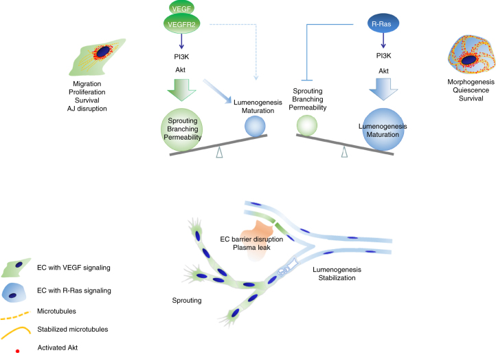
The formation of endothelial lumen is fundamental to angiogenesis and essential to the oxygenation of hypoxic tissues. The molecular mechanism underlying this important process remains obscure. Here, we show that Akt activation by a Ras homolog, R-Ras, stabilizes the microtubule cytoskeleton in endothelial cells leading to endothelial lumenogenesis. The activation of Akt by the potent angiogenic factor VEGF-A does not strongly stabilize microtubules or sufficiently promote lumen formation, hence demonstrating a distinct role for the R-Ras-Akt axis. We show in mice that this pathway is important for the lumenization of new capillaries and microvessels developing in ischemic muscles to allow sufficient tissue reperfusion after ischemic injury. Our work identifies a role for Akt in lumenogenesis and the significance of the R-Ras-Akt signaling for the patency of regenerating blood vessels.


Conflict of interest statement

The authors declare no competing financial interests.

Figures

Fig. 1
Fig. 1
R-Ras is required for microtubule stabilization and EC lumenogenesis in vitro. a Endothelial sprouts in 3-D fibrin gel culture were analyzed at day 5. Arrow, anastomosed adjacent sprouts forming a continuous lumen. b Mock-transduced control EC culture in higher magnification showed incomplete lumen formation in the sprouts (arrows). Lumen formation (arrowhead) was significantly accelerated and enhanced in R-Ras38V-transduced ECs. R-Ras knockdown by shRNA (shR-Ras) blocked tubular morphogenesis and lumen formation. c Size of individual lumen structure. The hallow structures of >5 µm length were considered as developing lumens. The area size of the individual lumen structure was determined by morphometry analysis and plotted on the graph. Sprouts grown from 10 EC-coated beads in two culture wells were analyzed for each group. Small vacuoles of <5 µm in length were not included in the analysis. Spouts without lumen were not examined. No lumen was formed by R-Ras-silenced ECs. p < 1 × 10−5. di R-Ras stabilizes endothelial microtubules. Immunofluorescence of the total (green) and acetylated α-tubulin (d) or delta 2-tubulin (e) (magenta). f The ratio of post-translationally modified α-tubulin (acetylated α-tubulin or delta 2-tubulin) to the total α-tubulin was quantified from immunofluorescence staining of the cells. **p < 0.001. g Immunofluorescence of α-tubulin and microtubule end-binding protein (EB1) to indicate the (+) ends of microtubules. Thin gray lines indicate the outlines of the cell membrane. Lower magnification images available in Supplementary Fig. 4. h In-gel staining of endothelial sprouts for total (green) and acetylated (red) α-tubulin in 3-D culture (day 7) is shown by confocal sectional images. Yellow color indicates co-staining. i Higher magnification to show the pattern of extending microtubules. Acetylated microtubule fibers are depicted by yellow lines in the schematic representation of an R-Ras38V-expressing sprout in cross-section. j Nocodazole was added at 10 µM to 5-day-old culture of mock or R-Ras38V-transduced EC sprouts. Images of the sprouts were taken before (Noc−) and 1.5 h after (Noc+) nocodazole treatment. Arrows indicate two discontinuous lumens in a control sprout and a complete uninterrupted lumen in an R-Ras38V+ sprout. Scale bars, 75 µm (a), 50 µm (b, h, j), 20 µm (d, e)
Fig. 2
Fig. 2
Akt mediates R-Ras-dependent microtubule stabilization and lumenogenesis. a R-Ras activates Akt isoforms. Ser473 phosphorylation of Akt1 and Akt2 were determined by western blot using phospho-specific antibody for each isoform as well as by immunoprecipitation of Ser473-phosphorylated Akt followed by western blot for each Akt isoform. b The effects of R-Ras on Akt Ser473 and GSK-3β Ser9 phosphorylation and α-tubulin acetylation were examined by western blot. c PI3K inhibition by LY294002 blocks the effects of R-Ras38V. d Endothelial sprouts in 3-D cultures of R-Ras38V-transduced, Rictor, or Raptor-silenced ECs. ECs were transduced with R-Ras38V followed by Rictor or Raptor knockdown using siRNA (siRictor or siRaptor) before embedded in fibrin gel. Arrows, R-Ras-induced lumenogenesis was unaffected by Raptor knockdown. e, f Constitutive activation of GSK-3β blocks R-Ras-dependent microtubule stabilization and lumenogenesis. ECs were first transduced with/without R-Ras38V and subsequently transduced with/without GSK-3β S9A. Acetylation of α-tubulin was analyzed by western blot (e) and immunofluorescence (f) and lumenogenesis examined in 3-D culture (f). gi Akt1 and Akt2 are essential to the R-Ras-mediated GSK-3β inhibition, microtubule stabilization, and lumenogenesis in vitro. Either Akt isoform was silenced in R-Ras38V-transduced ECs by siRNA (siAkt1 or siAkt2). GSK-3β phosphorylation and α-tubulin acetylation were examined by western blot (g) or western blot and immunofluorescence (g, h). The effect of Akt silencing on R-Ras-mediated EC lumenogenesis was examined in 3-D cultures (i). Arrowheads, severely disrupted lumen formation. Scale bars, 50 µm (d, f 3-D culture, h, i), 25 µm (f2-D culture)
Fig. 3
Fig. 3
VEGF and R-Ras signaling exert differential effects on Akt and microtubule. ECs were cultured in low-serum basal media (2% horse serum without growth factor supplements) for overnight and stimulated with (+) or without (−) 50 ng/ml VEGF-A for indicated time. Akt (Ser473) and GSK-3β (Ser9) phosphorylation and α-tubulin acetylation were analyzed by immunofluorescence (a) and/or western blot (b). c Levels of phosphorylated Akt, GSK-3β, and acetylated α-tubulin were quantitated by densitometry and normalized to the corresponding total protein levels. d Control or R-Ras-silenced ECs were stimulated with VEGF-A, and Akt/GSK-3β phosphorylation and α-tubulin acetylation were analyzed. e Western blots of phosphorylated Akt, GSK-3β, and acetylated α-tubulin were normalized to corresponding total protein levels. f, g Immunofluorescence of phospho-Akt (red) and total α-tubulin (green). ECs were cultured in basal media and stimulated with (+) or without (−) 50 ng/ml VEGF-A for 30 min (f). Mock or R-Ras38V-transduced ECs (g). Higher magnification of the boxed area is also shown. The graphs show the combined results of at least three independent experiments. Scale bars, 20 µm
Fig. 4
Fig. 4
Defective EC lumenogenesis and impaired muscle reperfusion in R-Ras deficiency. Hindlimb ischemia was induced in wild-type (WT) and R-Ras KO (KO) mice by left femoral artery ligation, and GC muscles were analyzed 14 days later. a Immunofluorescence of CD31 and PODXL to identify lumenized vessels. Arrows indicate lumen-less vessels formed in the ischemic GC muscles of R-Ras KO mice. b Transmission electron microscopy confirmed the absence of lumen structures in numerous R-Ras KO vessels developed in response to ischemia. Muscle were sectioned perpendicular to the muscle fibers. Arrow, a circulating erythrocyte indicates normal lumen formation in a wild-type vessel. c Analyses of Akt (Ser473) and GSK-3β (Ser9) phosphorylation and α-tubulin acetylation in ECs isolated from ischemic GC muscles at day 14. ECs were also isolated from a separate set of R-Ras KO mice, which received lentivirus injection into the GC muscles for EC-specific expression of R-Ras38V (R-Ras38VEC) via in vivo transduction. d α-Tubulin acetylation in the endothelium of intramuscular vessels. e Lectin perfusion (green) into intramuscular vessels (red) was determined in the whole-mounted GC muscle fascicles. Yellow color (green/red double-staining) indicates blood perfused vessels. Lectin perfusion % = lectin+CD31+ area/total CD31+ area × 100, p < 0.01, n = 5. f Analysis of hypoxia in GC muscles at day 7 by hypoxyprobe-1™ staining (brown). Thresholds were set empirically for identifying the area with strong, moderate, or weak staining and presented as % of total muscle area examined. p = 8 × 10−6, n = 5. g Muscle viability was assessed at day 14 by staining the slices of unfixed GC muscles with 2,3,5-triphenyltetrazolium chloride, which stains viable tissues in red. The infarct areas are unstained (pale yellow/white). h H&E staining of GC muscle sections. i Dystrophin immunostaining (green) of GC muscle cross-sections to quantify functional muscle fibers. The number of dystrophin+ muscle fibers/total muscle fibers (%) was determined in non-necrotic area. p < 10−4, n = 5. Scale bars, 25 µm (a, h), 2 µm (b), 10 µm (d), 50 µm (f), 2 mm (g)
Fig. 5
Fig. 5
RRAS gene delivery to ECs rescues vessel lumenogenesis and muscle reperfusion. a In vivo transduction and treatment schedule. The lentivirus carrying pLenti6/Cdh5-R-Ras38V expression vector was injected into GC muscles 3 days after ligation. GC muscle were analyzed at day 14. b Immunofluorescence of CD31 and PODXL to identify lumenized vessels in GC muscles after the lentivirus injection for EC-specific expression of R-Ras38V (R-Ras38VEC). PODXL positivity % (PODXL+CD31+ area/CD31+ area × 100) was determined to assess the fraction of lumenized vessel area. c Transmission electron microscopy of the GC muscles confirmed the increase in vessel lumenization upon R-Ras38VEC transduction. Arrow, a circulating erythrocyte found in the vessel lumen indicating normal lumen formation. d CD31 staining of whole-mounted GC muscle fascicles. e Analysis of vessel perfusion in whole-mounted GC muscle fascicles. Yellow color indicates lectin perfused vessels. f H&E staining of GC muscle sections. Arrows, necrotic areas. g Dystrophin immunostaining (green) of GC muscle cross-sections to quantify functional muscle fibers. The number of dystrophin+ muscle fibers/total muscle fibers (%) was determined in non-necrotic area. p < 10−4, n = 5. Scale bars, 25 µm (b), 3 µm (c), 100 µm (d), 50 µm (e), 150 µm (f)
Fig. 6
Fig. 6
R-Ras promotes formation of lumenized functional blood vessels via Akt. a In vivo transduction and treatment schedule. One day after lentivirus injection into GC muscles, R-Ras KO mice were treated with or without Akt inhibitor MK2206 by gavage every 2 days. GC muscle were analyzed at day 14. b Immunofluorescence of GC muscle cross-sections to examine α-tubulin acetylation in the endothelium of intramuscular vessels. *Blood cells in the lumen. c CD31 and PODXL immunofluorescence of the GC muscles that received lentivirus injection for EC-specific R-Ras38V D64A or R-Ras38V expression and subsequently treated with Akt inhibitor MK2206. *p < 0.01, n.s., not significant. d Analysis of vessel perfusion in whole-mounted GC muscle fascicles. Yellow color indicates lectin perfused vessels. **p < 0.02. Scale bars, 10 µm (b), 25 µm (c), 50 µm (d)
Fig. 7
Fig. 7
Distinct roles of VEGF vs. R-Ras-mediated Akt signaling in angiogenesis. VEGF and R-Ras signaling activate Akt in different manners. Akt accumulates in perinuclear region upon activation by VEGF. In contrast, R-Ras-dependent Akt activation results in the accumulation of activated Akt along the microtubule cytoskeleton as well as at the perinuclear region. The effect of the VEGF-Akt signaling is largely skewed toward vessel sprouting and permeability induction, and it is insufficient for driving vessel lumenization by itself (green). The R-Ras-Akt signaling, on the other hand, promotes lumenogenesis and supports the lumen structure with the stable cytoskeletal architecture of microtubules (blue). R-Ras also limits excessive, nonproductive endothelial sprouting, and block the VEGF-induced disruption of adherens junction (AJ) to maintain endothelial barrier integrity (Sawada et al.). Thus, the VEGF and R-Ras pathways are complementary to each other to generate functional blood vessels for tissue recovery from ischemia

References

    1. Herbert SP, Stainier DY. Molecular control of endothelial cell behaviour during blood vessel morphogenesis. Nat. Rev. Mol. Cell Biol. 2011;12:551–564. doi: 10.1038/nrm3176. - DOI - PMC - PubMed
    1. Nagy JA, Senger DR. VEGF-A, cytoskeletal dynamics, and the pathological vascular phenotype. Exp. Cell Res. 2006;312:538–548. doi: 10.1016/j.yexcr.2005.10.017. - DOI - PubMed
    1. Sigurbjornsdottir S, Mathew R, Leptin M. Molecular mechanisms of de novo lumen formation. Nat. Rev. Mol. Cell Biol. 2014;15:665–676. doi: 10.1038/nrm3871. - DOI - PubMed
    1. Kim DJ, Martinez-Lemus LA, Davis GE. EB1, p150Glued, and Clasp1 control endothelial tubulogenesis through microtubule assembly, acetylation, and apical polarization. Blood. 2013;121:3521–3530. doi: 10.1182/blood-2012-11-470179. - DOI - PMC - PubMed
    1. Janke C, Bulinski JC. Post-translational regulation of the microtubule cytoskeleton: mechanisms and functions. Nat. Rev. Mol. Cell Biol. 2011;12:773–786. doi: 10.1038/nrm3227. - DOI - PubMed

Publication types

MeSH terms

Substances