. 2017 Nov 23;7(1):16128.
doi: 10.1038/s41598-017-15867-8.

Chemokines protect vascular smooth muscle cells from cell death induced by cyclic mechanical stretch

Affiliations

Chemokines protect vascular smooth muscle cells from cell death induced by cyclic mechanical stretch

Jing Zhao et al. Sci Rep. .

Abstract

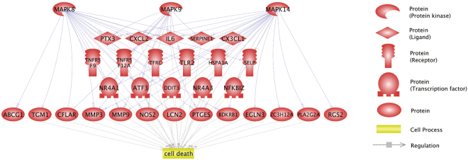
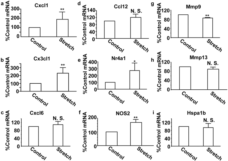
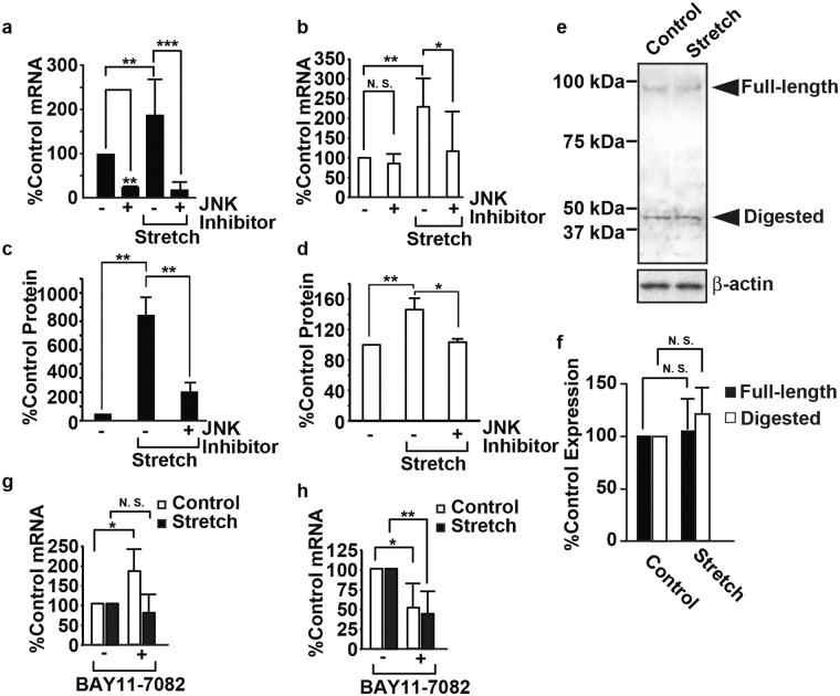
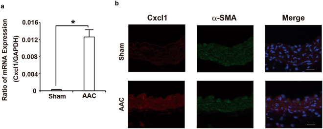
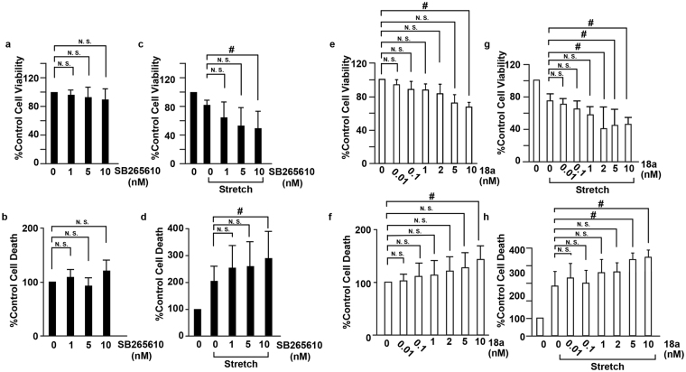
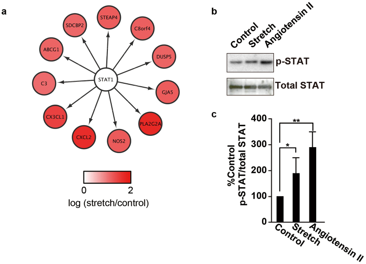
The pulsatile nature of blood flow exposes vascular smooth muscle cells (VSMCs) in the vessel wall to cyclic mechanical stretch (CMS), which evokes VSMC proliferation, cell death, phenotypic switching, and migration, leading to vascular remodeling. These responses have been observed in many cardiovascular diseases; however, the underlying mechanisms remain unclear. We have revealed that CMS of rat aortic smooth muscle cells (RASMCs) causes JNK- and p38-dependent cell death and that a calcium channel blocker and angiotensin II receptor antagonist decreased the phosphorylation of JNK and p38 and subsequently decreased cell death by CMS. In the present study, we showed that the expression of Cxcl1 and Cx3cl1 was induced by CMS in a JNK-dependent manner. The expression of Cxcl1 was also induced in VSMCs by hypertension produced by abdominal aortic constriction (AAC). In addition, antagonists against the receptors for CXCL1 and CX3CL1 increased cell death, indicating that CXCL1 and CX3CL1 protect RASMCs from CMS-induced cell death. We also revealed that STAT1 is activated in RASMCs subjected to CMS. Taken together, these results indicate that CMS of VSMCs induces inflammation-related gene expression, including that of CXCL1 and CX3CL1, which may play important roles in the stress response against CMS caused by hypertension.


Conflict of interest statement

The authors declare that they have no competing interests.

Figures

Figure 1
Figure 1
Molecular networks associated with JNK/p38 MAP kinase and cell death. The 91 genes dysregulated in RASMCs treated with cyclic mechanical stretch were subjected to bioinformatics analyses to identify genes that are related to both JNK and p38 MAP kinases and cell death. The regulations among the MAP kinases, dysregulated genes, and cell death are shown in the network.
Figure 2
Figure 2
Expression of transcripts of candidate genes in RASMCs subjected to CMS. RASMCs were subjected to CMS for four hours, and expressions of transcripts of Cxcl1 (a), Cx3cl1 (b), Cxcl6 (c), Ccl12 (d), Nr4a1 (e), NOS2 (f), Mmp9 (g), Mmp13 (h), and Hspa1b (i) were evaluated using the real-time RT-PCR method. The quantity of the transcripts is expressed as a percentage of the control, normalized with respect to GAPDH. Data are means ± SE (n = 4); *p < 0.05 and **p < 0.01 versus control, and N.S. indicates no significant difference.
Figure 3
Figure 3
Induction of CXCL1 and CX3CL1 in RASMCs subjected to CMS. RASMCs were incubated with SP600125 (20 μM) for 20 min and then subjected to CMS for four hours. Cells were harvested and analyzed by real-time RT-PCR with specific primers for Cxcl1 (a) or Cx3cl1 (b) or by immunoblotting using the anti-CX3CL1 antibody (e), and media were analyzed using ELISA for CXCL1 (c) or CX3CL1 (d). (f) The quantity of CX3CL1 expressed in RASMCs, as measured by scanning densitometry, is expressed as a percentage of the control, normalized with respect to beta-actin. Data are means ± SE (n = 3), and N.S. indicates no significant difference. RASMCs were incubated with BAY 11-7082 (5 µM) for 20 min and then subjected to CMS for four hours. Cells were harvested and analyzed by real-time RT-PCR with specific primers for Cxcl1 (g) or Cx3cl1 (h). The uncropped pictures of immuoblotting are shown in Supplemental Fig. S4 (a,b).
Figure 4
Figure 4
AAC-induced CXCL1 expression in the aorta. (a) Real-time RT-PCR analysis of Cxcl1 gene expression in the aorta of sham and AAC mice 6 h post-operation. (b) Double-color immunofluorescence images of CXCL1 (red) and a-SMA (green) in the aortas of sham and AAC mice 6 h post-operation. Representative results from four individual animals in each group are shown. Original magnification, ×400 (scale: 25 mm). All values represent means ± SEM (n4–6). *p < 0.05, AAC versus sham.
Figure 5
Figure 5
Inhibition of CXCL1 and CX3CL1 accelerated cell death induced by CMS. RASMCs were incubated with SB265610 (ad) or 18a (eh) at the indicated concentrations and then incubated under normal conditions (a,b,e,f) or subjected to CMS for four hours (c,d,g,h). After CMS, the cells were incubated for 24 h, and cell viability and cell death were evaluated by the MTT assay (a,c,e,g) and the release of LDH (b,d,f,h), respectively. Cell viability and cell death are expressed as a percentage of the control. Data are means ± SE (n = 6); *p < 0.05 versus control, and N.S. indicates no significant difference; ANOVA, Tukey’s HSD test.
Figure 6
Figure 6
Phosphorylation of Stat1 is induced by mechanical stretch in RASMCs. (a) The transcriptome analysis identified STAT1 as the key transcription factor regulating the expression of genes induced by CMS in RASMCs. (b) RASMCs were incubated under normal conditions, subjected to CMS for four hours or to 10 nM angiotensin II for 5 min (eh), and then harvested. Cell lysates were analyzed by immunoblotting using anti-phosphorylated (upper panel) and total STAT (lower panel) antibodies. (c) The quantity of phosphorylated STAT is expressed as a percentage of the control, normalized with respect to total STAT. Data are means ± SE (n = 3); *p < 0.05 and **p < 0.01 versus the control. The uncropped pictures of immuoblotting are shown in Supplemental Fig. S4 (c,d).

References

    1. Roberts WC. The hypertensive diseases. Evidence that systemic hypertension is a greater risk factor to the development of other cardiovascular diseases than previously suspected. Am. J. Med. 1975;59:523–32. doi: 10.1016/0002-9343(75)90260-0. - DOI - PubMed
    1. Intengan HD, Schiffrin EL. Vascular remodeling in hypertension: roles of apoptosis, inflammation, and fibrosis. Hypertension. 2001;38:581–7. doi: 10.1161/hy09t1.096249. - DOI - PubMed
    1. Sharifi AM, Schiffrin EL. Apoptosis in aorta of deoxycorticosterone acetate-salt hypertensive rats: effect of endothelin receptor antagonism. J Hypertens. 1997;15:1441–8. doi: 10.1097/00004872-199715120-00011. - DOI - PubMed
    1. Walsh KI, Smith RC, Kim HS. Vascular cell apoptosis in remodeling, restenosis, and plaque rupture. Circ. Res. 2000;87:184–8. doi: 10.1161/01.RES.87.3.184. - DOI - PubMed
    1. Díez J, et al. Altered regulation of smooth muscle cell proliferation and apoptosis in small arteries of spontaneously hypertensive rats. Eur. Heart J. 1998;19:G29–33. - PubMed

Publication types

MeSH terms