. 2017 Nov 9:8:812.
doi: 10.3389/fphar.2017.00812. eCollection 2017.

Shedding of Microvesicles from Microglia Contributes to the Effects Induced by Metabotropic Glutamate Receptor 5 Activation on Neuronal Death

Affiliations

Shedding of Microvesicles from Microglia Contributes to the Effects Induced by Metabotropic Glutamate Receptor 5 Activation on Neuronal Death

Martina Beneventano et al. Front Pharmacol. .

Abstract

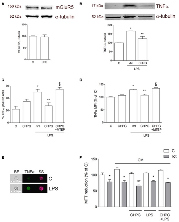
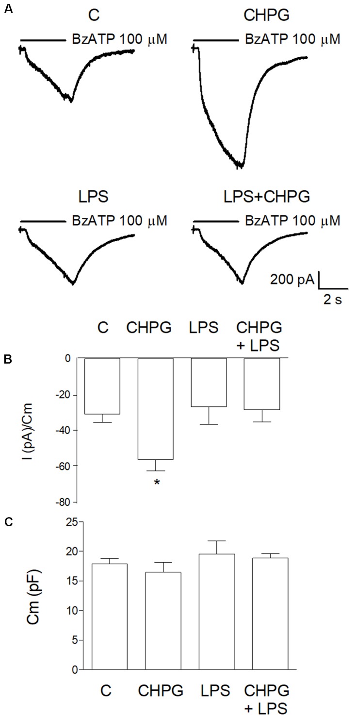
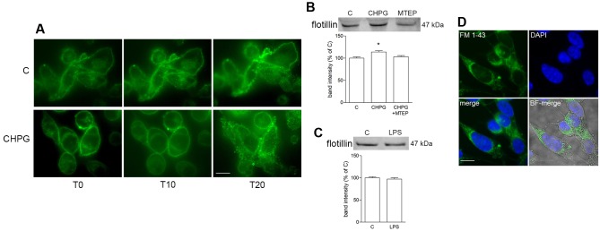
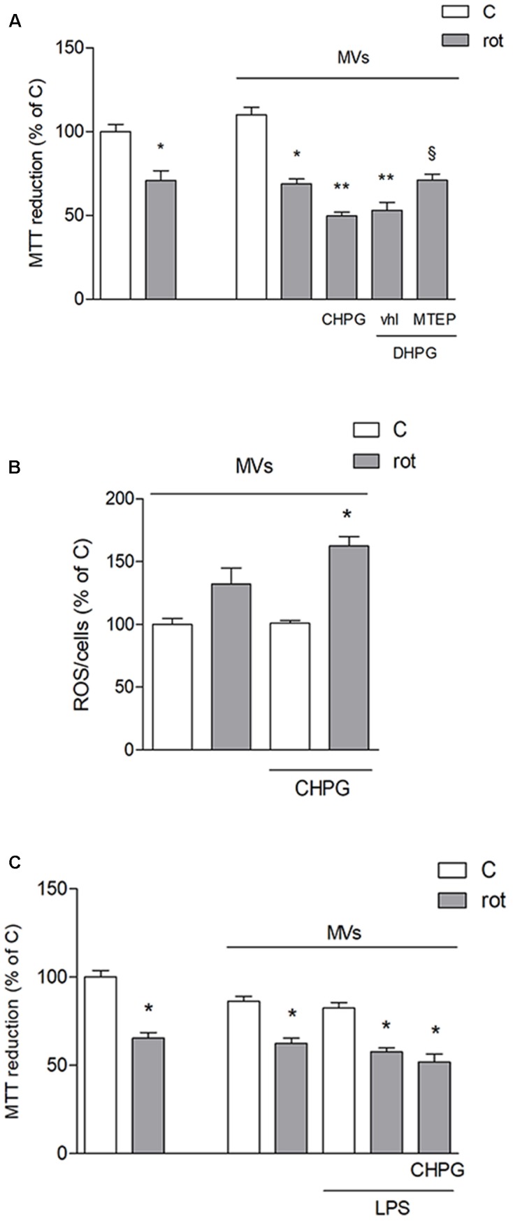
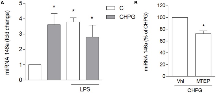
Metabotropic glutamate (mGlu) receptor 5 is involved in neuroinflammation and has been shown to mediate reduced inflammation and neurotoxicity and to modify microglia polarization. On the other hand, blockade of mGlu5 receptor results in inhibition of microglia activation. To dissect this controversy, we investigated whether microvesicles (MVs) released from microglia BV2 cells could contribute to the communication between microglia and neurons and whether this interaction was modulated by mGlu5 receptor. Activation of purinergic ionotropic P2X7 receptor with the stable ATP analog benzoyl-ATP (100 μM) caused rapid MVs shedding from BV2 cells. Ionic currents through P2X7 receptor increased in BV2 cells pretreated for 24 h with the mGlu5 receptor agonist CHPG (200 μM) as by patch-clamp recording. This increase was blunted when microglia cells were activated by exposure to lipopolysaccharide (LPS; 0.1 μg/ml for 6 h). Accordingly, a greater amount of MVs formed after CHPG treatment, an effect prevented by the mGlu5 receptor antagonist MTEP (100 μM), as measured by expression of flotillin, a membrane protein enriched in MVs. Transferred MVs were internalized by SH-SY5Y neurons where they did not modify neuronal death induced by a low concentration of rotenone (0.1 μM for 24 h), but significantly increased rotenone neurotoxicity when shed from CHPG-treated BV2 cells. miR146a was increased in CHPG-treated MVs, an effect concealed in MVs from LPS-activated BV2 cells that showed per se an increase in miRNA146a levels. The present data support a role for microglia-shed MVs in mGlu5-mediated modulation of neuronal death and identify miRNAs as potential critical mediators of this interaction.

Keywords: CHPG; extracellular vesicle; mGlu5; miRNA; microglia; neuroinflammation.


Figures

FIGURE 1
FIGURE 1
Metabotropic glutamate (mGlu5) receptor mediates anti-inflammatory effect on BV2 microglial cells. Western blot analysis of expression of mGlu5 receptor in BV2 cells treated with LPS (0.1 μg/ml) for 24 h (A). BV2 cells were activated with LPS (0.1 μg/ml for 6 h) after pretreatment with CHPG (200 μM for 24 h) and the selective mGlu5 receptor antagonist MTEP (100 μM added 30 min before CHPG). TNFα expression was evaluated by western blot (B) and flow cytometry (C,D). More specifically, assessment of the percentage of TNFα-positive cells (C) and the level of fluorescence expressed as mean fluorescent intensity (MFI; D) are shown. A representative image acquired by imaging flow cytometry showing cells in their shape and size (brightfield; BF), complexity (side scatter; SS), and TNFα immunostaining (green) is also reported (E). MTT assay on SH-SY5Y cells exposed to rotenone (100 nM for 24 h) in the presence of conditioned medium (CM) from BV2 cells activated with lipopolysaccharide (LPS; 0.1 μg/ml for 6 h) after pretreatment with CHPG (200 μM for 24 h) (F). Student’s t-test (A) and one-way ANOVA followed by Newman–Keuls test for significance (B–E) were applied. p < 0.05 vs. C; ∗∗p < 0.05 vs. LPS (Vhl); xp < 0.05 vs. CHPG+LPS; °p < 0.05 vs. CM rot.
FIGURE 2
FIGURE 2
CHPG treatment increases purinergic current through P2X7 receptors in basal but not LPS-activated conditions. In (A), representative traces of purinergic current recorded in BV2 cells in the following conditions: basal (C), after 24 h treatment with CHPG 200 μM, after 6 h treatment with LPS 0.1 μg/ml and after co-treatment of CHPG for 24 h and LPS added during the last 6 h. Currents were elicited after stimulation of P2X7 receptors with the selective agonist Bz-ATP, 100 μM. Summary of purinergic current elicited from cells stimulated as in (A) are reported in (B). Data are presented as current (pA) obtained by each cell and normalized to membrane capacitance (Cm), an estimation of cellular surface area. Bars are mean ± SEM (n = 11 for C; n = 8 for CHPG; n = 9 for LPS; and LPS + CHPG), p < 0.05 by one-way ANOVA followed by Newman–Keuls post hoc test. Summary of Cm measured for each cell in different conditions (C). Bars are mean ± SEM of corresponding cells used for bar diagram in (B).
FIGURE 3
FIGURE 3
Metabotropic glutamate 5-receptor activation increases microvesicles (MVs) release from BV2 microglial cells. BV2 cells were treated with DHPG (100 μM for 24 h) and labeled with the fluorescent dye FM1-43 (10 μM for 5 min). MVs formation following addition of Bz-ATP (100 μM) was monitored with time-lapse microscopy up to 20 min. Representative images are shown in (A). Western blot of flotillin of equal volume of MVs’ protein extracts from BV2 cells treated with CHPG (200 μM for 24 h) and MTEP (100 μM for 24 h; B), or with LPS (0.1 μg/ml for 6 h; C). Representative images of MVs shed from FM1-43-stained BV2 cells, transferred on top of SH-SY5Y cultures (D). Nuclei were stained with DAPI. Magnification = 40×(A), 63×(D). Scale bars = 20 μm (A) and 10 μm (D). p < 0.05 vs. control. One-way ANOVA followed by Newman–Keuls test for significance (B) and Student’s t-test (C) were applied to detect statistically significant differences.
FIGURE 4
FIGURE 4
Microvesicles shed from CHPG-treated microglia potentiate rotenone-induced neurotoxicity. In (A), MTT assay on SH-SY5Y cells exposed to rotenone (100 nM for 24 h) in the presence of MVs derived from BV2 cells treated with CHPG (200 μM for 24 h), DHPG (100 μM for 24 h), and MTEP (100 μM for 24 h) and transferred to SH-SY5Y cells after rotenone (100 nM for 24 h) exposure. Determination of ROS levels in SH-SY5Y cells exposed to rotenone (100 nM for 24 h) in the presence of MVs derived from CHPG-treated BV2 cells (B). MTT assay on SH-SY5Y cells (100 nM for 24 h) in the presence of MVs from LPS-activated BV2 cells (0.1 μg/ml for 6 h) pretreated with CHPG (200 μM for 24 h; C). p < 0.05 vs. control; ∗∗p < 0.05 vs. rot, xp < 0.05 vs. DHPG vhl. One-way ANOVA followed by Newman–Keuls test for significance was applied.
FIGURE 5
FIGURE 5
CHPG increases miRNA146a levels in BV2 cells-shed MVs. BV2 cells were activated with LPS (0.1 μg/ml for 6 h) after pretreatment with CHPG (200 μM, total exposure 24 h). MVs were isolated, miRNA extracted and levels of miRNA146a were analyzed by real time PCR (A). In (B), levels of miRNA146a were analyzed in MVs derived from MPTEP pretreated-cells (100 μM added 30 min before CHPG). Results are expressed as percentage of CHPG. One-way ANOVA followed by Newman–Keuls test (A) and Student’s t-test (B) were applied for significance. p < 0.05 vs. control (A); p < 0.05 vs. CHPG (B).

References

    1. Antonucci F., Turola E., Riganti L., Caleo M., Gabrielli M., Perrotta C., et al. (2012). Microvesicles released from microglia stimulate synaptic activity via enhanced sphingolipid metabolism. EMBO J. 31 1231–1240. 10.1038/emboj.2011.489 - DOI - PMC - PubMed
    1. Aronica E., van Vliet E. A., Mayboroda O. A., Troost D., da Silva F. H., Gorter J. A. (2000). Upregulation of metabotropic glutamate receptor subtype mGluR3 and mGluR5 in reactive astrocytes in a rat model of mesial temporal lobe epilepsy. Eur. J. Neurosci. 12 2333–2344. 10.1046/j.1460-9568.2000.00131.x - DOI - PubMed
    1. Awad H., Hubert G. W., Smith Y., Levey A. I., Conn P. J. (2000). Activation of metabotropic glutamate receptor 5 has direct excitatory effects and potentiates NMDA receptor currents in neurons of the subthalamic nucleus. J. Neurosci. 20 7871–7879. - PMC - PubMed
    1. Berthele A., Boxall S. J., Urban A., Anneser J. M., Zieglgansberger W., Urban L., et al. (1999). Distribution and developmental changes in metabotropic glutamate receptor messenger RNA expression in the rat lumbar spinal cord. Brain Res. Dev. Brain Res. 112 39–53. 10.1016/S0165-3806(98)00156-4 - DOI - PubMed
    1. Bianco F., Pravettoni E., Colombo A., Schenk U., Moller T., Matteoli M., et al. (2005). Astrocyte-derived ATP induces vesicle shedding and IL-1 beta release from microglia. J. Immunol. 174 7268–7277. 10.4049/jimmunol.174.11.7268 - DOI - PubMed

LinkOut - more resources