Review
. 2018 Mar;12(1):319-331.
doi: 10.1007/s12079-017-0429-z. Epub 2017 Nov 23.

The structure of the S-layer of Clostridium difficile

Affiliations
Review

The structure of the S-layer of Clostridium difficile

William J Bradshaw et al. J Cell Commun Signal. 2018 Mar.

Abstract

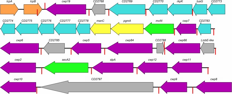
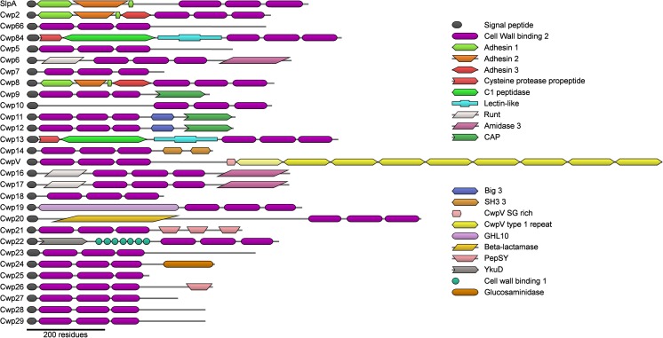
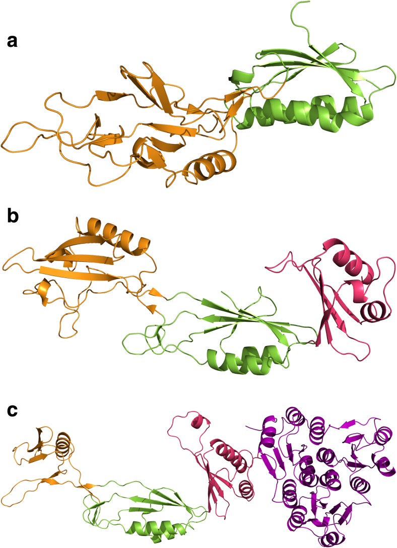
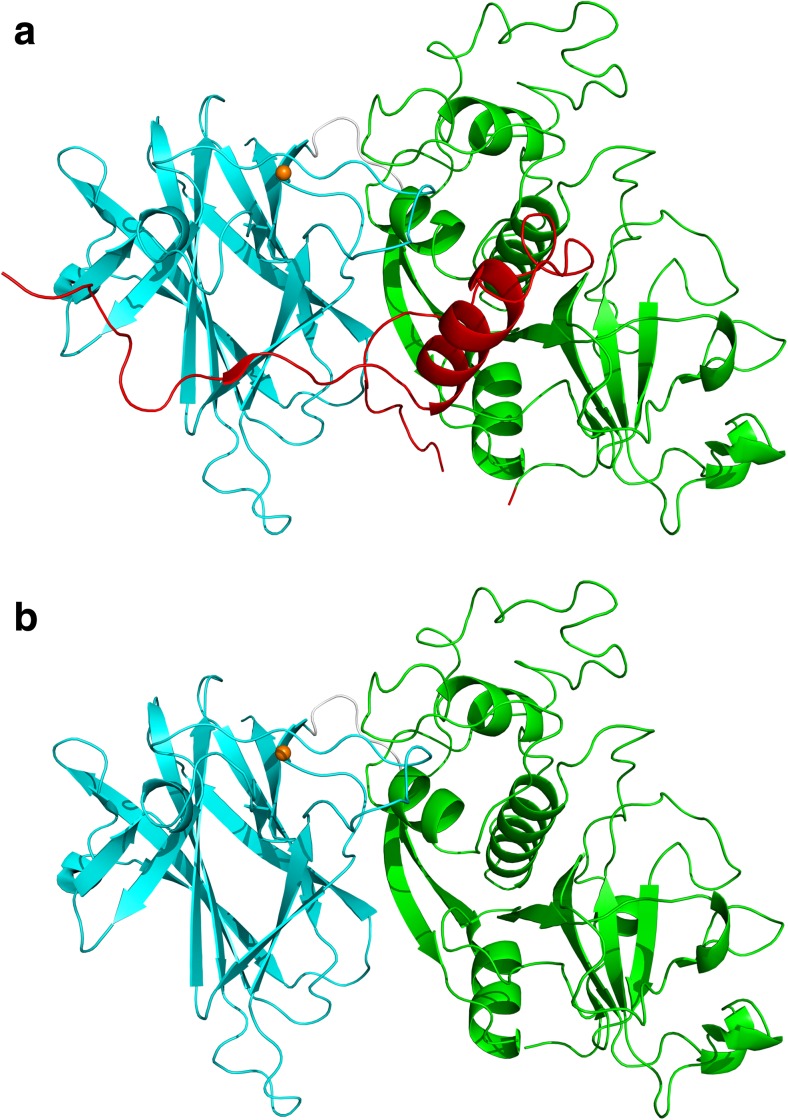
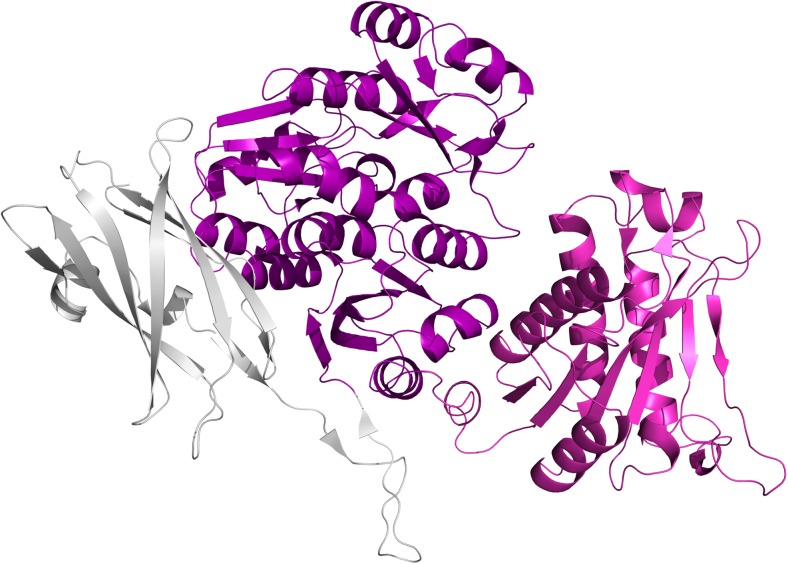
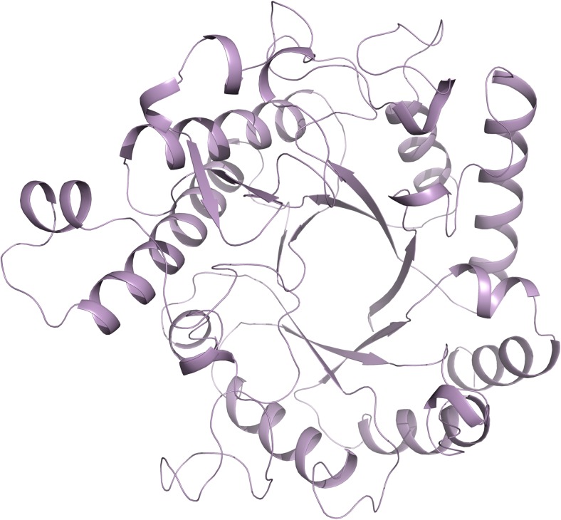
The nosocomially acquired pathogen Clostridium difficile is the primary causative agent of antibiotic associated diarrhoea and causes tens of thousands of deaths globally each year. C. difficile presents a paracrystalline protein array on the surface of the cell known as an S-layer. S-layers have been demonstrated to possess a wide range of important functions, which, combined with their inherent accessibility, makes them a promising drug target. The unusually complex S-layer of C. difficile is primarily comprised of the high- and low- molecular weight S-layer proteins, HMW SLP and LMW SLP, formed from the cleavage of the S-layer precursor protein, SlpA, but may also contain up to 28 SlpA paralogues. A model of how the S-layer functions as a whole is required if it is to be exploited in fighting the bacterium. Here, we provide a summary of what is known about the S-layer of C. difficile and each of the paralogues and, considering some of the domains present, suggest potential roles for them.

Keywords: Bacterial adhesion; C. difficile Infection; Cell wall protein; Clostridium Difficile; Colitis; S-layer.


Conflict of interest statement

The authors declare that they have no conflict of interest.

Figures

Fig. 1
Fig. 1
Schematic diagram of the S-layer of C. difficile. The lipid bilayer is shown in blue with the peptidoglycan in peach. Above this is a purple layer formed by the three cell wall binding domains of HMW SLP and paralogues. The surface exposed “functional” regions are shown on top, the majority of which are LMW SLP, shown in green. The S-layer also contains other proteins with a range of functions
Fig. 2
Fig. 2
The AP and slpA loci. The two adjacent loci, which respectively code for proteins involved in the production of PSII and proteins that attach to PSII, are shown. Genes coding for proteins with CWB2 domains are shown in purple, those involved in polysaccharide metabolism in cyan, attachment to peptidoglycan in peach, mannose biosynthesis in yellow and biopolymer export in green, other functions are in grey. CD2768 – hydrolase, CD2785 – membrane protein, CD2788 – GtrA-like membrane protein, CD2797 – calcium binding adhesin. Terminators predicted by Genome2D (Baerends et al. 2004) are shown in red
Fig. 3
Fig. 3
Putative domain representation of the 29 cwp genes found in the Clostridium difficile 630 genome. Each codes for three cell wall binding domains, while all except cwp18, cwp25, and potentially cwp7 appear to code for at least one other domain, which is likely to confer a specific function on the protein. Generated using DoMosaics with HMMER (Eddy ; Moore et al. 2014)
Fig. 4
Fig. 4
Adhesin structures. a LMW SLP: 3CVZ (Fagan et al. 2009). b Cwp2: 5NJL (Bradshaw et al. 2017a). c Cwp8: 5J7Q (Usenik et al. 2017). Cwp2 and Cwp8 assume similar folds with domain 2 rotated approximately 40°. Domain 2 of LMW SLP has significantly longer loop regions and is positioned differently to that of Cwp2 and Cwp8. LMW SLP is covalently bound to HMW SLP so it is likely that domain 3 of LMW SLP is at least somewhat different to that of Cwp2 and Cwp8. Domain colours follow those given in Fig. 3
Fig. 5
Fig. 5
The structure of Cwp84. a With the propeptide: 4CI7 (Bradshaw et al. 2014), b Without the propeptide: 4D59, 4D5A (Bradshaw et al. 2015). Cwp84 possesses a cysteine protease domain with a cathepsin L-like fold and a “lectin-like” domain of currently unknown function that closely interacts with the cysteine protease domain forming part of the active site groove. Domain colours follow those given in Fig. 3
Fig. 6
Fig. 6
The structure of Cwp6. 5J72 (Usenik et al. 2017). Cwp6 possesses central cell wall binding domains flanked by a Runt domain and an amidase 3 domain. The function of the usually metazoan runt domain is currently unknown. Domain colours follow those given in Fig. 3
Fig. 7
Fig. 7
The structure of Cwp19. 5OQ2, 5OQ3 (Bradshaw et al. 2017b). The GHL10 domain of Cwp19 has a typical TIM barrel fold formed by eight β-strands surrounded by eight α-helices. The active site is located centrally over the barrel

References

    1. Abraham EP, Chain E. An enzyme from bacteria able to destroy penicillin. Nature. 1940;146:873. doi: 10.1038/146837a0. - DOI - PubMed
    1. Altschul SF, Gish W, Miller W, Myers EW, Lipman DJ. Basic local alignment search tool. J Mol Biol. 1990;215(3):403–410. doi: 10.1016/S0022-2836(05)80360-2. - DOI - PubMed
    1. Anjuwon-Foster BR, Tamayo R. A genetic switch controls the production of flagella and toxins in Clostridium difficile. PLoS Genet. 2017;13(3):e1006701. doi: 10.1371/journal.pgen.1006701. - DOI - PMC - PubMed
    1. Baerends RJ, Smits WK, de Jong A, Hamoen LW, Kok J, Kuipers OP (2004) Genome2D: a visualization tool for the rapid analysis of bacterial transcriptome data. Genome Biol 5(5):R37 - PMC - PubMed
    1. Barkin JA, Sussman DA, Fifadara N, Barkin JS. Clostridium difficile infection and patient-specific antimicrobial resistance testing reveals a high metronidazole resistance rate. Dig Dis Sci. 2017;62(4):1035–1042. doi: 10.1007/s10620-017-4462-9. - DOI - PubMed