. 2017 Dec;82(6):951-960.
doi: 10.1002/ana.25104. Epub 2017 Dec 7.

Targeting hypersensitive corticostriatal terminals in restless legs syndrome

Affiliations

Targeting hypersensitive corticostriatal terminals in restless legs syndrome

Gabriel Yepes et al. Ann Neurol. 2017 Dec.

Abstract

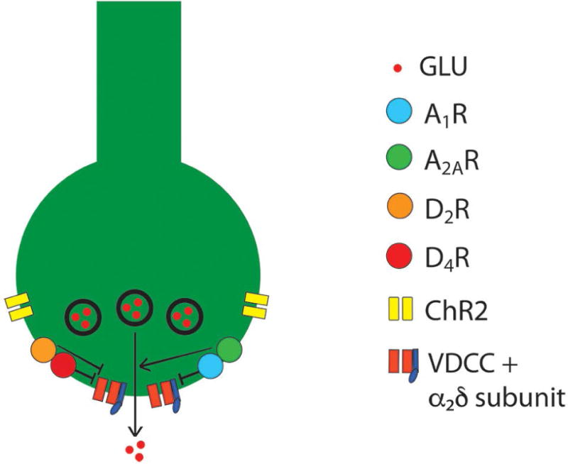
Objective: The first aim was to demonstrate a previously hypothesized increased sensitivity of corticostriatal glutamatergic terminals in the rodent with brain iron deficiency (BID), a pathogenetic model of restless legs syndrome (RLS). The second aim was to determine whether these putative hypersensitive terminals could constitute a significant target for drugs effective in RLS, including dopamine agonists (pramipexole and ropinirole) and α2 δ ligands (gabapentin).

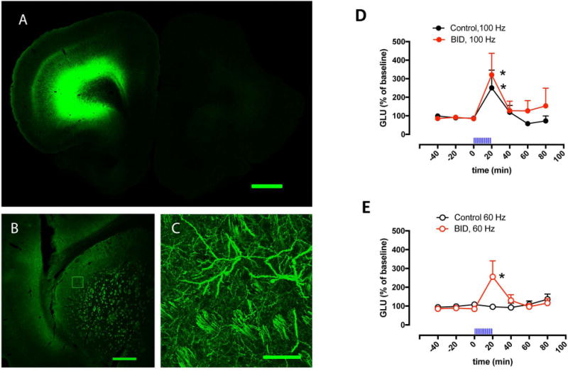
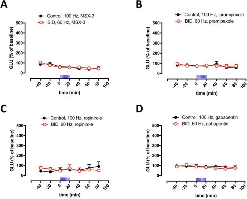
Methods: A recently introduced in vivo optogenetic-microdialysis approach was used, which allows the measurement of the extracellular concentration of glutamate upon local light-induced stimulation of corticostriatal glutamatergic terminals. The method also allows analysis of the effect of local perfusion of compounds within the same area being sampled for glutamate.

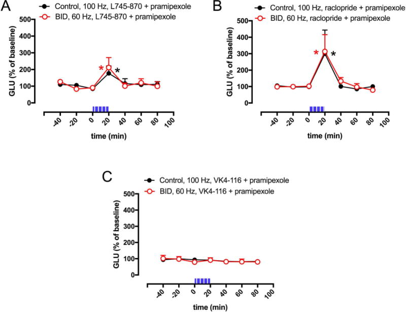
Results: BID rats showed hypersensitivity of corticostriatal glutamatergic terminals (lower frequency of optogenetic stimulation to induce glutamate release). Both hypersensitive and control glutamatergic terminals were significant targets for locally perfused pramipexole, ropinirole, and gabapentin, which significantly counteracted optogenetically induced glutamate release. The use of selective antagonists demonstrated the involvement of dopamine D4 and D2 receptor subtypes in the effects of pramipexole.

Interpretation: Hypersensitivity of corticostriatal glutamatergic terminals can constitute a main pathogenetic mechanism of RLS symptoms. Selective D4 receptor agonists, by specifically targeting these terminals, should provide a new efficient treatment with fewer secondary effects. Ann Neurol 2017;82:951-960.


Conflict of interest statement

Potential Conflict of Interests

The authors declare no potential conflict of interests

Figures

Figure 1
Figure 1. Effect of BID on optogenetically-induced corticostriatal glutamate release
(A–C) Confocal laser microscopy of coronal brain sections showing the localization of ChR2-EYFP after unilateral AA microinjection in the agranular motor cortex. (A) Unilateral expression of ChR2-EYFP in the agranular motor cortex; coronal section at 3.0 mm anterior from bregma; scale bar, 1 mm. (B) Expression of ChR2-EYFP in the ipsilateral lateral striatum; coronal section at 0 mm anterior from bregma; scale bar, 0.5 mm. (C) Superposition of 5 adjacent confocal planes (5 μm-think planes of a 25 μm-think section) from the framed field in (B), showing corticostriatal terminals; scale bar, 0.05 mm. (D,E) Effect of local optogenetic stimulation at high-frequency (100 Hz, D) and low-frequency (60 Hz, E) on the extracellular levels of glutamate in the lateral striatum of BID rats (red plot) and controls (black plot); time ‘0’ represents the values of samples prior to stimulation; the period of stimulation (20 min) is represented as a train of vertical lines; results are expressed as means + S.E.M. of percentage of the average of three values before stimulation (n = 9–12 per group). *: p<0.05, as compared to value of the last sample before the stimulation, respectively (paired t test).
Figure 2
Figure 2. Inhibition of optogenetically-induced corticostriatal glutamate release by MSX-3, pramipexole, ropinirole and gabapentin
(A–D) Effect of perfusion of the adenosine A2AR antagonist MSX-3, the dopamine receptor agonists pramipexole and ropinirole and the α2δ ligand gabapentin (1 μM in all cases) on optogenetically-induced glutamate release in the lateral striatum of BID rats (red plot) and controls (black plot); time ‘0’ represents the values of samples prior to stimulation; the period of stimulation (20 min) is represented as a train of vertical lines; results are expressed as means + S.E.M. of percentage of the average of three values before stimulation (n = 6–9 per group). *: p<0.05, as compared to value of the last sample before the stimulation, respectively (paired t test).
Figure 3
Figure 3. Effect D2-like receptor antagonists on pramipexole-mediated inhibition of optogenetically-induced corticostriatal glutamate release
(A–C) Effect of co-perfusion of pramipexole (1 μM) with the selective D4R antagonist L745–870, the non-selective D2R-D3R antagonist raclopride or the D3R antagonist VK4–116 (10 μM in all cases) on optogenetically-induced glutamate release in the lateral striatum of BID rats (red plot) and controls (black plot); time ‘0’ represents the values of samples prior to stimulation; the period of stimulation (20 min) is represented as a train of vertical lines; results are expressed as means + S.E.M. of percentage of the average of three values before stimulation (n = 7–9 per group). *: p<0.05, as compared to value of the last sample before the stimulation, respectively (paired t test).
Figure 4
Figure 4. Schematic representation of a corticostriatal glutamatergic terminal and their modulatory dopamine and adenosine receptors
Dopamine and adenosine modulate corticostriatal glutamate release by acting on A1R-A2AR and D2R-D4R heteromers. The A1R-A2AR heteromer act as an adenosine concentration-dependent switch, by which a low adenosine concentration activates preferentially A1R, which produces inhibition of glutamate release, and a high adenosine concentration also activates A2AR, which shuts down A1R signaling and promotes and A2AR-mediated stimulation of glutamate release. The D2R-D4R heteromer provides a dopamine concentration-dependent stepwise inhibitory mechanism of glutamate release, that depends on the higher affinity of dopamine for the D4R and on a D4R-mediated-increase of D2R signaling. The BID-dependent increased in the excitability of the glutamatergic terminal to release glutamate seems to depend on functional downregulation of A1R and upregulation of A2AR, which can be counteracted by A2AR antagonists D2R or D4R agonists and α2δ ligands (see text). The function of voltage-dependent calcium channels (VDCC), which activation promotes vesicular fusion and neurotransmitter release, is regulated by Gi-coupled receptors (ßγ-mediated inhibition), including A1R, D2R and D4R, as well as by accessory α2δ subunits, the targets of gabapentin-like compounds. ChR2: channelrhodopsin 2 (see text).

Similar articles

Cited by

References

    1. Allen RP, Walters AS, Montplaisir J, et al. Restless legs syndrome prevalence and impact: REST general population study. Arch Intern Med. 2005;165:1286–1292. - PubMed
    1. Allen RP, Stillman P, Myers AJ. Physician-diagnosed restless legs syndrome in a large sample of primary medical care patients in western Europe: Prevalence and characteristics. Sleep Med. 2010;11:31–37. - PubMed
    1. Ferri R, Cosentino FI, Manconi M, et al. Increased electroencephalographic high frequencies during the sleep onset period in patients with restless legs syndrome. Sleep. 2014;37:1375–1381. - PMC - PubMed
    1. Ferri R, Rundo F, Zucconi M, et al. An evidence-based analysis of the association between periodic leg movements during sleep and arousals in restless legs syndrome. Sleep. 2015;38:919–924. - PMC - PubMed
    1. Earley CJ, Connor J, Garcia-Borreguero D, et al. Altered brain iron homeostasis and dopaminergic function in restless legs syndrome (Willis-Ekbom Disease) Sleep Med. 2014;15:1288–1301. - PubMed

MeSH terms