. 2017 Dec;82(6):961-971.
doi: 10.1002/ana.25105. Epub 2017 Dec 7.

Increasing motor neuron excitability to treat weakness in sepsis

Affiliations

Increasing motor neuron excitability to treat weakness in sepsis

Paul Nardelli et al. Ann Neurol. 2017 Dec.

Abstract

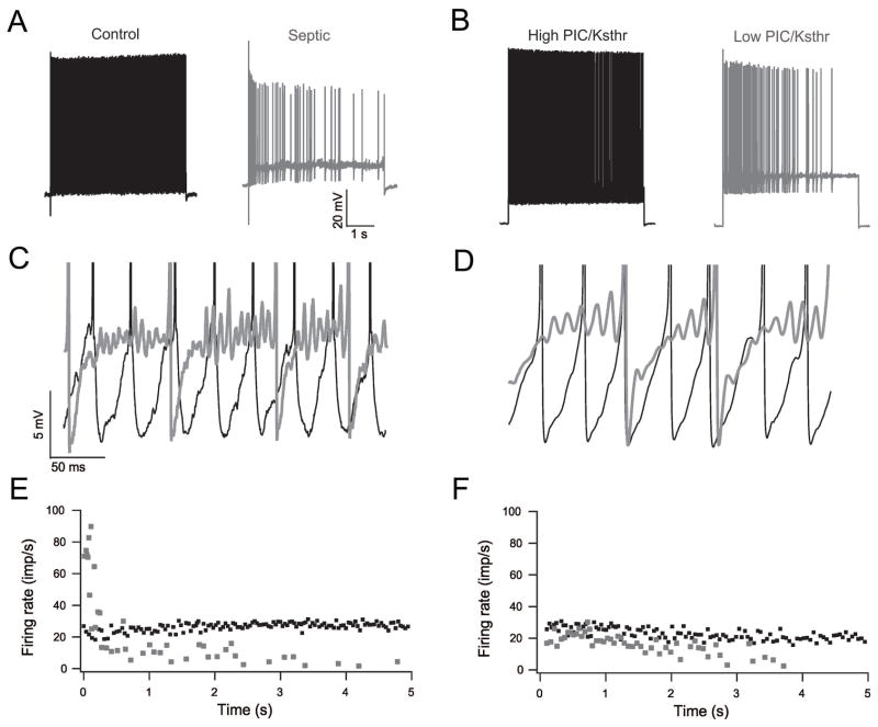
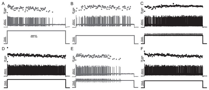
Objective: Weakness induced by critical illness (intensive care unit acquired weakness) is a major cause of disability in patients and is currently untreatable. We recently identified a defect in repetitive firing of lower motor neurons as a novel contributor to intensive care unit acquired weakness. To develop therapy for intensive care unit acquired weakness, it was necessary to determine the mechanism underlying the defect in repetitive firing.

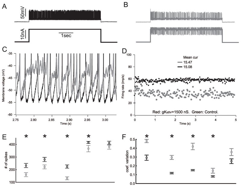
Methods: Both computer simulation and in vivo dynamic voltage clamp of spinal motor neurons in septic rats were employed to explore potential mechanisms underlying defective repetitive firing.

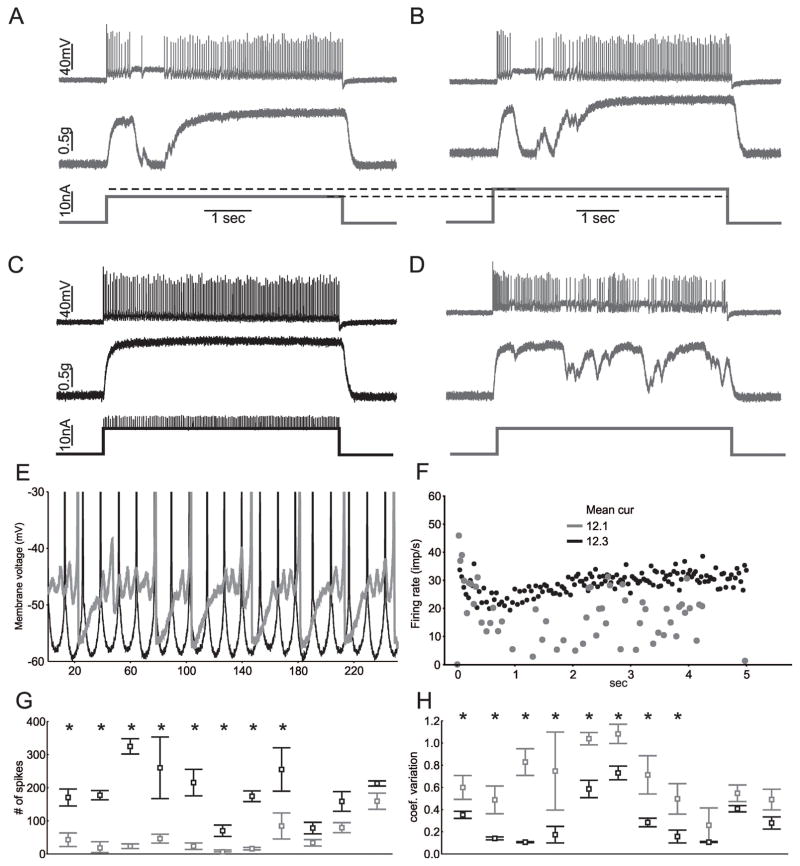
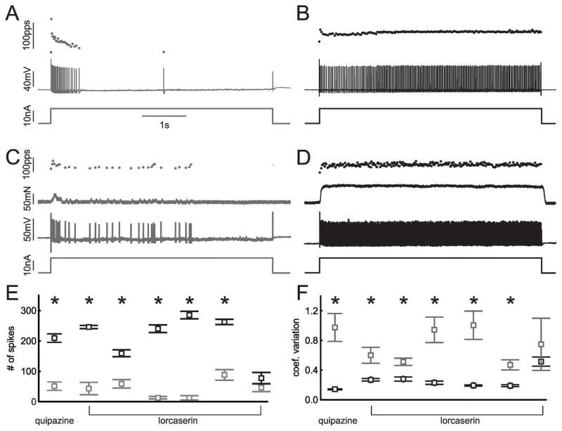
Results: Our results suggest alteration in subthreshold voltage-activated currents might be the mechanism underlying defective repetitive firing. It has been shown previously that pharmacologic activation of serotonin receptors on motor neurons increases motor neuron excitability, in part by enhancing subthreshold voltage-activated inward currents. Administration of a U.S. Food and Drug Administration-approved serotonin agonist (lorcaserin) to septic rats greatly improved repetitive firing and motor unit force generation.

Interpretation: Our findings suggest activation of serotonin receptors with lorcaserin may provide the first ever therapy for intensive care unit acquired weakness in patients. Ann Neurol 2017;82:961-971.


Conflict of interest statement

Potential Conflicts of Interest

The authors declare no potential conflicts of interest.

Figures

Figure 1
Figure 1
Reducing the PIC/Ksthr ratio in a motor neuron model reproduces sepsis-induced oscillations, reduced firing rate and increased variability of firing rate. A) Shown are comparison of action potential traces of control (black) and septic (gray) motor neurons. Firing of the septic motor neuron is slow and irregular. B) Action potential traces from modeled motor neuron with high PIC/Ksthr ratio (black) and low PIC/Ksthr ratio (gray). When PIC/Ksthr ratio is low, firing of the motor neuron is slow and irregular. C) Superimposed blow-ups focusing on the voltage range near action potential threshold for the real control (black) and septic (gray) traces from the trials shown in A. In the trace of the motor neuron from the control rat there are no subthreshold oscillations in membrane potential such that firing is rapid and regular. In the trace from the septic rat there are subthreshold oscillations in membrane potential such that firing is irregular. D) Superimposed blow-ups focusing on the voltage range near action potential threshold for the modeled motor neurons. In the trace of the modeled motor neuron with high PIC/Ksthr ratio (black) firing is rapid and regular. In the trace from the modeled motor neuron with low PIC/Ksthr ratio (gray) subthreshold oscillations are present. E) Plot of the instantaneous firing rate during the trials of the real motor neurons shown in A. The control motor neuron fires more rapidly and consistently at a similar level of current injection above rheobase current. F) Plot of the instantaneous firing rate during the trials of the modeled motor neurons shown in B. When the PIC/Ksthr ratio is reduced the mean firing rate is reduced and firing rate is more variable at the same level of current injection above rheobase current.
Figure 2
Figure 2
Decreasing the PIC/Ksthr ratio in motor neurons from untreated rats causes defects in firing similar to those triggered by sepsis. A) The action potential (top) and current injection (bottom) traces recorded intracellularly from a motor neuron in an untreated rat. B) Traces from the same motor neuron using dynamic clamp to reduce the PIC/Ksthr ratio. Instead of firing continuously at a high rate, as shown in A, the motor neuron firing rate is irregular with brief pauses. C) Superimposed blow-ups focusing on the voltage range near action potential threshold for the traces from the trials shown in A (black) and B (gray). When the PIC/Ksthr ratio is normal (no dynamic clamp) there are no subthreshold oscillations in membrane potential such that firing is rapid and regular. When the ratio is reduced, there are subthreshold oscillations in membrane potential (gray) such that firing is irregular. D) Plot of the instantaneous firing rate during the trials shown in A and B. When the PIC/Ksthr ratio is reduced the mean firing rate is reduced from near 60 to below 40 and the instantaneous firing rate is more variable despite the mean current injection being higher than when PIC/Ksthr ratio is normal. E) Plot of the number of spikes fired during the 5s current injection for 5 motor neurons before (black) and after (gray) reduction of PIC/Ksthr ratio by dynamic clamp. Multiple trials were run with and without dynamic clamp for each motor neuron and the mean data plotted. In all cases the mean current injection was matched for the comparison of dynamic clamp to standard current injection. F) Plot of the coefficient of variation of firing rate before and after reduction of PIC/Ksthr ratio by dynamic clamp. In E and F, * indicates a statistically significant difference (p < .05) for the motor neuron studied.
Figure 3
Figure 3
Increasing the PIC/Ksthr ratio is sufficient to improve firing and force production of motor units in septic rats. A) The action potentials, motor unit force and current injection traces for a single motor neuron during a 5s injection of current in vivo in a rat that had been septic for two days. There is an abnormal pause in firing of the motor neuron that causes motor unit force to fall to zero. B) Increasing current injection into the motor neuron does not improve firing. C) Dynamic clamp was used to add PIC and this normalizes firing and force production. Mean current injection was similar in B and C (12.1 vs 12.3 nA). D) Following the trial in C, a square current pulse was injected and again there are pauses in firing that cause muscle force to drop. E) Superimposed blow-ups focusing on the voltage range near action potential threshold for the trials shown in B (gray) and C (black). When the PIC/Ksthr ratio is increased with dynamic clamp, oscillations are eliminated and firing becomes fast and regular. F) Plot of the instantaneous firing rate during the trials shown in C and D. When the PIC/Ksthr ratio is increased the mean firing rate is increased and less variable. G) Plot of the number of spikes fired during the 5s current injection for 11 motor neurons before (gray) and after (black) increase of PIC/Ksthr ratio. Multiple trials were run for each motor neuron with the same current injection and the mean data plotted. In all cases the mean current injection was matched for the comparison of dynamic clamp to standard current injection. H) Plot of the coefficient of variation of firing rate before and after increasing the PIC/Ksthr ratio by dynamic clamp. In G and H, * indicates a statistically significant difference for the motor neuron studied.
Figure 4
Figure 4
Activation of 5HT receptors improve firing of motor neurons in septic rats in vivo. A) Motor neuron firing in a rat two days after induction of sepsis. B) Firing of the same motor neuron to the same current injection 15 minutes after injection of 10 mg/kg quipazine. C and D are records from a motor neuron from a septic rat before and 15 minutes after injection of 3 mg/kg of lorcaserin. E) Plot of the number of spikes fired during the 5s current injection for 1 motor neuron before (gray) and after (black) 10 mg/kg quipazine and 6 motor neurons before and after 3 mg/kg lorcaserin. Multiple trials were run for each motor neuron with the same current injection before and at least 15 minutes after drug injection and the mean data plotted. In all cases the mean current injection was matched for the comparison of firing before and after administration of drug. F) Plot of the coefficient of variation of firing rate before and after quipazine and lorcaserin. In E and F, * indicates a statistically significant difference for the motor neuron studied.
Figure 5
Figure 5
Lowering the PIC/Ksthr ratio in the motor neuron impaled is sufficient to reverse the improvement in motor neuron firing induced by treatment with lorcaserin. A and B) Shown are the current injection, action potentials, and instantaneous firing rates for a motor neuron during two 5s injections of high (A) and low (B) current in vivo in a rat that had been septic for two days. At both high and low levels of current injection the motor neuron fires erratically and cannot sustain firing throughout the 5s current injection. C) Dynamic clamp was used to increase the PIC/Ksthr ratio and this increases firing rate and lessens variability of firing rate throughout the 5s current injection. D) A further record from the same motor neuron 20 minutes after injection of 3 mg/kg lorcaserin. Motor neuron firing is rapid, and steady throughout the 5s injection of a square current pulse. E) Lowering the PIC/Ksthr ratio with dynamic clamp reverses the effect of lorcaserin such that firing becomes slow and the rate more variable. F) When dynamic clamp is turned off, the effect of lorcaserin treatment is again evident as firing is rapid and steady throughout the current injection.

Similar articles

Cited by

References

    1. Friedrich O, Reid MB, Van den Berghe G, et al. The Sick and the Weak: Neuropathies/Myopathies in the Critically Ill. Physiol Rev. 2015;95:1025–1109. - PMC - PubMed
    1. Stevens RD, Marshall SA, Cornblath DR, et al. A framework for diagnosing and classifying intensive care unit-acquired weakness. Critical Care Medicine. 2009;37:S299–308. - PubMed
    1. Latronico N, Bolton CF. Critical illness polyneuropathy and myopathy: a major cause of muscle weakness and paralysis. Lancet Neurol. 2011;10:931–941. - PubMed
    1. van den Berghe G, Wouters P, Weekers F, et al. Intensive insulin therapy in the critically ill patients. N Engl J Med. 2001;345:1359–1367. - PubMed
    1. Truong AD, Fan E, Brower RG, Needham DM. Bench-to-bedside review: mobilizing patients in the intensive care unit--from pathophysiology to clinical trials. Crit Care. 2009;13:216. - PMC - PubMed

MeSH terms