. 2018 Mar:193:13-30.
doi: 10.1016/j.trsl.2017.10.008. Epub 2017 Nov 21.

Development of mannose functionalized dendrimeric nanoparticles for targeted delivery to macrophages: use of this platform to modulate atherosclerosis

Affiliations

Development of mannose functionalized dendrimeric nanoparticles for targeted delivery to macrophages: use of this platform to modulate atherosclerosis

Hongliang He et al. Transl Res. 2018 Mar.

Abstract

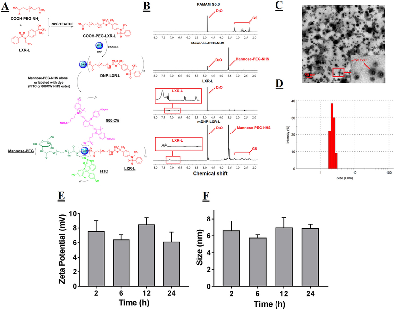
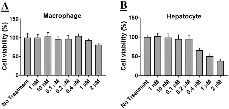
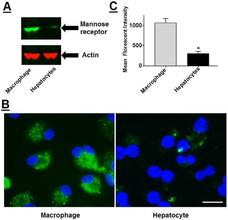
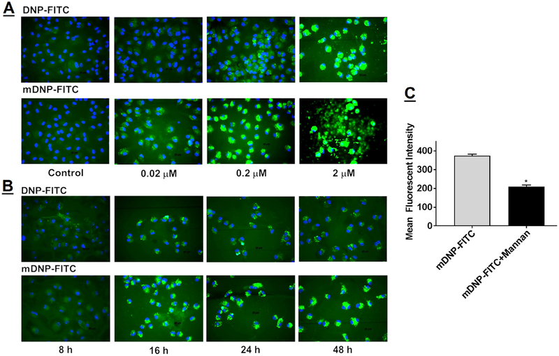
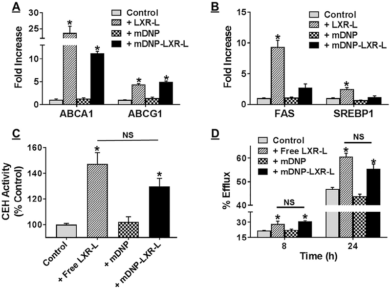
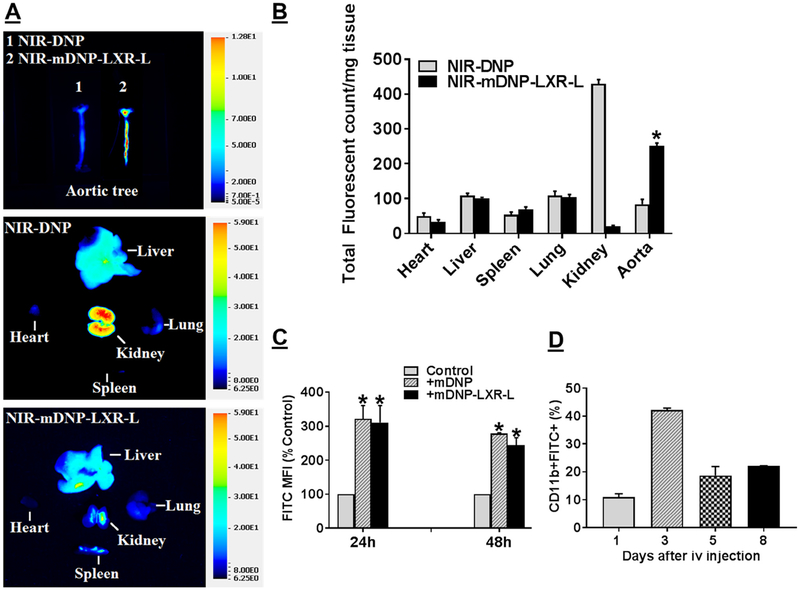
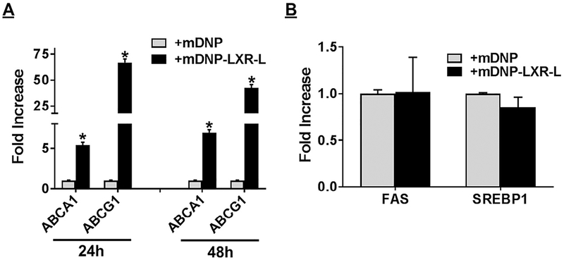
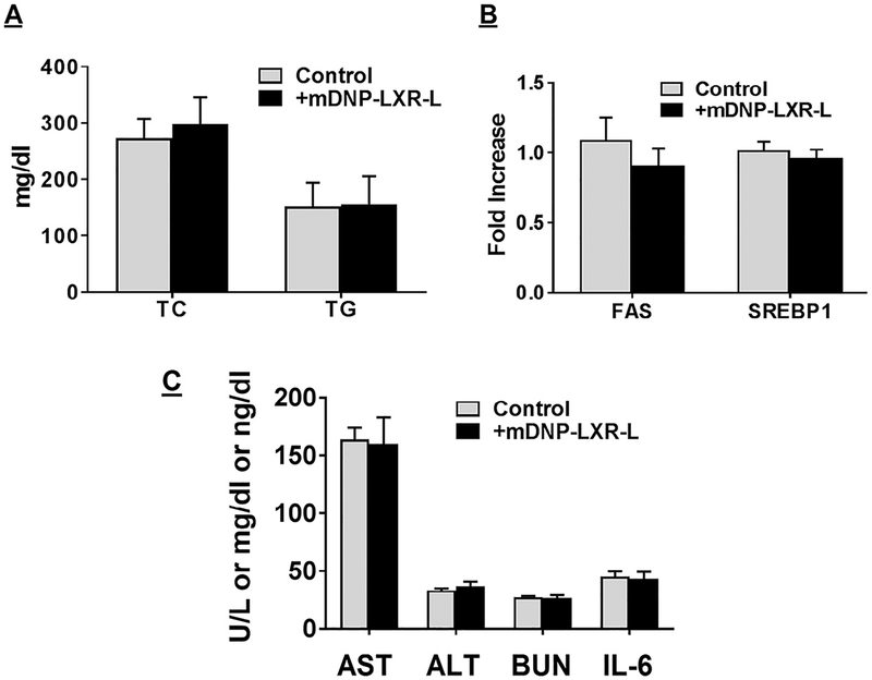
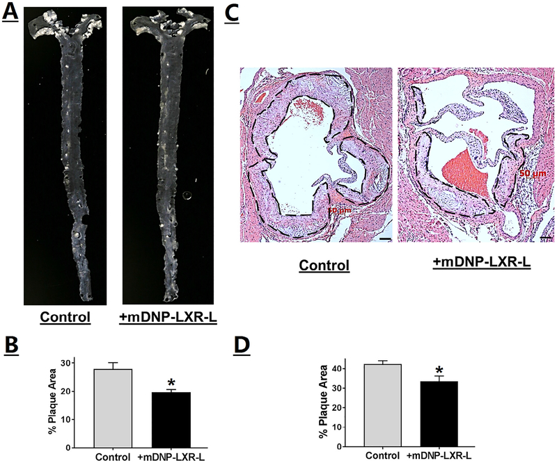
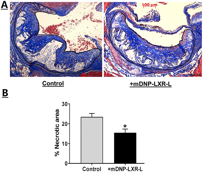
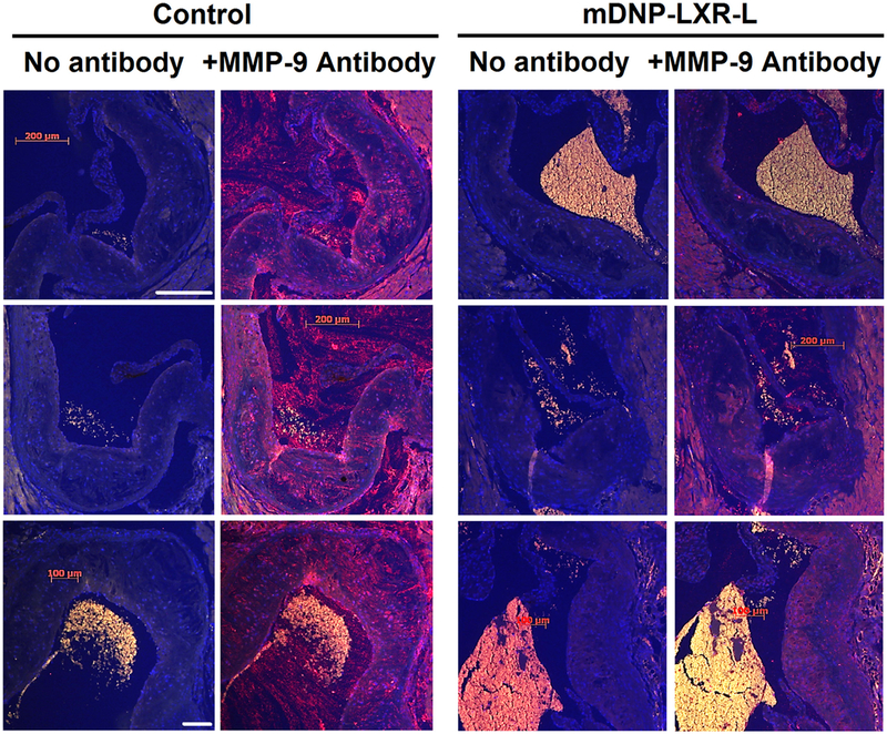
Dysfunctional macrophages underlie the development of several diseases including atherosclerosis where accumulation of cholesteryl esters and persistent inflammation are 2 of the critical macrophage processes that regulate the progression as well as stability of atherosclerotic plaques. Ligand-dependent activation of liver-x-receptor (LXR) not only enhances mobilization of stored cholesteryl ester but also exerts anti-inflammatory effects mediated via trans-repression of proinflammatory transcription factor nuclear factor kappa B. However, increased hepatic lipogenesis by systemic administration of LXR ligands (LXR-L) has precluded their therapeutic use. The objective of the present study was to devise a strategy to selectively deliver LXR-L to atherosclerotic plaque-associated macrophages while limiting hepatic uptake. Mannose-functionalized dendrimeric nanoparticles (mDNP) were synthesized to facilitate active uptake via the mannose receptor expressed exclusively by macrophages using polyamidoamine dendrimer. Terminal amine groups were used to conjugate mannose and LXR-L T091317 via polyethylene glycol spacers. mDNP-LXR-L was effectively taken up by macrophages (and not by hepatocytes), increased expression of LXR target genes (ABCA1/ABCG1), and enhanced cholesterol efflux. When administered intravenously to LDLR-/- mice with established plaques, significant accumulation of fluorescently labeled mDNP-LXR-L was seen in atherosclerotic plaque-associated macrophages. Four weekly injections of mDNP-LXR-L led to significant reduction in atherosclerotic plaque progression, plaque necrosis, and plaque inflammation as assessed by expression of nuclear factor kappa B target gene matrix metalloproteinase 9; no increase in hepatic lipogenic genes or plasma lipids was observed. These studies validate the development of a macrophage-specific delivery platform for the delivery of anti-atherosclerotic agents directly to the plaque-associated macrophages to attenuate plaque burden.


Figures

Fig 1.
Fig 1.
Synthesis and characterization of mDNP-LXR-L. (A) Schematic to show the steps involved in the synthesis. LXR-L was PEGylated and then coupled with DNP G5.0 to form DNP-LXR-L. Mannose-PEG-NHS was conjugated with DNP-LXR-L to get mDNP-LXR-L. To facilitate in vitro and in vivo imaging, mDNP-LXR-L was labeled with either FITC or 800CW. (B) The 300 MHz 1H NMR spectrum of pure PAMAM G5, mannose-PEG-NHS, LXR-L, and mDNP-LXR-L. In the spectrum of mDNP-LXR-L, multiple protons peaks between 2.2 and 3.4 ppm belong to protons from PAMAM G5; a singlet peak at 3.6 ppm belongs to the repeat units of PEG in mannose-PEG-NHS and peaks in the red rectangle stand for the LXR-L. (C) Morphology of mDNP-LXR-L visualized by TEM. (D) Hydrodynamic size of mDNP-LXR-L measured by DLS. (E) Changes on zeta potential of mDNP-LXR-L in colloidal state over 24 hours. (F) Changes on size of mDNP-LXR-L in colloidal state over 24 hours. DLS, Differential light scattering; FITC, fluorescein isothiocyanate; LXR-L, liver-x-receptor ligand; mDNP, mannose-functionalized dendrimeric nanoparticles; NHS, N-hydroxysuccinimide; NMR, Nuclear Magnetic Resonance; PAMAM, polyamidoamine dendrimer; TEM, Transmission Electron Microscopy.
Fig 2.
Fig 2.
mDNP-LXR-L does not affect cell viability: (A) Freshly isolated MPMs (0.8 × 10⁶ cells/well) were plated in 48-well plates and growth medium was changed after 4 hours. After 24 hours, the growth medium was replaced with fresh medium containing increasing concentrations of mDNP-LXR-L and incubated for additional 24 hours. Cell viability was determined using WST-1 assay. (B) Freshly isolated hepatocytes (0.3 × 10⁶ cells/well) were plated in 48-well plates, and growth medium was changed after 4 hours. After 24 hours, the growth medium was replaced with fresh medium containing increasing concentrations of mDNP-LXR-L and incubated for additional 24 hours. Cell viability was determined using WST-1 assay. Data are expressed as % cell viability compared with the no treatment control (mean ± SD, n = 6). LXR L, liver-x-receptor ligand; mDNP, mannose-functionalized dendrimeric nanoparticles; MPM, mouse peritoneal macrophages; WST-1, Water soluble tetrazolium-1.
Fig 3.
Fig 3.
Specific uptake of mDNP-FITC by macrophages: (A) Total protein extracts prepared from freshly isolated MPMs as well as primary mouse hepatocytes were subjected to Western blot analyses to evaluate the expression of mannose receptor. Blots were stripped and reprobed with β-actin as the housekeeping gene. (B) MPMs as well as hepatocytes were plated in 2-well chamber slides as described under Methods section and incubated with mDNPFITC (0.2 μM) for 24 hours. Cells were washed with PBS, fixed with buffered formalin, and imaged using Carl Zeiss inverted fluorescent microscope. (C) MPMs as well as hepatocytes were plated in 6-well tissue culture dishes and incubated with mDNP-FITC (0.2 μM). After 24 hours, cells were harvested and cell-associated FITC fluorescence was determined by FACS. Data (mean ± SD, n = 6) are shown as mean fluorescent intensities. FACS, Fluorescence activated cell sorting; FITC, fluorescein isothiocyanate; mDNP, mannose-functionalized dendrimeric nanoparticles; MPM, mouse peritoneal macrophages; PBS, phosphate-buffered saline.
Fig 4.
Fig 4.
Concentration- and time-dependent increase in the uptake of mDNP-FITC by MPMs. (A) Freshly isolated MPMs were plated in 2-well chamber slides and incubated with increasing concentration of either FITC (green)-labeled DNP (DNP-FITC) or with FITC-labeled mannose functionalized DNP (mDNP-FITC) for 24 hours. (B) Freshly isolated MPMs were plated in 2-well chamber slides and incubated with 0.2 μM of either DNP-FITC or with mDNP-FITC for 8–48 hours, as indicated. Cells were washed with PBS, permeabilized, counterstained with nuclear stain DAPI (blue), and imaged as described under Methods section. Representative images from 3 independent experiments are shown. (C) MPMs were plated in 24-well plates and incubated with mDNP-FITC (0.2 μM) in the absence or presence of mannose receptor antagonist, mannan (0.1 mM). After 24 hours, the cells were harvested and cell-associated FITC fluorescence was determined by FACS. Data (mean ± SD, n = 6) are shown as mean fluorescent intensities. DAPI, 4’, 6-diamino-2-phenylindole; FACS, Fluorescence activated cell sorting; FITC, fluorescein isothiocyanate; mDNP, mannosefunctionalized dendrimeric nanoparticles; MPM, mouse peritoneal macrophages; PBS, phosphate-buffered saline.
Fig 5.
Fig 5.
mDNP-LXR-L delivered LXR ligand appropriately increases gene expression leading to increased FC efflux. MPMs (A) or primary hepatocytes (B) were plated in 6-well culture dishes and incubated with either LXR ligand T0901317 or mDNP or mDNP-LXR-L (0.2 ^CM) for 24 hours; untreated cells were used as controls. Expression of LXR target genes (ABCA1 and ABCG1 in MPMs and FAS and SREBP1 in hepatocytes) was evaluated by QPCR using total RNA as described under Methods section. Data (mean ± SD, n = 3) are expressed as fold increase over controls. (C) MPMs e plated in 6-well plates were exposed to either LXR ligand T0901317 or mDNP or mDNP-LXR-L (0.2 μM) for 24 hours. Cells were washed once with cold PBS and harvested in cold buffer on ice as described under Methods section. Following disruption of cells by sonication and removal of cell debris, CEH activity was measured using a radiometric assay. CEH activity (mean ± SD, n = 3) is expressed as % untreated controls. (D) MPMs, plated in 24-well plate, were loaded and labeled with [3H]-cholesterol as described under Methods section. During the 24 hours equilibration, cells were exposed to either LXR ligand T0901317 or mDNP or mDNP-LXR-L (0.2 μM). FC efflux to medium containing 10% FBS was monitored for 8 and 24 hours. Data (mean ± SD, n = 6) are expressed as % efflux. *P < 0.05 and NS—not significant. CEH, cholesteryl esters hydrolase; FBS, fetal bovine serum; LXRL, liver-x-receptor ligand; mDNP, mannose-functionalized dendrimeric nanoparticles; MPM, mouse peritoneal macrophages; PBS, phosphate-buffered saline; QPCR, quantitative or real-time polymerase chain reaction.
Fig 6.
Fig 6.
In vivo uptake of mDNP by tissues and arterial plaque-associated macrophages: LDLR−/− mice (both sexes) with established atherosclerotic plaques were injected (i.v.) with either NIR-DNP or NIR-mDNP-LXR-L in sterile PBS. (A and B) After 24 hours, mice were euthanized, and major organs (heart, liver, spleen, lung, and kidney) as well as entire aorta from the heart to the iliary bifurcation was removed and cleaned to remove all the adventitious tissue. After opening to expose the arterial plaques, the aortas were imaged to assess uptake of fluorescent mDNP. Representative images are shown in A and quantification is shown in B. Data (mean ± SD, n = 6) are presented as total fluorescence per milligram tissue. (C) For assessing the uptake of mDNP-FITC by aortic plaque-associated macrophages, Western diet-fed LDLR−/− mice were either injected with PBS, or mDNP-FITC or mDNP-LXR-L-FITC. Control animals were injected with PBS alone. Following euthanasia after 24 or 48 hours, aortic arch from each mouse was quickly dissected and digested to obtain single cell suspension as described under Methods section. Cells were then stained for CD11b and analyzed by flow cytometry. Mean fluorescent Intensity, a measure of FITC-labeled DNP uptake is shown as % control (mean ± SD, n = 4). *P < 0.05. (D) In a separate experiment, time-dependent retention of FITC-labeled mDNP-LXLR-L by atherosclerotic plaque-associated macrophages after a single i.v. injection was monitored. Data (mean ± SD, n = 3) are shown as % of CD11b+FITC+ cells in the total cells isolated. FITC, fluorescein isothiocyanate; LXR-L, liver-x-receptor ligand; mDNP, mannose-functionalized dendrimeric nanoparticles; NIR, Near infrared.
Fig 7.
Fig 7.
Delivery of LXR ligand using mDNP-LXR-L increases expression of LXR target genes in plaque-associated macrophages but does not affect expression of lipogenic genes in the liver. Mannose functionalized DNP (mDNP) with or without conjugated LXR ligand in sterile PBS were injected via the tail vein of LDLR−/− atherosclerotic mice of both sexes. Control animals were injected with PBS alone. Following euthanasia, aortic arch and liver from each mouse was quickly dissected and total RNA was isolated. Gene expression of the indicated genes in aortic arch associated macrophages (A) and liver (B) was assessed by QPCR as described under Methods section. Data (mean ± SD, n = 3) are expressed as fold increase over controls. *P < 0.05. LXR, liver-x-receptor; mDNP, mannose-functionalized dendrimeric nanoparticles; PBS, phosphate-buffered saline; QPCR, Quantitative or real-time polymerase chain reaction.
Fig 8.
Fig 8.
Specific delivery of LXR ligand by mDNP-LXR-L does not affect plasma lipid composition and hepatic gene expression. Atherosclerotic LDLR−/− mice of both sexes were given 4 weekly i.v. injections of mDNP-LXR-L. Plasma and liver were collected at the time of euthanasia and analyzed as described under Methods section. (A) Plasma total cholesterol (TC) and triglyceride (TG) levels are shown as mg/dL (mean ± SD, n = 6). (B) Expression (relative to untreated controls) of indicated genes in liver was determined and data are presented as mean ± SD, n = 6. (C) Plasma levels of AST, ALT (U/L), BUN (mg/dL), and IL-6 (ng/dL) and data are presented as mean ± SD, n = 6. ALT, alanine transaminase; BUN, blood urea nitrogen; IL-6, Interleukin-6; LXR-L, liver-x-receptor ligand; mDNP, mannose-functionalized dendrimeric nanoparticles.
Fig 9.
Fig 9.
Specific delivery of LXR ligand by mDNP-LXR-L attenuates plaque development. LDLR−/− mice were fed a Western-type high-fat high-cholesterol diet for 12 weeks and divided into 2 experimental groups, and received weekly injections of either PBS (control) or mDNP-LXR-L (200 μg in 100 μL sterile PBS, labeled treated) via the tail vein. Mice were euthanized after 4 weeks of treatment. (A) Representative en face images of the aortas. (B) Quantification of the area occupied by the plaque in the aortic arch (white opaque areas in the images) was performed using Axiovision software and the data are presented as % plaque area (mean ± SD, n = 6, both sexes). (C) Hearts were fixed in buffered formalin, embedded in paraffin, and 5-μm sections were stained with H&E. Images were acquired using Carl Zeiss inverted microscope. Representative images of the aortic root showing the plaque development in the aortic valve leaflets (marked by black dashed lines). (D) Quantification of the plaque area. Data are expressed as % plaque area per leaflet (mean ± SD, n = 6, both genders). *P < 0.05. H&E, hematoxylineosin; LXR-L, liver-x-receptor ligand; mDNP, mannose-functionalized dendrimeric nanoparticles; PBS, phosphate-buffered saline.
Fig 10.
Fig 10.
Specific delivery of LXR ligand by mDNP-LXR-L attenuates plaque necrosis. (A) Serial sections (5 μm) of the aortic root from paraffin-embedded hearts (from mice described in Fig 9) were stained with Mason’s trichrome stain. Images were acquired using Carl Zeiss inverted microscope. Representative images of the aortic root showing the necrotic areas (white blank areas) in the aortic valve leaflets. (B) Quantification of the necrotic area. Total plaque was outlined (solid black line) and necrotic white areas were identified using Axiovision software (shown by dotted line) and % necrotic area calculated. Data are expressed as % necrotic area per plaque (mean ± SD, n = 9). *P < 0.05. LXR-L, liver-x-receptor ligand; mDNP, mannose-functionalized dendrimeric nanoparticles
Fig 11.
Fig 11.
Specific delivery of LXR ligand by mDNP-LXR-L attenuates expression of pro-inflammatory and NF-κB target gene MMP-9. Serial sections (5 μm) of the aortic root from mice described in Fig 9 (n = 6) were stained for MMP-9 as described under Methods section; specificity of MMP-9 staining was confirmed by staining the sections in the absence of MMP-9 antibody (labeled—No antibody). Images were acquired using Carl Zeiss inverted microscope under same exposure parameters. Three representative images for each condition are shown. Control—heart sections from mice with no i.v. injections; +mDNP-LXR-L—Heart sections from mice given 4 weekly injections of mDNP-LXR-L. LXR-L, liver-x-receptor ligand; mDNP, mannose-functionalized dendrimeric nanoparticles; MMP, matrix metalloproteinase; NF-κB, nuclear factor kappa B.

Similar articles

Cited by

References

    1. He H, Ghosh S, Yang H. Nanomedicines for dysfunctional macrophage-associated diseases. J Control Release 2017;247:106–26. - PMC - PubMed
    1. Martinez-Pomares L The mannose receptor. J Leukoc Biol 2012;92:1177–86. - PubMed
    1. Shen J, Hilgenbrink AR, Xia W, et al. Folate receptor-β constitutes a marker for human proinflammatory monocytes. J Leukoc Biol 2014;96:563–70. - PMC - PubMed
    1. Miyanishi M, Tada K, Koike M, et al. Identification of tim4 as a phosphatidylserine receptor. Nature 2007;450:435–9. - PubMed
    1. Park D, Tosello-Trampont A-C, Elliott MR, et al. Bai1 is an engulfment receptor for apoptotic cells upstream of the elmo/dock180/rac module. Nature 2007;450:430–4. - PubMed

Publication types